In vitro method for disassmbly/reassembly of papillomavirus virus-like particles (VLPs). Homogeneous VLP and cavsomere compositions produced by said methods: use thereof as vehicle for improved purification, and delivery of active agents
a technology of papillomavirus and vlps, which is applied in the field of high-efficiency means, can solve the problems of only slightly effective disassembly conditions and inhibit the reassembly of capsomeres into vlps, and achieve the effects of stabilizing vlps, moderately inhibiting reassembly, and equal ionic strength
- Summary
- Abstract
- Description
- Claims
- Application Information
AI Technical Summary
Benefits of technology
Problems solved by technology
Method used
Image
Examples
example 1
Quantitative Disassembly of HPV-11 VLPs
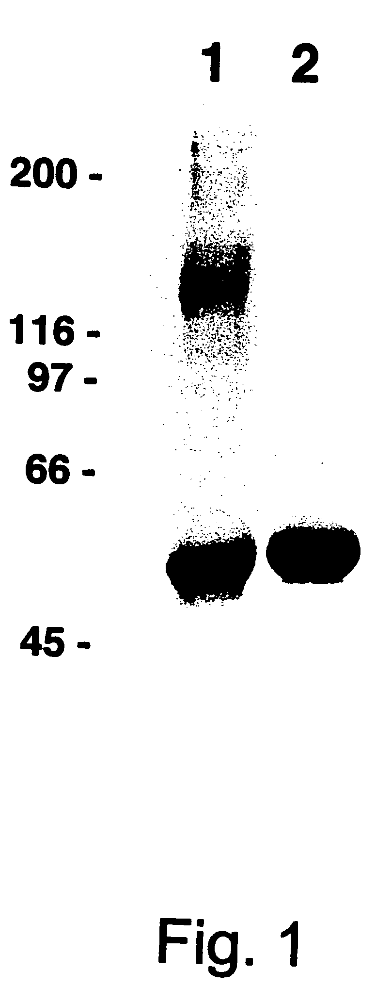
[0143]Relatively large quantities of HPV-11 L1 VLPs were prepared as starting material for the study of VLP disassembly and reassembly. HPV-11 L1 VLPs were isolated from recombinant baculovirus-infected High Five® cells by —CsCl and sucrose gradient centrifugation. The calculated purity of these L1 preparations, based on densitometric analysis of SDS / PAGE, ranged between 70-90% (see FIG. 1, lane 2). In addition, in linear sucrose gradients most of the protein migrated as expected for a mixture of individual and clumped VLPs (FIG. 4a), and in the electron microscope a mixture of intermediate and full-size (50-55 nm) particles were apparent (FIG. 5a).
[0144]The covalent and non-covalent interactions which stabilize the assembled L1 VLPs are not entirely known, but earlier work on papillomavirus VLPs and related polyomavirus virions and VLPs suggested the importance of ionic strength, divalent cations (Brady et al, I ViroL, 23:717-724 (1977); Salun...
example 2
Characterization of Disassembled HPV-11 VLPs
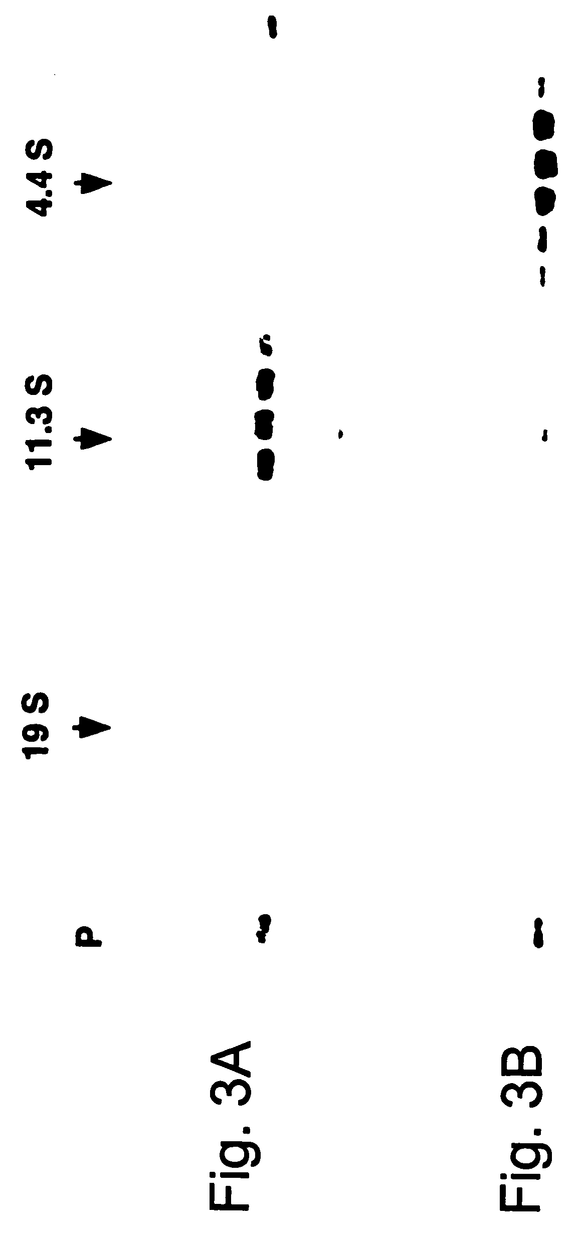
[0147]Following long-term exposure to high concentrations of reducing agent, the purified VLPs appear to be broken down to the level of capsomeres. As shown in FIG. 3a, the disassembled VLPs generated by incubation with 5% βME for 16 hours at 4° C. migrated on 5-20% linear sucrose gradients with an average sedimentation coefficient of 11.3±1.5 S (n=5), determined relative to sedimentation standards. Larger species, with a calculated sedimentation coefficient of 16-18 S (perhaps dimeric capsomeres), and even pelleted materials were occasionally observed. However, less than 10% of the L1 was detected at the top of the gradient (expected position for L1 monomer) or in the pellet (expected position for intact VLPs or aggregated capsomeres), suggesting that the purified VLP starting material was largely disassembled to the level of individual capsomeres upon prolonged reduction. This conclusion is supported by electron microscopic analysis of V...
example 3
Quantitative Reassembly of HPV-11 VLPs
[0150]VLP reassembly from HPV-11 capsomeres occurred upon removal of reducing agent, either by dialysis or column chromatography. Starting wit a homogeneous preparation of soluble capsomeres, prolonged dialysis in the absence of reducing agents consistently yielded a defined population of reassembled VLPs (FIGS. 4c and 5c,d). The reassembled VLPs retained the structural epitopes recognized by monoclonal antibodies H11.F1 and H11.A3 (FIG. 6c).
[0151]For reassembly, capsomeres (1-5 ml at 0.5-1.0 mg / ml total protein) were dialyzed versus 4×1 L PBS-0.5M NaCl at 4° C. for >24 hrs; the elevated salt concentration was designed to stabilize the VLPs. Whereas the addition of chelating agents did not appreciably enhance the ability of reducing agents to disassemble VLPs (Table 1), the presence of 2 mM EDTA moderately interfered with reassembly, yielding VLPs which migrated on a 10-65% linear sucrose gradient as a fairly discrete population of 150 S particl...
PUM
| Property | Measurement | Unit |
|---|---|---|
| diameter | aaaaa | aaaaa |
| ionic strength | aaaaa | aaaaa |
| ionic strength | aaaaa | aaaaa |
Abstract
Description
Claims
Application Information
 - R&D
- Intellectual Property
- Life Sciences
- Materials
- Tech Scout
- Unparalleled Data Quality
- Higher Quality Content
- 60% Fewer Hallucinations
Browse by: Latest US Patents, China's latest patents, Technical Efficacy Thesaurus, Application Domain, Technology Topic, Popular Technical Reports.
© 2025 PatSnap. All rights reserved.Legal|Privacy policy|Modern Slavery Act Transparency Statement|Sitemap|About US| Contact US: help@patsnap.com


