Integrated arc anode x-ray source for a computed tomography system

a computed tomography and arc anode technology, applied in tomography, material analysis using wave/particle radiation, instruments, etc., can solve problems such as the inability of current x-ray tubes to generate image artifacts affecting the system

Inactive Publication Date: 2008-04-29
GE MEDICAL SYST GLOBAL TECH CO LLC
View PDF9 Cites 21 Cited by
  • Summary
  • Abstract
  • Description
  • Claims
  • Application Information

AI Technical Summary

Benefits of technology

[0011]The present invention enables large detector volumetric imaging free from cone-beam artifacts caused through point x-ray source limitations. The present invention also addresses high speed scanning because source is not limited by anode bearing reliability as it utilizes a stationary anode. The system also provides the aforementioned scanning with 360° rotation coverage (in contrast to prior schemes, which were limited to “half scans”).

Problems solved by technology

During rotation, however, image artifacts are generated that affect the system when the cone angle reaches larger ranges.
Current x-ray tubes do not have this capability.

Method used


Image

  • Integrated arc anode x-ray source for a computed tomography system
  • Integrated arc anode x-ray source for a computed tomography system
  • Integrated arc anode x-ray source for a computed tomography system

Examples


Embodiment Construction

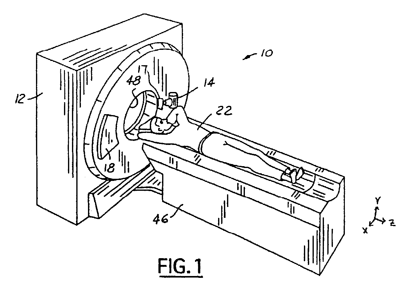
[0018]The present invention is illustrated with respect to a computed tomography (CT) scanning system 10, particularly suited to the medical field. The present invention is, however, applicable to various other uses that may require CT scanning, as will be understood by one skilled in the art.

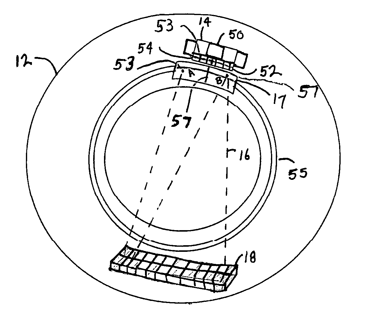
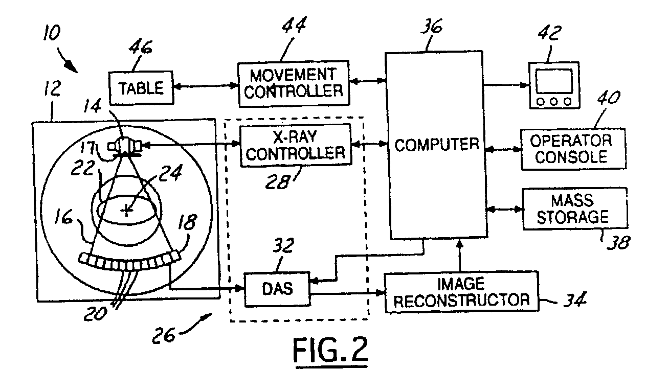
[0019]Referring to FIGS. 1 and 2, a computed tomography imaging system 10, including a gantry 12 surrounding a patient 22 on a table 46 within a patient bore 48, is illustrated. The gantry 12 has an x-ray source 14 coupled thereto that projects a beam of x-rays 16 from a stationary arc anode 17 toward a detector array 18 on the opposite side of the gantry 12.

[0020]The system10 further includes a control mechanism 26 having an x-ray controller 32 and a data acquisition system (DAS) 32. The system 10 still further includes control components, such as a movement controller 44, a host computer 36, an operator console 40, a monitor 42, an image reconstructor 34, and a mass storage 38, all of which w...


PUM

PropertyMeasurementUnit
heightaaaaaaaaaa
reflectionaaaaaaaaaa
computed tomographyaaaaaaaaaa

Abstract

An imaging system includes a gantry rotating around an object on a table. A cathode emitter is coupled to the gantry and generates an electron beam. An anode, also coupled to the gantry, is stationary with respect to the cathode emitter and generates an x-ray flux in response to the electron beam. A detector is coupled to the gantry and is adapted to receive the x-ray flux and generate therefrom a detector signal.

Description

BACKGROUND OF INVENTION[0001]The present invention relates generally to imaging systems and more particularly to volumetric computed tomography.[0002]A computed tomography or CT scan is a method of taking pictures of the inside of the body using an ultra-thin x-ray beam. As the x-ray beam passes through the body, it is absorbed by bones, tissues and fluid within the body, thereby varying resultant beam intensity. The intensity of the attenuated x-ray beam emerging from the body is measured by a device that converts x-ray beam photons into electrical signals. These signals are converted into a detailed picture.[0003]A typical CT scanner, such as a third or fourth generation CT scanner, includes a gantry having an annular frame for rotatably supporting an annular disk about a rotation or scanning axis of the scanner. The disk includes a central opening large enough to receive a patient extending along the scanning axis, and the disk is rotated about the patient during a scanning proce...

Claims


Application Information

Patent Timeline
Patent Type & Authority Patents(United States)
IPC IPC(8): G01N23/00A61B6/03G21K5/02H01J35/08H01J35/30H05G1/02H05G1/60
CPCA61B6/032A61B6/4028H01J35/30H01J2235/068
Inventor LOUNSBERRY, BRIAN D.
Owner GE MEDICAL SYST GLOBAL TECH CO LLC
Why Eureka
Try Eureka