Eureka AIR delivers breakthrough ideas for toughest innovation challenges, trusted by R&D personnel around the world.

Scatter correction for time-of-flight positron emission tomography data

a positron emission tomography and time-of-flight technology, applied in the field of nuclear medicine, can solve the problems of poor spatial resolution, lower sensitivity, and offset of the snr gain provided by the tof measurement of about 500 ps resolution

Active Publication Date: 2008-07-08
SIEMENS MEDICAL SOLUTIONS USA INC
View PDF2 Cites 28 Cited by
  • Summary
  • Abstract
  • Description
  • Claims
  • Application Information

AI Technical Summary

Benefits of technology

[0029]The present invention solves the existing need in the art by providing an accurate scatter correction method for TOF-PET, thereby permitting quantitative image reconstruction from TOF-PET data without artifacts caused by inclusion of scatter data.

Problems solved by technology

TOF scanners developed in the early 1980s were used for research and clinical applications, but the SNR gain provided by the TOF measurements of about 500 ps resolution was offset by poorer spatial resolution and lower sensitivity due to the low stopping power of the BaF2 and CsF scintillation crystals used in such scanners.
Consequently, those TOF systems could not compete successfully with conventional (non-TOF) BGO scanners.
However, it is to be expected that scatter correction techniques developed for conventional PET may not be adequate for TOF PET, particularly those model-based algorithms that simulate scatter contributions.

Method used


Image

  • Scatter correction for time-of-flight positron emission tomography data
  • Scatter correction for time-of-flight positron emission tomography data
  • Scatter correction for time-of-flight positron emission tomography data

Examples


Embodiment Construction

[0038]The present invention will now be described and disclosed in greater detail. It is to be understood, however, that the disclosed embodiments are merely exemplary of the invention and that the invention may be embodied in various and alternative forms. Therefore, specific structural and functional details disclosed herein are not to be interpreted as limiting the scope of the claims, but are merely provided as an example to teach one having ordinary skill in the art to make and use the invention.

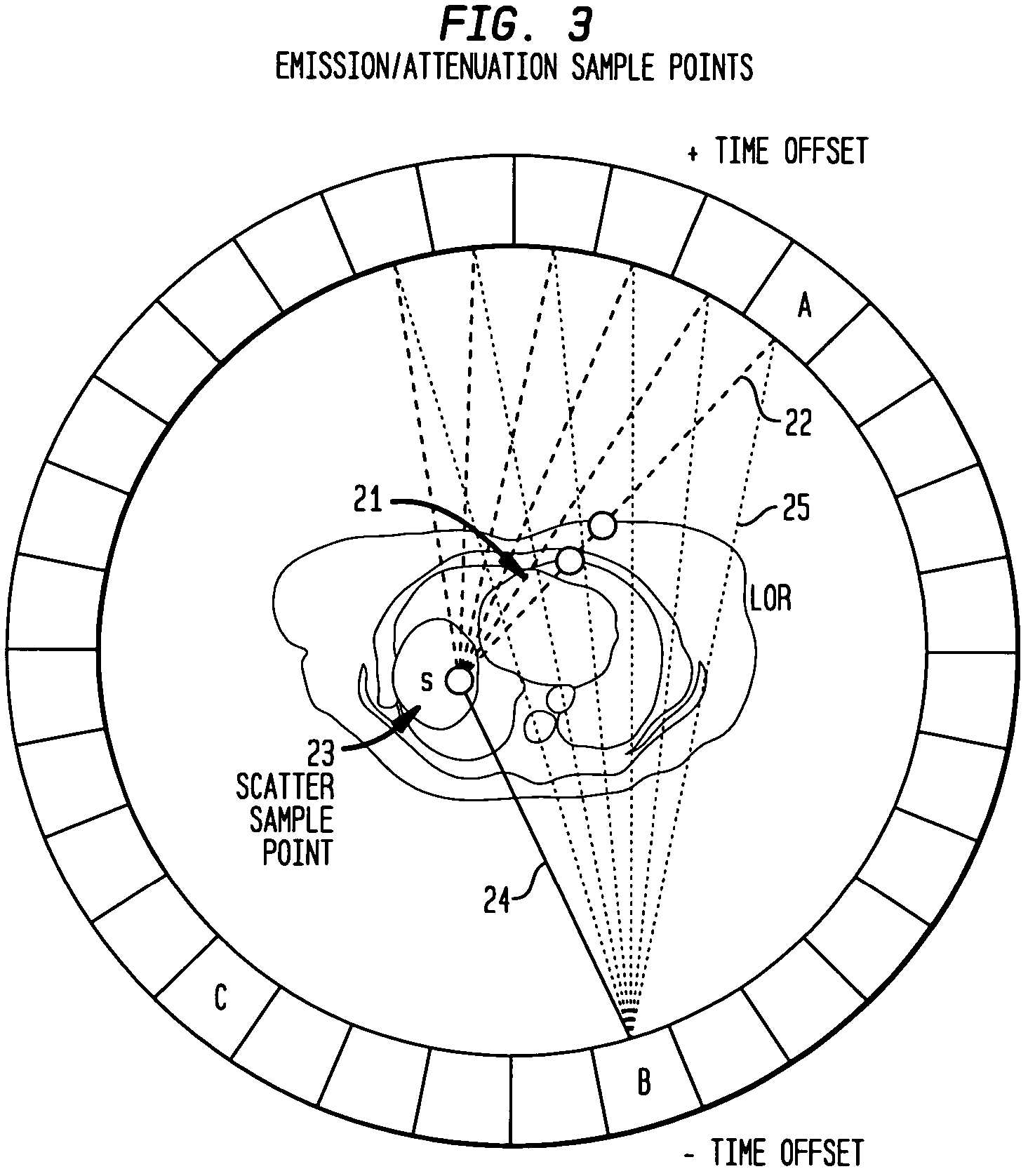
[0039]As explained above, in conventional PET, the integrals through the emission image along rays of travel from a scatter point to a detector, such as ∫SBλ(s)ds, do not depend on the LOR with which they are associated. Since each such ray is associated with many LORs, as illustrated in FIG. 3, the numerical computation of the scatter rate can be made more efficient by computing each ray integral only once, and then simply reusing this value whenever the scatter contribution to an asso...


PUM


Abstract

Correction of time-of-flight (TOF) PET data for scattered radiation explicitly models the TOF of the annihilation photon pairs along their individual scattered paths, yielding a distinct, accurate estimated scatter contribution for each time offset bin of the measured TOF data. This is accomplished by extending the single scatter simulation algorithm to include a new detection efficiency function εTOF,n.

Description

CLAIM OF PRIORITY FROM RELATED APPLICATION[0001]This application claims priority under 35 U.S.C. § 119(e) from Provisional Application Ser. No. 60 / 726,711, filed Oct. 14, 2005.FIELD OF THE INVENTION[0002]The present invention generally relates to nuclear medicine, and systems for obtaining nuclear medicine images. In particular, the present invention relates to systems and methods for reconstructing nuclear medicine images from time-of-flight (TOF) positron emission tomography (PET) data.BACKGROUND OF THE INVENTION[0003]Nuclear medicine is a unique medical specialty wherein radiation is used to acquire images which show the function and anatomy of organs, bones or tissues of the body. Radiopharmaceuticals are introduced into the body, either by injection or ingestion, and are attracted to specific organs, bones or tissues of interest. Such radiopharmaceuticals produce gamma photon emissions which emanate from the body and are captured by a scintillation crystal, with which the photo...

Claims


Application Information

Patent Timeline
Patent Type & Authority Patents(United States)
IPC IPC(8): G01T1/164
CPCA61B6/037G01T1/2985
Inventor WATSON, CHARLES C.
Owner SIEMENS MEDICAL SOLUTIONS USA INC
Why Eureka
Eureka Blog
Learn More