
Internal image antibodies for optical imaging and therapy

Inactive Publication Date: 2008-10-07
MALLINCKRODT INC
View PDF25 Cites 5 Cited by
  • Summary
  • Abstract
  • Description
  • Claims
  • Application Information

AI Technical Summary

Benefits of technology

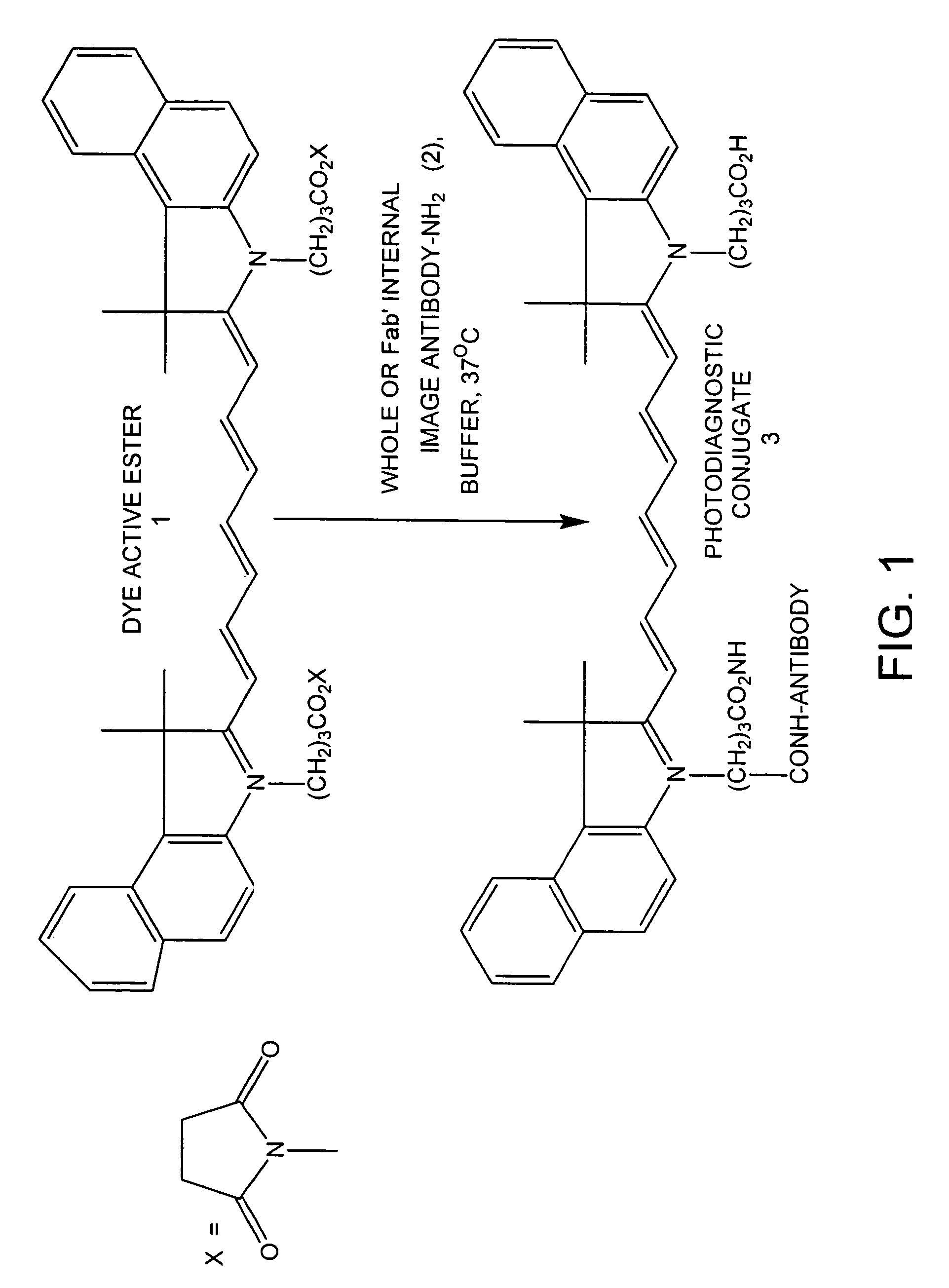
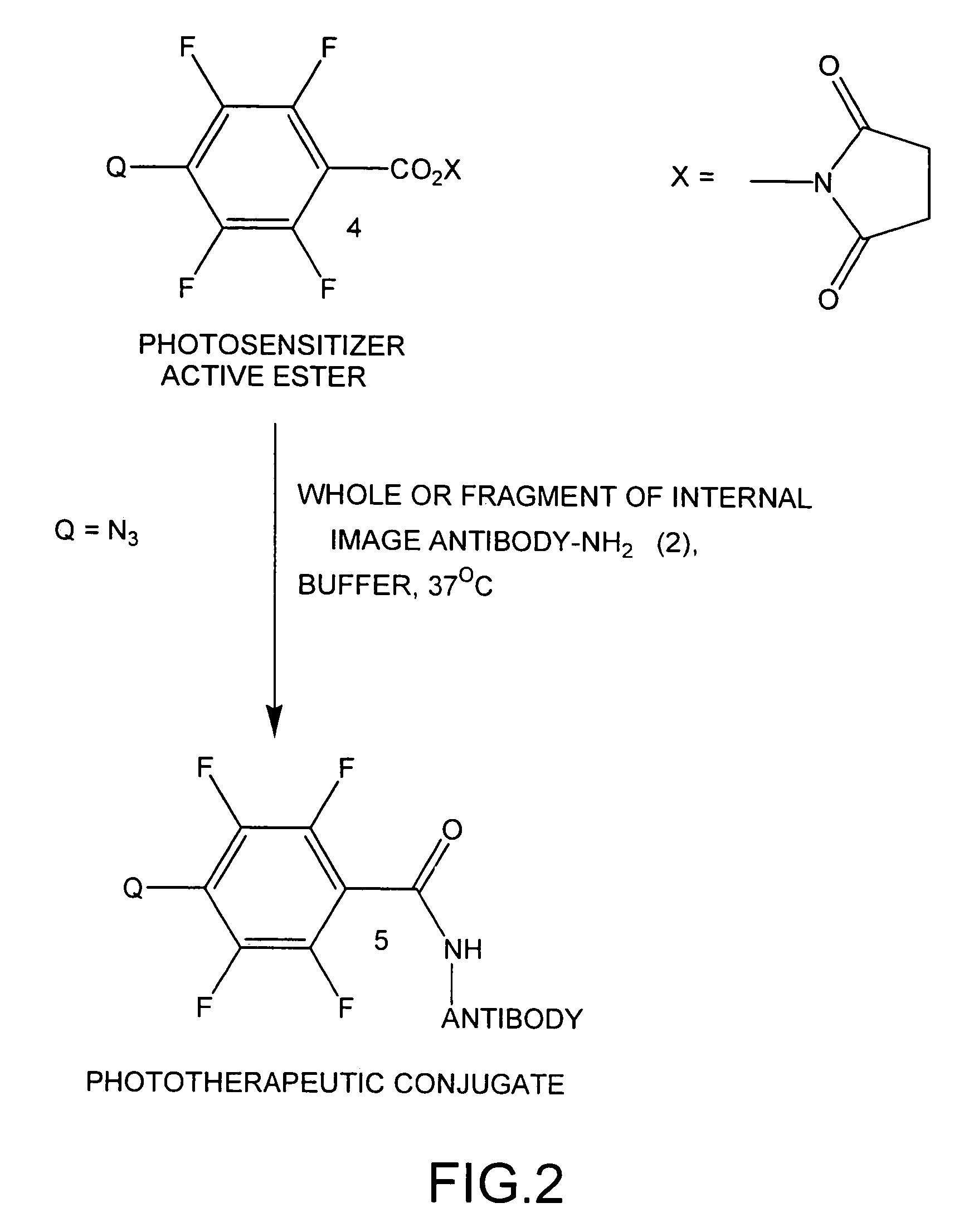
[0014]In another embodiment, the invention discloses novel bioconjugates of internal image antibodies or antibody fragments attached to one or more photoactive molecules for phototherapeutic purposes. In this embodiment, PM is a photosensitizer (PS), which either by itself or associated with a precursor (Q), produces reactive intermediates. In this embodiment, the conjugate has the general formula:
[0015]The invention also discloses a method for performing a diagnostic procedure using internal image antibody bioconjugates. In the method, a ligand is selected that binds to a biological receptor, such as the receptor for steroids, cardiac glycosides, somatostatin, bombesin, cholecystokinen, neurotensin, and heat stable toxin. The receptor-binding ligand may be one or more drugs, hormones, peptides, carbohydrates, nucleosides, peptidomimetics, glycomimetics, or biosynthetic intermediates. The ligand used as the first generation antigen may be the complete ligand, or may be a certain region of the ligand such a fragment that binds the receptor with high affinity, etc. The receptor-binding ligand is prepared as a first generation antigen, optionally conjugating the receptor-binding ligand to a

Problems solved by technology

This observation, however, is not general and it is not possible at this time to predict a priori the binding properties of small molecule bioconjugates with a high degree of confidence.
This is often not possible for various reasons, including the lack of stability of many biological receptors.
Thus, most receptor targeting with anti-receptor antibodies have been elusive.

Method used


Image

  • Internal image antibodies for optical imaging and therapy
  • Internal image antibodies for optical imaging and therapy
  • Internal image antibodies for optical imaging and therapy

Examples


example 1

Preparation of Digoxin Internal Image Antibody Fab′ Fragments and Conjugation to an Optical Diagnostic Agent.

[0046](a) Fusion of Mouse Myeloma Cells with the Spleen Cells of AJ Mice Immunized with Balb-C Mouse Anti-Digoxin Antibody.

[0047]Monoclonal antibodies were produced using standard hybridoma technology, as is known to one of ordinary skill in the art. Briefly, two AJ mice were immunized with murine (Balb-C) monoclonal anti-digoxin antibody (Medex Laboratories). A booster injection was given three weeks after the primary immunization and the spleens were removed after three days. Mouse myeloma and spleen cells were washed three times with Dulbecco's Modified Eagle Medium (DME) and suspended in DME (10 ml). A 5 mL portion of each of these cell suspensions was mixed and centrifuged. The supernatant was discarded and the pellet was treated with 1 mL polyethylene glycol (added over a 45 second period), 3 mL DME (added over a 30 second period), and an additional 9 mL of DME was adde...

example 2

Preparation of Digoxin Internal Image Antibody Fab′ Fragments and Conjugation to a Phototherapeutic Agent.

[0058]The Fab′ fragments (2) obtained in Example 1, step (c) are used for conjugation to a phototherapeutic agent. The conjugation procedure is carried out in a manner similar to the one published previously for human serum albumin (Achilefu et al., Investigative Radiology, 2000, 35, 479-485), which is expressly incorporated by reference herein in its entirety.

[0059]With reference to FIG. 2, a mixture of the Fab′ fragment (2) and about 20 fold molar excess of the photosensitizer active ester (4), is incubated in PBS for four hours. The phototherapeutic conjugate (5) is purified by Sephadex G-25 size exclusion chromatography using PBS as the eluant, and is subsequently lyophilized.

example 3

Preparation of Whole Digoxin Internal Image Antibodies and Conjugation to an Optical Diagnostic Agent.

[0060]Whole digoxin internal image antibodies obtained in Example 1, steps (a) and (b) were used for conjugation to a photodiagnostic agent. The conjugation procedure was carried out in a manner similar to the one published previously for human serum albumin (Pandurangi et al., Journal of Organic Chemistry, 1998, 63, 9019-9030).


PUM

PropertyMeasurementUnit
Dimensionless propertyaaaaaaaaaa

Abstract

Methods using internal image antibodies for photodiagnosis and / or phototherapy. The internal image antibodies are conjugated with a photoactive molecule such as a dye or photosensitizer, to target specific regions, such as biological receptors. The photoactive molecules are then activated for diagnosis or therapy. Advantageously, the internal image antibody is specific for a biological receptor, but does not require isolation of the receptor to prepare the antibody, and provides the desired specificity and selectivity for targeted diagnosis or therapy.

Description

RELATED APPLICATION[0001]This application is a Divisional of and claims priority to U.S. patent application Ser. No. 09 / 919,243 filed Jul. 31, 2001, now abandoned and expressly incorporated by reference herein in its entirety.FIELD OF THE INVENTION[0002]The invention relates generally to diagnosis and therapy using photoactive compounds. More particularly, the invention relates to bioconjugates comprising internal image antibodies or their fragments attached to photoactive molecules.BACKGROUND OF THE INVENTION[0003]The use of visible, near-infrared (NIR), and low-energy ultraviolet light in clinical practice is growing rapidly. Compounds absorbing or emitting in the visible, NIR, or long wavelength (UV-A, >350 nm) region of the electromagnetic spectrum are potentially useful for diagnostic techniques such as tomographic or planar imaging, endoscopic examination, optoacoustic imaging and sonofluorescene imaging.[0004]Notwithstanding the importance of photodiagnostic applications, ...

Claims


Application Information

Patent Timeline
IPC IPC(8): A61K39/395C07K16/42A61K41/00A61K49/00A61P7/02A61P9/10A61P35/00A61P43/00C07K16/28C12P21/08
CPCA61K49/0032A61K49/0058C07K16/4208A61K41/0071A61K2039/505A61P35/00A61P43/00A61P7/02A61P9/10
Inventor RAJAGOPALAN, RAGHAVANBUGAJ, JOSEPH E.ACHILEFU, SAMUEL I.DORSHOW, RICHARD B.
Owner MALLINCKRODT INC
Learn More