Site-specific labelling of proteins

a protein and site-specific technology, applied in the field of protein labeling methods, can solve the problems of limiting the number of colours that can be used to “tag” a protein in vivo, affecting the biological and cellular activities of the target protein, and limiting the strategy of labelling a target protein

Inactive Publication Date: 2008-11-11
NAT UNIV OF SINGAPORE
View PDF1 Cites 62 Cited by
  • Summary
  • Abstract
  • Description
  • Claims
  • Application Information

AI Technical Summary

Problems solved by technology

Although this technique may be a quite powerful method of visualizing proteins in vivo, the fusion of GFP or other fluorescent proteins with a target protein may affect the target protein's biological and cellular activities, due to the relatively large size of the fusion (i.e. 27 KDa for GFP).
Furthermore, there are currently few fluorescent proteins available, thereby limiting the number of colours that can be used to “tag” a protein in vivo.
As well, this strategy is limited to labelling a target protein only with protein molecular labels, typically fluorophores, but not other molecular probes such as small molecule probes.
The introduction of such a motif has the potential to disrupt the native folding of the target protein.
Furthermore, this method may result in background labelling, making it difficult to detect labelled target protein if expressed at low levels.
This approach also results in the introduction of a macromolecular fusion into the target protein, potentially perturbing the native function of the protein.
However, these methods require the introduction of non-natural occurring functionalities into the target biomolecule in vivo, and thus are useful for only limited applications, for example cell surface engineering and labelling of membrane proteins.
These methods may be complicated to use in that they require genetic manipulation of the host cellular expression system used to express the target protein.

Method used


Image

  • Site-specific labelling of proteins
  • Site-specific labelling of proteins
  • Site-specific labelling of proteins

Examples


example 1

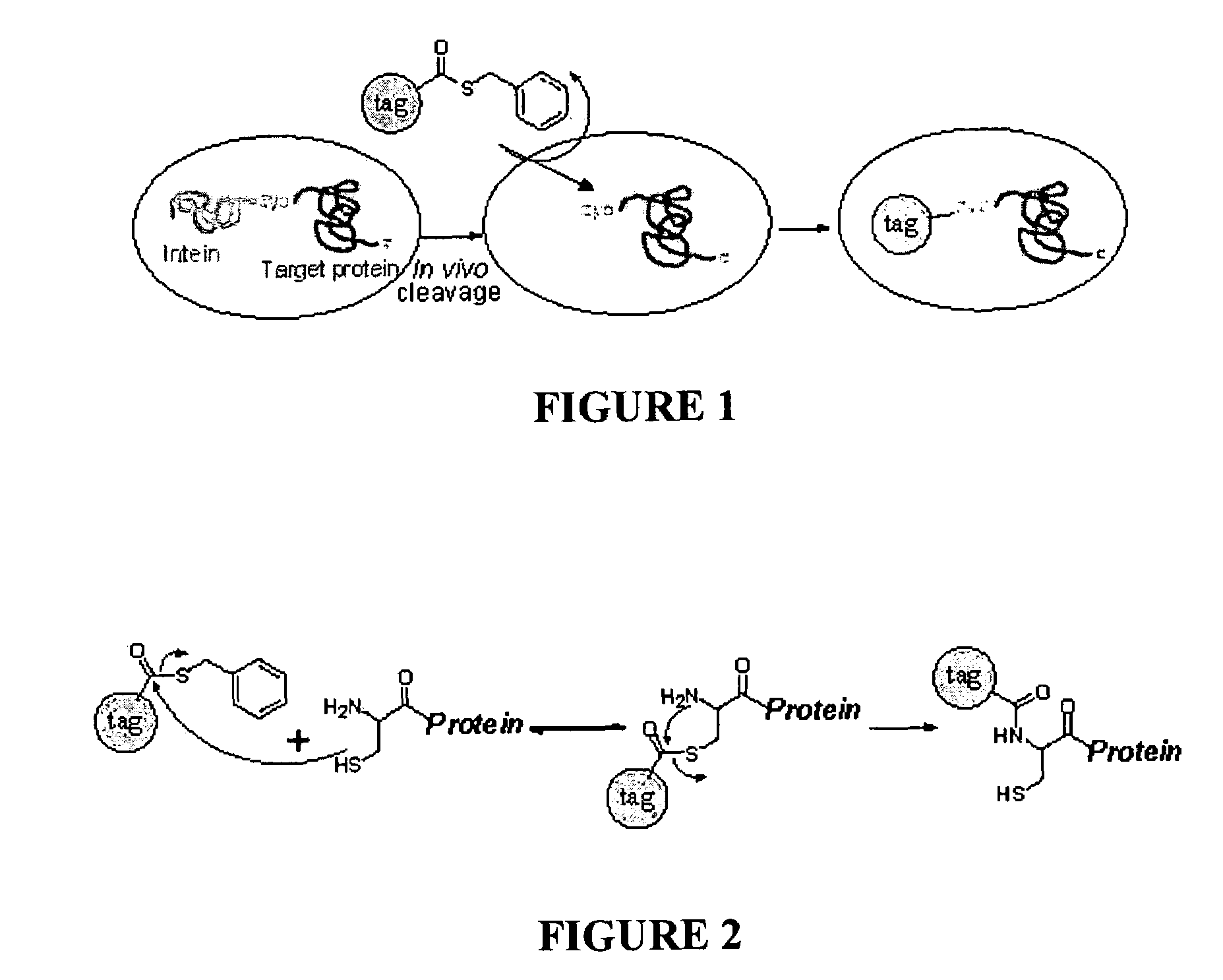
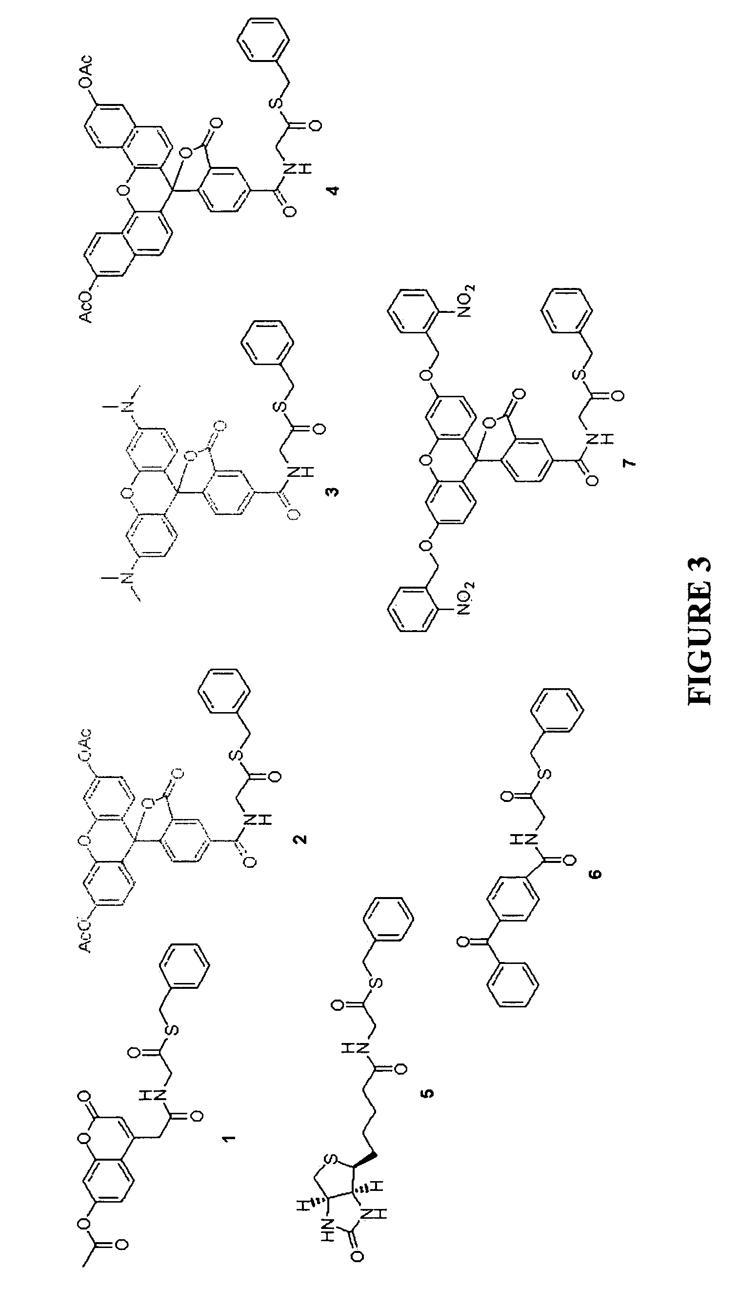
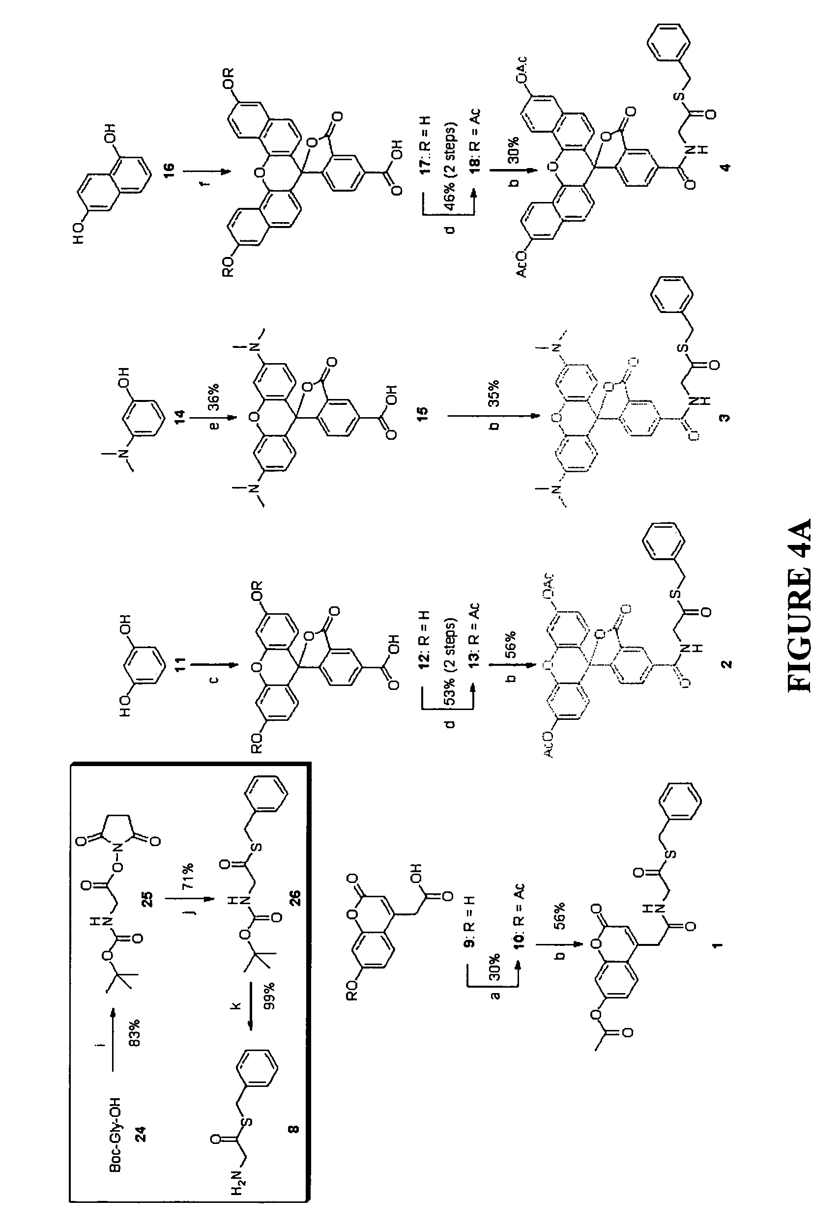
Synthesis of Membrane-Permeable Thioester Containing Probes

[0073]A total of 7 different thioester-containing probes were synthesized for in vivo protein labelling at N-terminal cysteines, the various structures of which are depicted in FIG. 3. Probes 1 to 4 are fluorophore-containing thioesters; 5 and 6 are biotin- and benzophenone-containing probes, respectively; 7 is a “caged” molecule of 2, in which the fluorescence is designed to be “turned on” selectively upon photolysis. All probes were designed to be cell-permeable: acetates of different fluorophores were incorporated in 1, 2 and 4 to increase their cell permeability. The fluorophore in 3, tetramethylrhodamine (TMR), as well as the biotin and benzophenone moieties in 5 and 6, respectively, were previously shown to be cell-permeable (R. P. Haugland, Handbook of fluorescent probes and research products, 9th Ed, Molecular Probes: Eugene, Oreg., 2002). The hydrophobic, benzyl-based thioester moiety in all 7 probes should further...

example 2

In vitro Labelling of Proteins Expressing an N-Terminal Cysteine

[0098]A model protein, EGFP (enhanced green fluorescent protein), engineered to contain an N-terminal cysteine, was incubated with probes 2, 3, 4 and 5 individually to assess the labelling strategy.

[0099]Probes were prepared as 200 μM stocks (25× in DMSO) and stored at −20° C. In a typical labelling reaction, 6 μl of each probe (final concentration: 8 μM) was added to 50 μl of pure protein (1 mg / ml) dissolved in 1×PBS (final concentration of protein: ˜2 nM), with or without 1 mM DTT, and the reaction was topped up with 1×PBS to a final volume of 150 μl. DTT was added to experiments, where live cells were used, to reduce background labelling. At specific time intervals, 15 μl of the reaction was withdrawn and quenched by addition of 1.7 μl of 100 mM cysteine to the reaction mixture (final concentration of cysteine: 10 mM), followed by denaturation with SDS-PAGE loading dye at 95° C. for 3 min. The loading dye also serve...

example 3

In vivo Labelling of N-Terminal Cysteine Proteins in Bacterial Cells

[0105]The strategy was next applied to the labelling of N-terminal cysteine proteins expressed inside live bacterial cells.

[0106]Two model proteins, EGFP (enhanced green fluorescent protein) and GST (gluthathione-S-transferase), were PCR amplified from pEGFP (Clontech, USA) and pGEX-4T1 (Pharmacia Biotech, USA), respectively, and cloned into the pTWIN1 / 2 expression vector (NEB, USA) downstream of the C-terminus the Ssp DnaB intein tag. The genes were inserted between the first SapI site and the PstI site on the vector, with a cysteine introduced by PCR as the first amino acid of the target protein, generating pTWIN1-EGFP and pTWIN2-GST, respectively. PCR primers used for pTWIN1-EGFP were 5′-GGT GGT TGC TCT TCC AAC TGC AGA GCC ATG GTG AGC AAG GGC-3′ [SEQ ID NO:1] and 5′-GGT GGT CTG CAG TTA CTT GTA CAG CTC GTC-3′ [SEQ ID NO:2]. PCR primers used for pTWIN2-GST were 5′-GGT GGT TGC TCT TCC AAC TGC AGA GCC ATG TCC CCT ATA...


PUM

PropertyMeasurementUnit
timeaaaaaaaaaa
volumeaaaaaaaaaa
concentrationaaaaaaaaaa

Abstract

An in vivo method for labelling a protein having an N-terminal cysteine is disclosed. A probe comprising a thioester group and a detectable label is introduced into a cell expressing the N-terminal cysteine protein, allowing for the N-terminal cysteine to cleave the thioester bond and resulting in the label being covalently attached to the N-terminus of the protein by an amide bond.

Description

CROSS-REFERENCE TO RELATED APPLICATION[0001]The present invention claims priority from U.S. provisional patent application No. 60 / 515,418, filed on Oct. 30, 2003, which is hereby fully incorporated herein by reference.FIELD OF THE INVENTION[0002]The present invention relates generally to methods for labelling proteins.BACKGROUND OF THE INVENTION[0003]Studying the dynamic movement and interactions of proteins inside living cells is critical for a better understanding of cellular mechanisms and functions. Traditionally this has been done by in vitro labelling of proteins with fluorescent and other molecular probes, followed by transfer of the labelled protein into a live cell for real time monitoring, using advanced imaging techniques including confocal microscopy (G. T. Hermanson, Bioconjugate Techniques, Academic Press, San Diego, Calif., 1996).[0004]Recent advances in genetic engineering have made it possible to directly generate fluorescently labelled proteins in living cells, or ...

Claims


Application Information

Patent Timeline
Patent Type & Authority Patents(United States)
IPC IPC(8): C07K1/13C07K14/47G01N33/53G01N33/58
CPCG01N33/58
Inventor YAO, SHAO QINYEO, SU-YIN DAWN
Owner NAT UNIV OF SINGAPORE
Why Eureka
Try Eureka