Reflectometry instrument and method for measuring macular pigment

a technology of reflectometry and macular pigment, which is applied in the field of reflectometry instruments, can solve the problems of affecting vision, affecting vision, and affecting the ability of people with macular degeneration to read, recognize faces, and drive safely on unfamiliar surfaces, and achieves the effects of impairing or damaging eyesight and vision, and only providing coarse resolution to surrounding parts of the macula

Active Publication Date: 2008-12-23
ZEAVISION LLC
View PDF14 Cites 33 Cited by
  • Summary
  • Abstract
  • Description
  • Claims
  • Application Information

AI Technical Summary

Benefits of technology

[0026]According to one aspect of the present invention, a reflectometry instrument is provided to measure the macular pigment of a macula of a human eye. The reflectometry instrument includes a light source, a spectrometer, a first lens, and a second lens. The light source emits an illumination beam in the direction toward the macula. The spectrometer measures a detection beam where the detection beam is a portion of the illumination beam reflected from the macula and is indicative of the amount of macular pigment in the macula. The first lens, which includes an anti-reflection coating, is adapted to transmit the illumination beam to the macula and also transmits the detection beam from the macula to the spectrometer. A second lens is adapted to transmit the illumination beam to the macula and also is adapted to transmit the detection beam from the macula to the spectrometer. The second lens is disposed adjacent to the first lens and includes an anti-reflection coating. The illumination beam and the detection beam remain separated when the illumination beam and the detection beam pass through the first lens and the second lens. The anti-reflection coating and beam separation helps to minimize the leaking of backscattered light from the illumination beam into the detection beam.
[0027]According to another aspect of the present invention, a reflectometry instrument is provided to measure the macular pigment of a macula of a human eye. The reflectometry instrument includes a light source, a spectrometer, a first lens, and a second lens. The light source is adapted to emit an illumination beam in a direction toward the macula. The spectrometer measures a detection beam where the detection beam is a portion of the illumination beam reflected from the macula and is indicative of the amount of macular pigment in the macula. The first lens is adapted to transmit the illumination beam to the macula at a location offset from the center of the first lens. The first lens is also adapted to transmit the detection beam from the macula to the spectrometer at another location offset from the center of the lens. The second lens, disposed adjacent to the first lens, is adapted to transmit the illumination beam to the macula at a location offset from the center of the second lens. The second lens is further adapted to transmit the detection beam from the macula to the spectrometer at another location offset from the center of the second lens. The illumination beam and the detection beam are spatially separated when the illumination beam and the detection beam pass through the first lens and the second lens. The offset from the central axes of the lenses and the beam separation helps to minimize the leaking of backscattered light from the illumination beam into the detection beam.
[0028]According to yet another aspect of the present invention, a method of determining the amount of macular pigment in the macula of a human eye is disclosed. The method includes the act of passing an illumination beam through a lens system having a first lens and a second lens. The illumination beam passes through the first lens and the second lens offset from the centers of the first lens and second lens. In response to passing through the lens system, the method further includes directing the illumination beam onto the macula so as to produce a detection beam exiting from the eye. The method additionally includes the act of passing the detection beam through the lens system offset from the centers of the first lens and second lens. The detection beam and the illumination beam avoid the central regions at the first lens and the second lens. The specular reflections of the illumination beam are minimized in the detection beam. The method further includes receiving the detection beam at a spectrometer and measuring the characteristics of the detection beam. The characteristics may include the total macular pigment amount, the macular pigment amounts of zeaxanthin and lutein, and / or the amounts of pigment in the patient's lens.

Problems solved by technology

People who suffer from macular degeneration often lose the ability to read, recognize faces, drive, or walk safely on unfamiliar routes.
The surrounding portions of the macula can only provide coarse resolution.
This process occurs so rapidly that a person does not notice it happening, and does not pay attention to how a complete visual image and impression is being assembled and updated from combinations of fine and coarse resolution images.
It includes any progressive disorder or disease that causes the macula to gradually degenerate, to a point that substantially impairs or damages eyesight and vision.
In addition to their involvement in the macula and macular degeneration, zeaxanthin and lutein also are present in other eye structures (including the eye lens), and undesirably low levels of those two carotenoids appear to be correlated with higher risks of disorders such as cataracts.
However, that single minor structural difference, between zeaxanthin versus lutein, has profound effects on the traits, performance, and tissue concentrations of those two different molecules, in both plants and animals.
Since it is relatively straight, it cannot fit properly into the circular light-harvesting structures that help carry out photosynthesis, in plants.
Oxidative radicals are important damaging agents in any cells and tissues that are being bombarded by high levels of UV radiation, since UV radiation often breaks bonds that involve oxygen atoms, in ways that create unpaired electrons where the broken bonds previously existed.
In addition, it is not known whether lutein is consistently deposited, into human cell membranes, in a membrane-spanning orientation.
However, the conversion process cannot convert lutein into the normal stereoisomer of zeaxanthin found in plants and in the diet (the 3R,3′R stereoisomer).
For example, many instruments require the eye to be dilated before use, which can be uncomfortable to the patient and add extra time and cost to the procedure.

Method used


Image

  • Reflectometry instrument and method for measuring macular pigment
  • Reflectometry instrument and method for measuring macular pigment
  • Reflectometry instrument and method for measuring macular pigment

Examples


Embodiment Construction

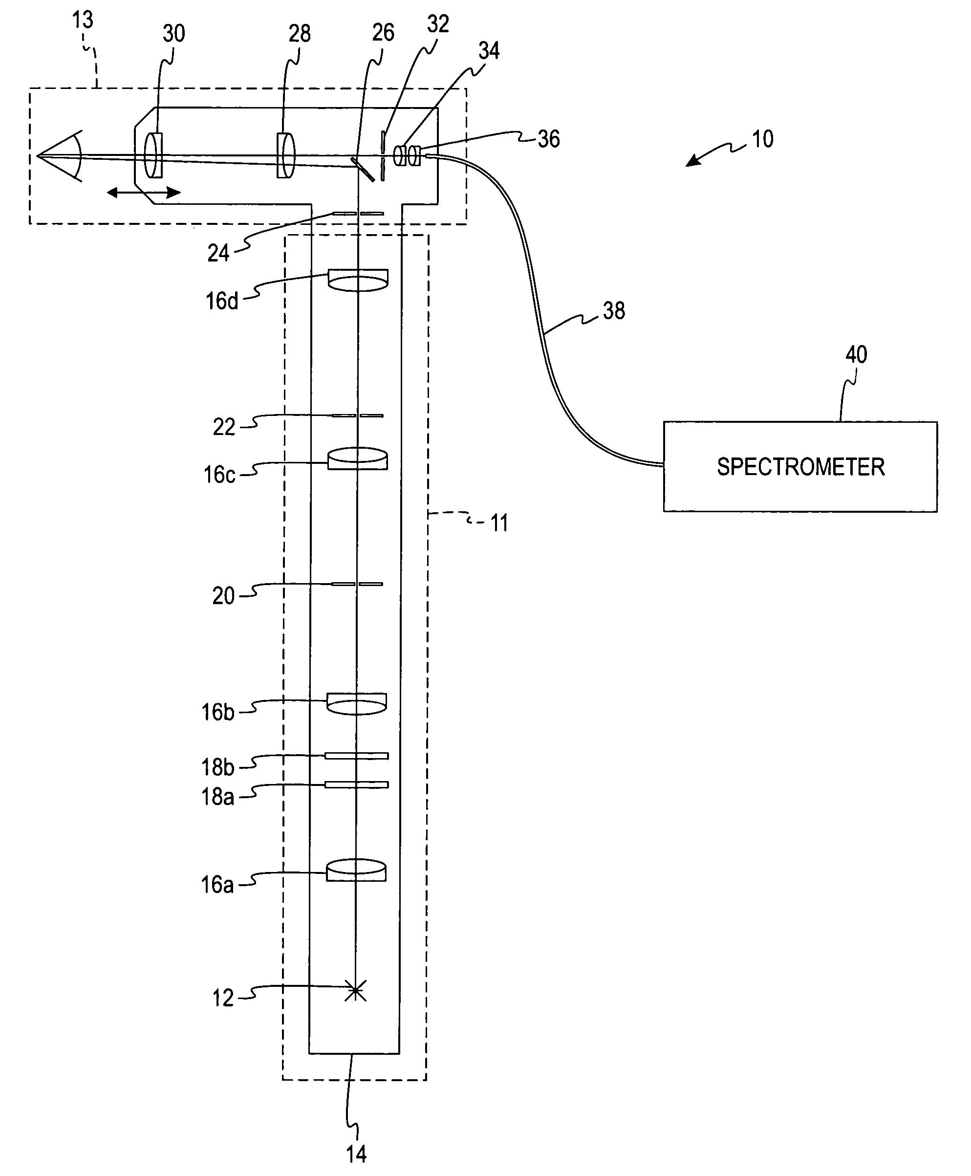
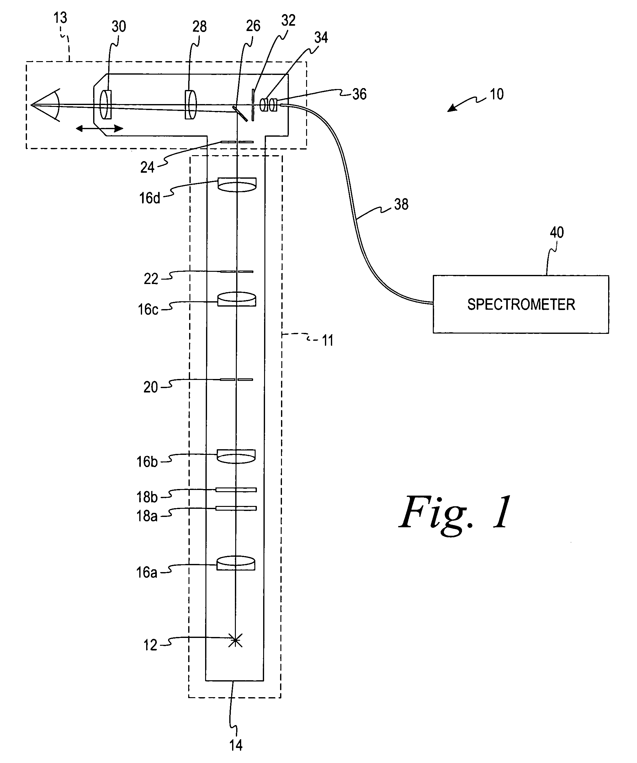
[0040]FIG. 1 illustrates a reflectometry instrument 10 adapted to measure the characteristics of a human eye. The reflectometry instrument 10 will be described in reference to two main portions—a source system 11 and a beam separation system 13. The source system 11 includes a light source 12, a plurality of source lenses 16, filters 18, a filament mask 20, and a retinal stop 22. The source system 11 generates an illumination beam having certain characteristics that will be transmitted to the patient's eye. As mentioned above, the reflectometry instrument 10 may be used on an undilated pupil, making the system much easier to use and decreasing the time required to test a patient.

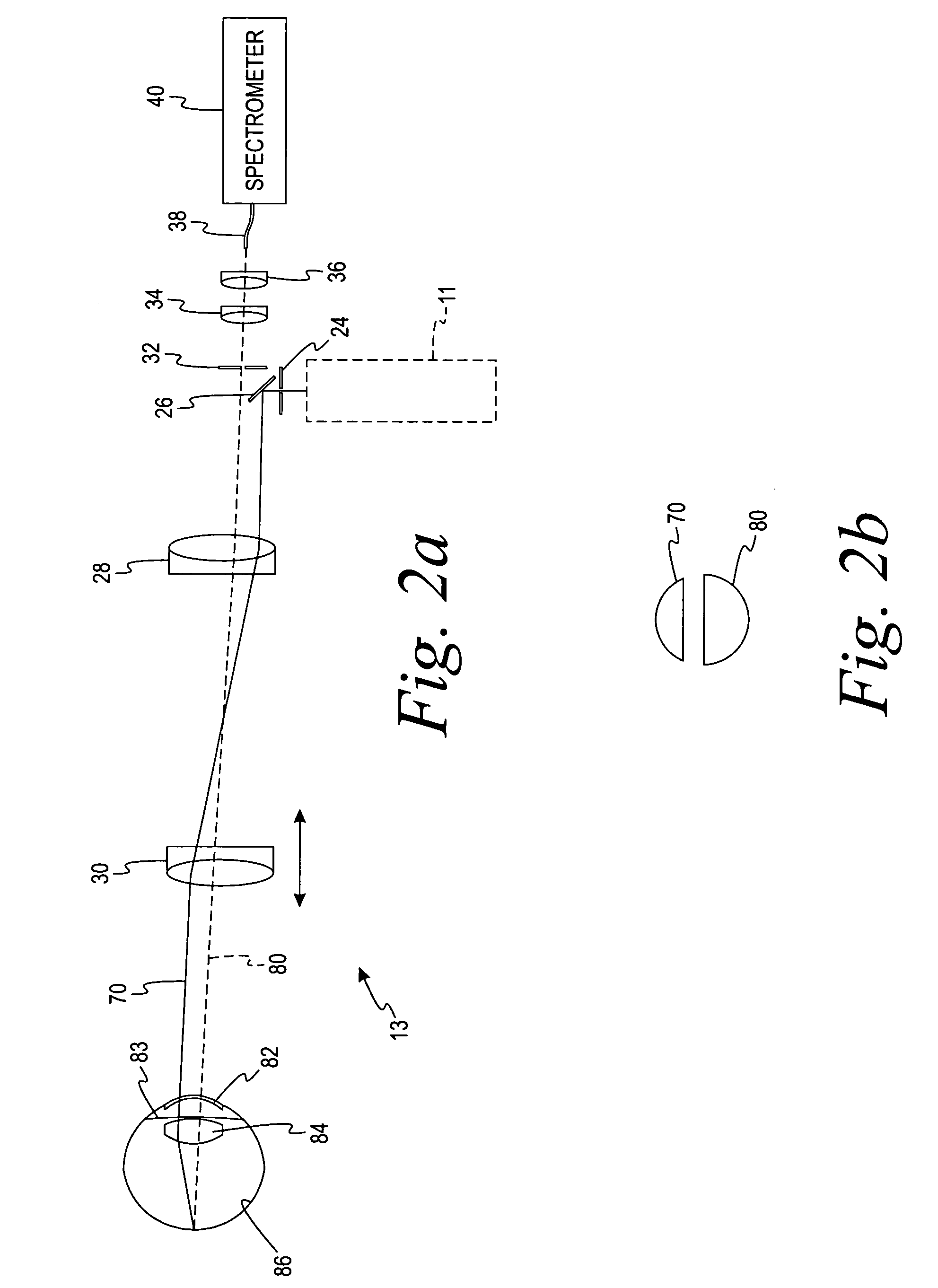
[0041]The beam separation system 13 includes an illumination pupil mask 24, a mirror 26, a first lens 28, a second lens 30, a detection pupil mask 32, a third lens 34, and a fourth lens 36. The beam separation system 13 is used for providing an illumination beam to the patient's eye and receiving a detection...


PUM


Abstract

A reflectometry instrument includes a light source, a spectrometer, and a first and second lens. The light source emits an illumination beam to the macula. The spectrometer measures a detection beam that is a portion of the illumination beam reflected from the eye and is indicative of the eye characteristics (e.g. macular pigment). The first and second lenses transmit the illumination beam to the macula and transmit the detection beam from the macula to the spectrometer. The instrument is used on an undilated pupil and minimizes unwanted reflections by at least one of the following: the first and second lenses include anti-reflection coatings; the illumination and detection beams pass through the first and second lenses at locations offset from their centers; and the illumination and the detection beams remain separated when passing through the first and second lenses. Zeaxanthin, lutein, and the total macular pigment levels are measured by the instrument.

Description

FIELD OF THE INVENTION[0001]The present invention relates to a reflectometry instrument that measures characteristics of the patient's eye, such as macular pigment, with a high degree of accuracy and without dilating the patient's pupil.BACKGROUND OF THE INVENTION[0002]The retina is the layer of nerve cells at the back of the eye, which convert light into nerve signals that are sent to the brain. In humans, and in other primates (but not in most other mammals, or other types of animals), the retina has a small yellowish area in the center of the field of vision. That yellowish area is called the “macula.” It provides fine-resolution vision in the center of the visual field and is essential to good vision. People who suffer from macular degeneration often lose the ability to read, recognize faces, drive, or walk safely on unfamiliar routes.[0003]The surrounding portions of the macula can only provide coarse resolution. This physiological feature limits and controls the number of nerv...

Claims


Application Information

Patent Timeline
Patent Type & Authority Patents(United States)
IPC IPC(8): A61B3/10
CPCA61B3/1176A61B3/156A61B3/1208A61B3/12
Inventor VAN DE KRAATS, JANVAN NORREN, DIRKBERENDSCHOT, TOS T. J. M.
Owner ZEAVISION LLC
Why Eureka
Try Eureka