
Methods to engineer stratified cartilage tissue

Inactive Publication Date: 2009-01-13
RUSH UNIV MEDICAL CENT +1
View PDF72 Cites 34 Cited by
  • Summary
  • Abstract
  • Description
  • Claims
  • Application Information

AI Technical Summary

Benefits of technology

[0020]The present methods can also involve contacting one or more test agents with one or more cells or tissues selected from the group consisting of: (a) the first group of chondrogenic cells; (b) the one or more additional groups of chondrogenic cells; (c) the first group of chondrogenic cells with cell-associated matrix; (d) the one or more addi

Problems solved by technology

As there is no blood supply to the cartilage matrix, cartilage has a limited ability to heal once damaged and often undergoes degenerative pathological changes.
Effectively treating cartilage injuries is further complicated because a complete understanding of the mechanisms and natural history of cartilage injuries and the healing and regeneration of injured cartilage is lacking.
This lack of knowledge has both large human and economic costs because cartilage damage affects millions of people every year in the United States alone.
Efforts to restore cartilage or promote cartilage repair have been largely unsuccessful for several reasons.
However, the cartilage produced by this procedure is fibrocartilage, which has relatively poor mechanical properties and does not reproduce the complex physical and chemical properties of articular cartilage.
The major disadvantages of this technique are donor-site morbidity (in the case of autograft) and risk of disease transmission (in the case of allograft).
Additionally, tissue rejection can occur with allografts which compromises the surgical result.
Each of the current methods of cartilage repair has substantial limitations.
The major drawbacks of this type of approach are: (1) difficulty in attaining or maintaining the chondrocyte phenotype; (2) unknown biological effects of the scaffold material on the implanted and native chondrocytes and other joint tissues; and (3) limited attachment of the engineered tissue construct to the defect bed.
Likewise these approaches utilize a homogenous population of chondrocytes isolated generically from cartilage tissue and do not accurately imitate the layered structure or function of natural cartilage.

Method used


Image

  • Methods to engineer stratified cartilage tissue
  • Methods to engineer stratified cartilage tissue
  • Methods to engineer stratified cartilage tissue

Examples


examples

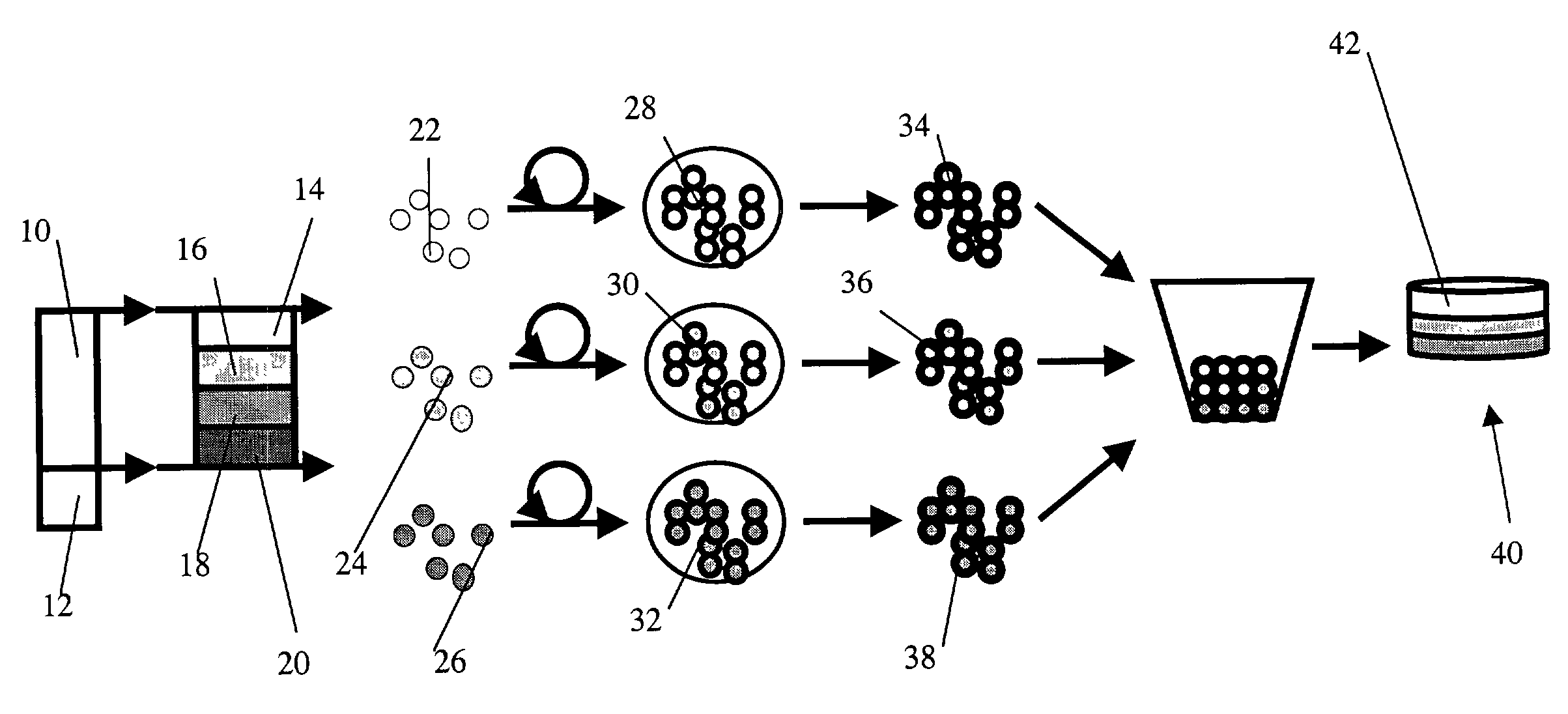
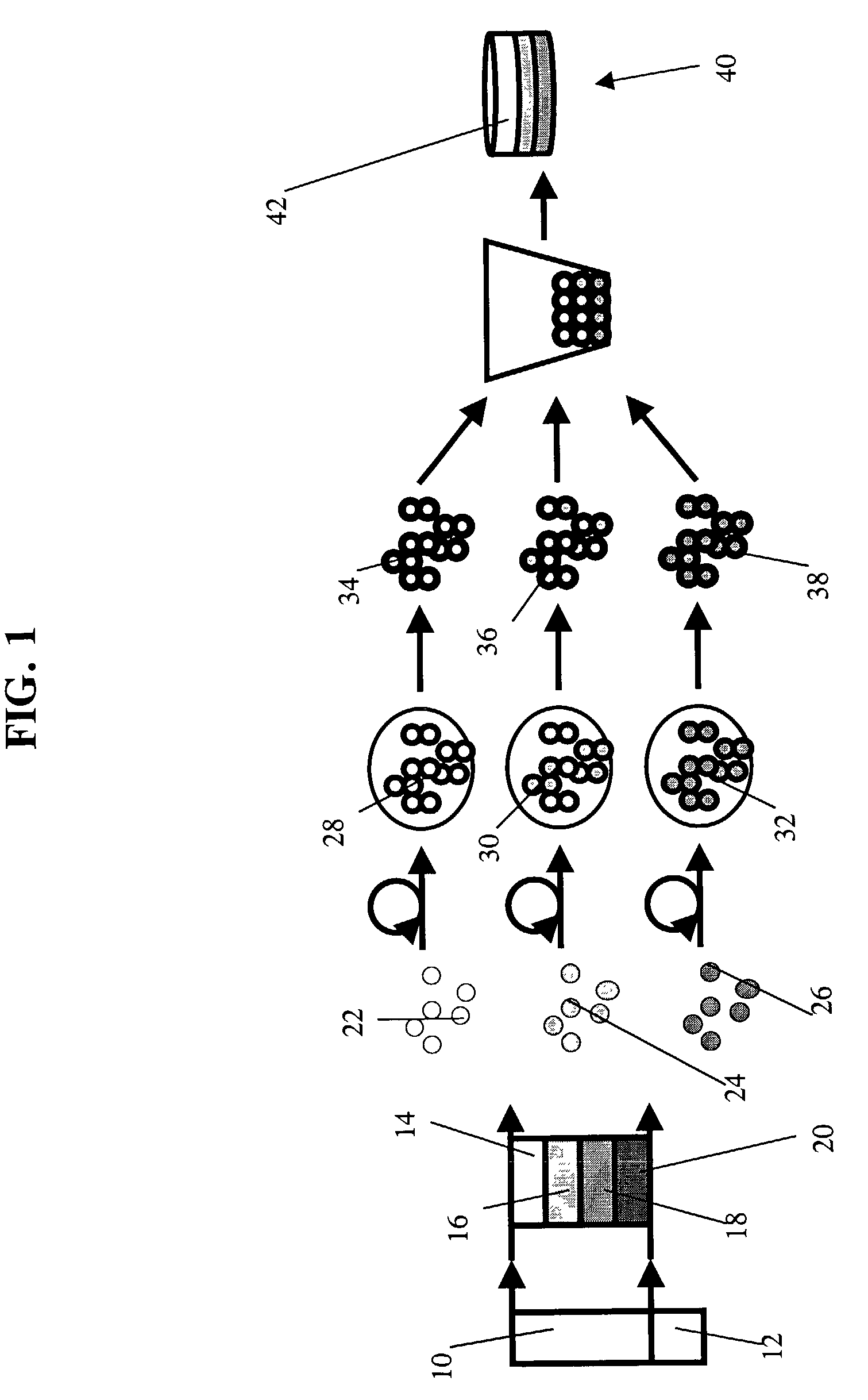
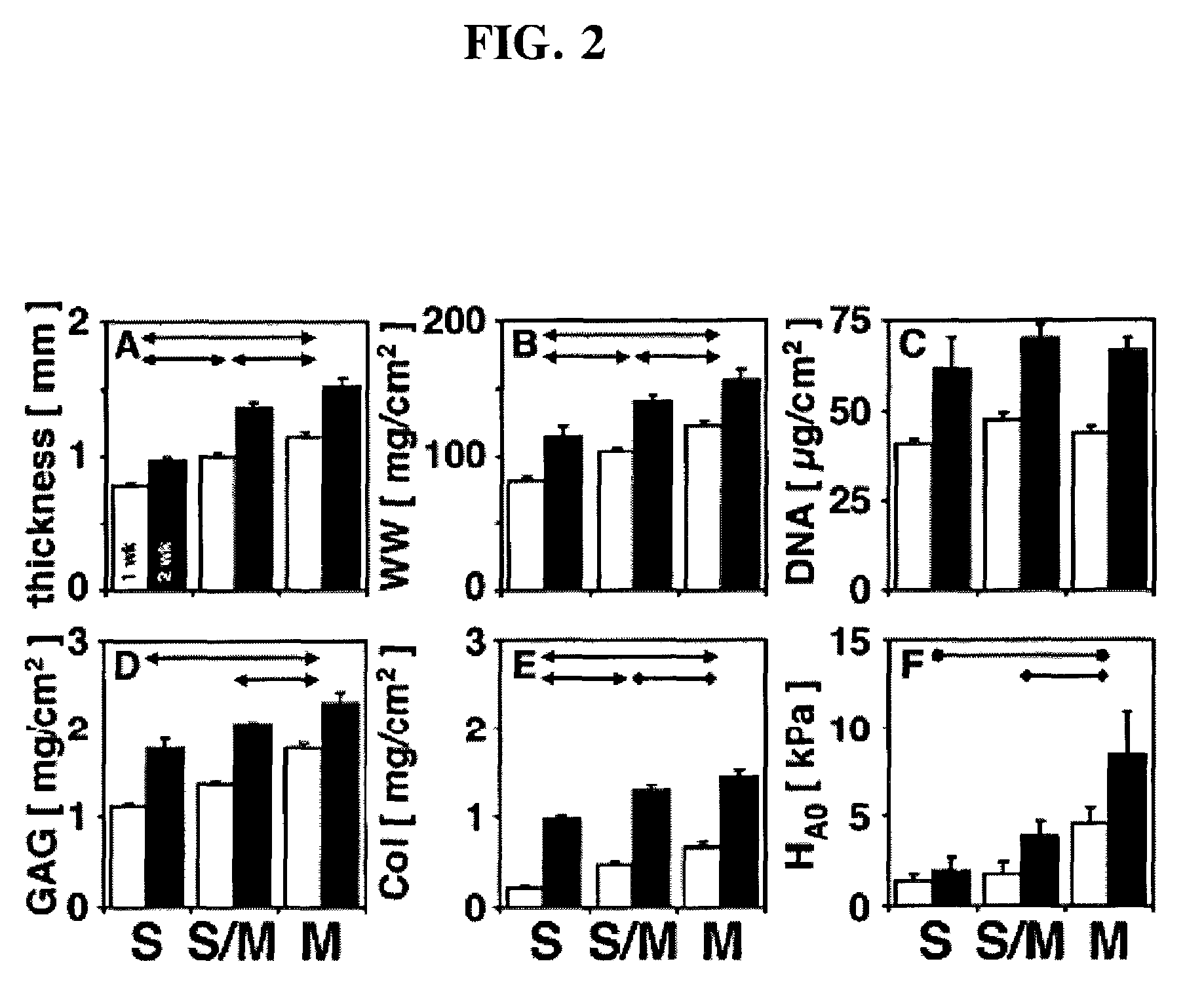
[0075]Experimental Protocol: Articular cartilage slices from the superficial (0-200 μm) and middle (400-1600 μm) zones were harvested from the femoropatellar groove of bovine calf knees (1-3 wk old, 6 animals). Superficial and middle zone chondrocyte subpopulations were isolated by sequential digestion for 1 hour with 0.2% pronase (Sigma-Aldrich, St. Louis, Mo.) and 12 hours with 0.025% collagenase P (Roche Diagnostics, Indianapolis, Ind.). Mok, et al., J. Biol. Chem. 269, 33021-7 (1994). Chondrocytes were suspended in 1.2% alginate (Keltone LV, Kelco, Chicago, Ill.) in 150 mM NaCl at 4×10⁶ cells / ml, expressed from a 22 gauge needle into a 102 mM CaCI2 solution to polymerize the alginate as small beads. Hauselmann, et al., Matrix 12, 116-29 (1992). The alginate beads were then transferred to a T-175 flask containing DMEM / F12 with additives (10% fetal bovine serum, 25 μg / ml ascorbate, 100 U / ml penicillin, 100 μg / ml streptomycin, 0.25 μg / ml Fungizone, 0.1 mM MEM non-essential amino ac...


PUM

PropertyMeasurementUnit
Permeabilityaaaaaaaaaa
Cohesive energyaaaaaaaaaa

Abstract

The present invention is directed to a cultured, stratified cartilage tissue that comprises a tissue-engineered, cohesive cartilage construct comprised of two or more cartilage layers, wherein each cartilage layer comprises chondrogenic cells having a chondrocytic phenotype corresponding to chondrocytes present in: a superficial-tangential zone of natural cartilage; a middle-transitional zone of natural cartilage; a deep-radial zone of natural cartilage; or a calcified cartilage zone of natural cartilage.

Description

[0001]This application claims priority to U.S. patent applications Ser. No. 60 / 322,888, filed Sep. 15, 2001 and 60 / 382,438, filed May 22, 2002, the entire contents of both of which are hereby entirely incorporated by reference.STATEMENT REGARDING FEDERALLY SPONSORED RESEARCH[0002]This invention was made with government support under Grant Number BES-9987353 awarded by the National Science Foundation. The government has certain rights in the invention.FIELD OF INVENTION[0003]The present invention relates to bioengineered cartilage tissues. More particularly this invention relates to stratified engineered cartilage tissue which closely replicates natural cartilage tissue and systems and methods for its use.BACKGROUND OF THE INVENTION[0004]Cartilage is a complex avascular tissue that is organized into several distinct layers or zones, the superficial zone, middle zone, deep zone and a calcified cartilage zone, each of which is made up of chondrocyte cells surrounded by extra-cellular m...

Claims


Application Information

Patent Timeline
IPC IPC(8): A61F2/28A61F2/02A61L27/00A61K35/12A61K35/32A61L27/38C12N5/077
CPCA61K35/32A61L27/3612A61L27/3645A61L27/3817A61L27/3843A61L27/3891A61L27/3895C12N5/0655A61F2/30756A61K35/12C12N2500/32C12N2500/38C12N2509/00A61L2430/06
Inventor SAH, ROBERT L.LI, KELVIN W.KLEIN, TRAVIS J.SCHUMACHER, BARBARA L.MASUDA, KOICHITHONAR, EUGENE J-M. A.
Owner RUSH UNIV MEDICAL CENT
Learn More