Method for detecting regulatory T cells using expression of folate receptor 4 as indicator, method for treating diseases using the detection method, pharmaceutical composition for immunostimulation, and method for treating diseases using the composition

a technology of folate receptor and expression of t cells, applied in the direction of drug compositions, instruments, immunostimulation, etc., can solve the problems of inability to clinically use a t cell population from which tsub>reg /sub>cells have been removed, inability to express the immune response of activated t cells, and inability to isolate or remove tsub>reg /sub>cells, etc., to achieve the effect of re a t cell expression of regulatory t cell expression of folate receptor 4 expression of folate receptor 4 expression of folate receptor 4 expression of folate receptor 4 expression of folate receptor 4 expression of folate receptor 4 expression of folate receptor 4 expression of folate receptor 4 expression of folate receptor 4 expression of folate receptor 4 expression of folate receptor 4 expression of folate receptor 4 expression of folate receptor 4 expression of folate receptor 4 expression of folate receptor 4 method, method of treatment method, method of treatment method, method of drug substance substance,

Active Publication Date: 2009-02-10
KYOTO UNIV +1
View PDF0 Cites 4 Cited by
  • Summary
  • Abstract
  • Description
  • Claims
  • Application Information

AI Technical Summary

Benefits of technology

[0007]Still another object of the present invention is to provide a pharmaceutical composition that effectively expresses the immune response of activated T cells by reducing the number of Treg cells in vivo, and a method for activating immunity and for treating disease using the pharmaceutical composition.
[0008]The present inventors conducted extensive research for achieving the above objects and found that Treg cells are expressed on the surface of folate receptor 4 (folate receptor δ, folate binding protein 3) at higher levels than activated T cells and naive T cells, and therefore Treg cells can be distinguished from activated T cells and naive T cells using the expression of folate receptor 4 on the surface of the cell as an indicator. Furthermore, an anti-folate receptor 4 antibody or a folate receptor 4-binding fragment, which is used for specifically detecting Treg cells, has an effect for reducing Treg cells in vivo, stimulating immunity activity in vivo, and in particular an effect for treating tumors or infectious diseases. The present invention was accomplished based on these findings and by adding improvements thereto.

Problems solved by technology

However, because CD25 and GITR are expressed not only on Treg cells but also on activated T cells at high levels, it is known that CD25 and GITR cannot be used with high accuracy as indicators for distinguishing between Treg cells and activated T cells.
However, since Foxp3 is a transcription factor, which is not expressed on the surface of cells, it is impossible to clinically use a T cell population from which Treg cells have been removed or isolated Treg cells in a live state by employing a method wherein Treg cells are isolated or removed using Foxp3 as an indicator.
As described above, it is impossible to isolate or remove Treg cells by detecting and distinguishing the Treg cells from activated T cells in a live state using prior art techniques.
Furthermore, there is no report to date regarding not only a technique for isolating or depleting live Treg cells but also a technique by which the number of Treg cells is selectively reduced in vivo.

Method used


Image

  • Method for detecting regulatory T cells using expression of folate receptor 4 as indicator, method for treating diseases using the detection method, pharmaceutical composition for immunostimulation, and method for treating diseases using the composition
  • Method for detecting regulatory T cells using expression of folate receptor 4 as indicator, method for treating diseases using the detection method, pharmaceutical composition for immunostimulation, and method for treating diseases using the composition
  • Method for detecting regulatory T cells using expression of folate receptor 4 as indicator, method for treating diseases using the detection method, pharmaceutical composition for immunostimulation, and method for treating diseases using the composition

Examples


reference example 1

Preparation of Mouse Hybridoma Cells that Produce Anti-Folate Receptor 4 Antibody

[0102]First, CD25+CD4+ T cell strains for serving as an immunogen were prepared by the following method. Spleen / lymph-node cells derived from normal BALB / c mice were placed in an RPMI culture medium containing culture supernatant (a 8-fold dilution) of hybridoma J11d (anti-24CD antibody-producing cell, purchased from the American Type Culture Collection) and culture supernatant (a 10-fold dilution) of hybridoma 3.155 (anit-CD8 antibody-producing cell, purchased from the American Type Culture Collection), and allowed to stand on ice for 30 minutes. The spleen / lymph-node cells in the RPMI culture medium were put into 10 cm dishes (2 dishes per mouse) coated with anti-rat IgG caprine antibody (product of ICN Pharmaceuticals, 5 ml / dish of anti-rat IgG caprine antibody diluted to 5 μg / ml), and incubated at 4° C. for 30 minutes. The suspension cells were then collected as cells rich in CD4+ cells by a panning...

reference example 2

Preparation of Anti-Folate Receptor 4 Antibody (TH6 antibody)

[0105]The mouse hybridoma TH6 cells (5×10⁶) obtained in Reference Example 1 were administered to the abdominal cavities of SCID mice (product of Clea Japan, Inc.), which are immunodeficient mice, and abdominal dropsy was extracted on day 10 and after from the injection. The extracted abdominal dropsy was subjected to centrifugation, cell components and the like were removed therefrom by being passed through a 0.45 μm filter (product of Millipore), and then the monoclonal antibody (TH6 antibody) was purified using Protein G column (product of Amersham Biosciences KK). Elution of monoclonal antibody from the column was conducted using 0.1 M glycine-HCl at pH 2.7. Thus-obtained solution of monoclonal antibody was subjected to dialysis with PBS using a dialysis membrane (Spectrum Laboratories, MWCO 12-14000) and filtration using 0.2 μm filter (product of Millipore), obtaining purified antibody.

reference example 3

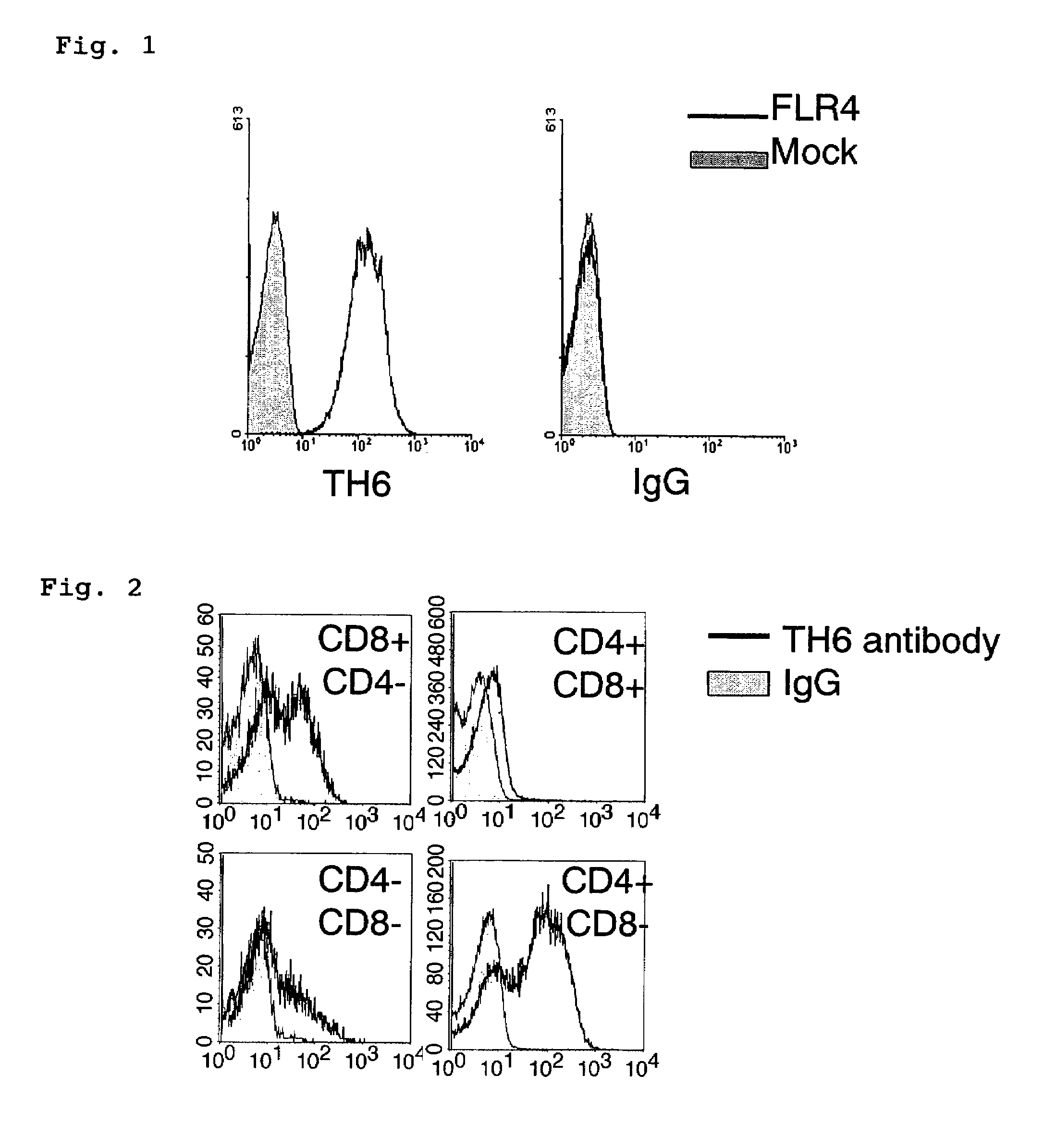
Reaction Specificity of Anti-Folate Receptor 4 Antibody (TH6 Antibody)

[0106]A pMXS-IG vector (provided by the Institute of Medical Science, the University of Tokyo) obtained by incorporating full-length cDNAs of mouse folate receptor 4 (FLR4) into Plat-E cells that are subclone of human HEK293T cells (provided by the Institute of Medical Science, the University of Tokyo) or an empty pMXS-IG vector was subjected to transfection using Fugene 6 (Roche Molecular Biochemicals). Biotin-labeled TH6 antibody (labeled using an Amersham biotinization kit) or biotin-labeled IgG2b (product of PharMingen Company) was added to the cells (1×10⁸ cells / ml) in such a manner that the content of the biotin-labeled TH6 antibody or biotin-labeled IgG2b became 1 μg / ml, and reacted on ice for 30 minutes. After washing, PE-labeled streptavidin (product of PharMingen Company) was added thereto in such a manner that the amount of the PE-labeled streptavidin became 0.5 μg / ml, reacted on ice for 30 minutes, and...


PUM

PropertyMeasurementUnit
axial lengthaaaaaaaaaa
pHaaaaaaaaaa

Abstract

An object of the present invention is to provide a technique to distinguish between Treg cells and activated T cells in a live state. Another object of the present invention is to provide a pharmaceutical composition for immunostimulation that can reduce the number of Treg cells in vivo and effectively express the immune response of activated T cells. In the method of the present invention, Treg cells are detected from test cells containing (i) regulatory T cells and (ii) at least one type of cell selected from the group consisting of naive T cells and activated T cells, wherein expressions of folate receptor 4 on the surfaces of cells are measured and Treg cells are detected using the expressions as an indicator. The present invention uses anti-folate receptor 4 antibody or folate receptor 4-binding fragment as an active ingredient contained in a pharmaceutical composition for immunostimulation.

Description

TECHNICAL FIELD[0001]The present invention relates to a method for detecting regulatory T cells, a method for isolating regulatory T cells, a method for treating diseases using these detection and isolation methods, and a reagent for detecting regulatory T cells.[0002]The present invention also relates to a pharmaceutical composition for immunostimulation that can effectively express the immune response of activated T cells by reducing the number of regulatory T cells, and an immunostimulation method using the composition.BACKGROUND ART[0003]CD4+CD25+ T cells are called regulatory T cells (hereunder referred to as “Treg cells”), and it is known that such T cells have an immunosuppressive activity and play an important role in maintaining immunological tolerance (for example, see Sakaguchi, S., et al., 1995, “Immunologic self-tolerance maintained by activated T cells expressing IL-2 receptor alpha-chains (CD25). Breakdown of a single mechanism of self-tolerance causes various autoimm...

Claims


Application Information

Patent Timeline
Patent Type & Authority Patents(United States)
IPC IPC(8): A01N63/00A61K39/395A61P35/00A61P37/04C12N5/07C12N5/09C12Q1/04G01N33/53
CPCC07K16/28G01N33/56972A61K2039/505A61P35/00A61P37/04A61P37/06
Inventor SAKAGUCHI, SHIMONHIROTA, KEIJIYAMAGUCHI, TOMOYUKI
Owner KYOTO UNIV
Why Eureka
Try Eureka