Electrical apparatus and system with improved tissue capture component

a tissue capture and electric device technology, applied in the field of electric devices and systems with improved tissue capture components, can solve the problems of insufficient specimen material for diagnosis, inability to allow a more advanced pathological investigation, and a risk of false negative, so as to improve the capture component, enhance the tip region, and reduce the widthwise extent

Inactive Publication Date: 2009-02-24
COVIDIEN AG
View PDF9 Cites 182 Cited by
  • Summary
  • Abstract
  • Description
  • Claims
  • Application Information

AI Technical Summary

Benefits of technology

[0027]The present invention is directed to an electrosurgical apparatus and system for cutting about and retrieving a tissue volume with an improved capture component. This capture component generally is fashioned with a plurality of thin elongate leafs which extend from a base portion to tip regions having cable guide outlets. A pursing cable assembly configured with a plurality of electrosurgically energizable stainless steel multi-strand braided cables is supported by the multi-leaf structure in a manner wherein the cables are maneuvered through the guide outlets to establish a cutting leading edge which envelopes a target tissue volume as the moving leafs progressively assume the configuration of a tissue capturing cage. Each cable extends rearwardly within the associated instrument to connection with a slidable terminator component. By blocking the movement of the terminator component as the cable assembly defines a maximum diametric extent, a pursing action ensues as the tips of the leafs converge toward the instrument axis. With the present invention, a forwardly disposed stainless steel guide support region of each leaf is formed with a diminished widthwise extent and is enclosed within an extruded polymeric cable guide. This not only provides an improved leaf cable guide function but permits enhanced flexure at the tip region of each leaf. That enhanced flexure promotes a steeper angle of attack during the pursing activity to, in turn, improve the cutting profile of the advancing and pursing cutting cable leading edge.
[0028]The improved leaf forward region flexibility is accommodated for with respect to determining the completion of a capture maneuver by the provision of a capture stop within the instrument itself which blocks leaf drive and evokes a motor stall condition which is utilized as a signal to terminate the capture activity. In addition to providing a very positive termination of capture signal, utilization of the capture stop avoids any unwanted curling or flexure at the leaf tip regions following their reaching a full pursing orientation.
[0029]The cable guides are formed as polytetrafluoroethylene (Teflon) extrusions and thus, exhibit a surface friction characteristic which is improved over the corresponding surface characteristic of earlier utilized stainless steel capture component leafs. Utilization of such extruded cable guides into which the forward stainless steel leaf components are inserted substantially lessens the complexity of fabrication of the capture component with an attendant improvement in manufacturing costs.

Problems solved by technology

With fine needle aspiration, there remains a risk of false-negative results, which most often occur in cases involving extremely fibrotic tumor.
In addition, after the procedure has been performed there may be insufficient specimen material for diagnosis.
Rather fragmented portions of tissue are withdrawn which do not allow a more advanced pathological investigation.
This limitation also is observed with respect to large core needle biopsies.
However, they still do not provide optimum pathological information.
In addition, while less expensive than open surgical biopsy, the ABBI has proven expensive compared to other biopsy techniques, and it has been noted that the patient selection for ABBI is limited by the size and location of the tumor, as well as by the presence of very dense parenchyma around the tumor.
Studies undertaken with respect to the employment of this instrument in the recovery of tissue samples from very dense tissue including fibrous tissue have revealed that excessive drive motor current values may be encountered as the cables of its capture component are tensioned.
Manufacture of the capture component leafs and associated tubular cable guides has been successful but necessarily complex.

Method used


Image

  • Electrical apparatus and system with improved tissue capture component
  • Electrical apparatus and system with improved tissue capture component
  • Electrical apparatus and system with improved tissue capture component

Examples


Embodiment Construction

[0053]In the discourse to follow, the above-discussed EN-BLOC® system is described in order to facilitate an understanding of the general design and operation of the capture component of the instrument and system at hand. As the description unfolds, the improvements to the capture component and its associated drive are detailed.

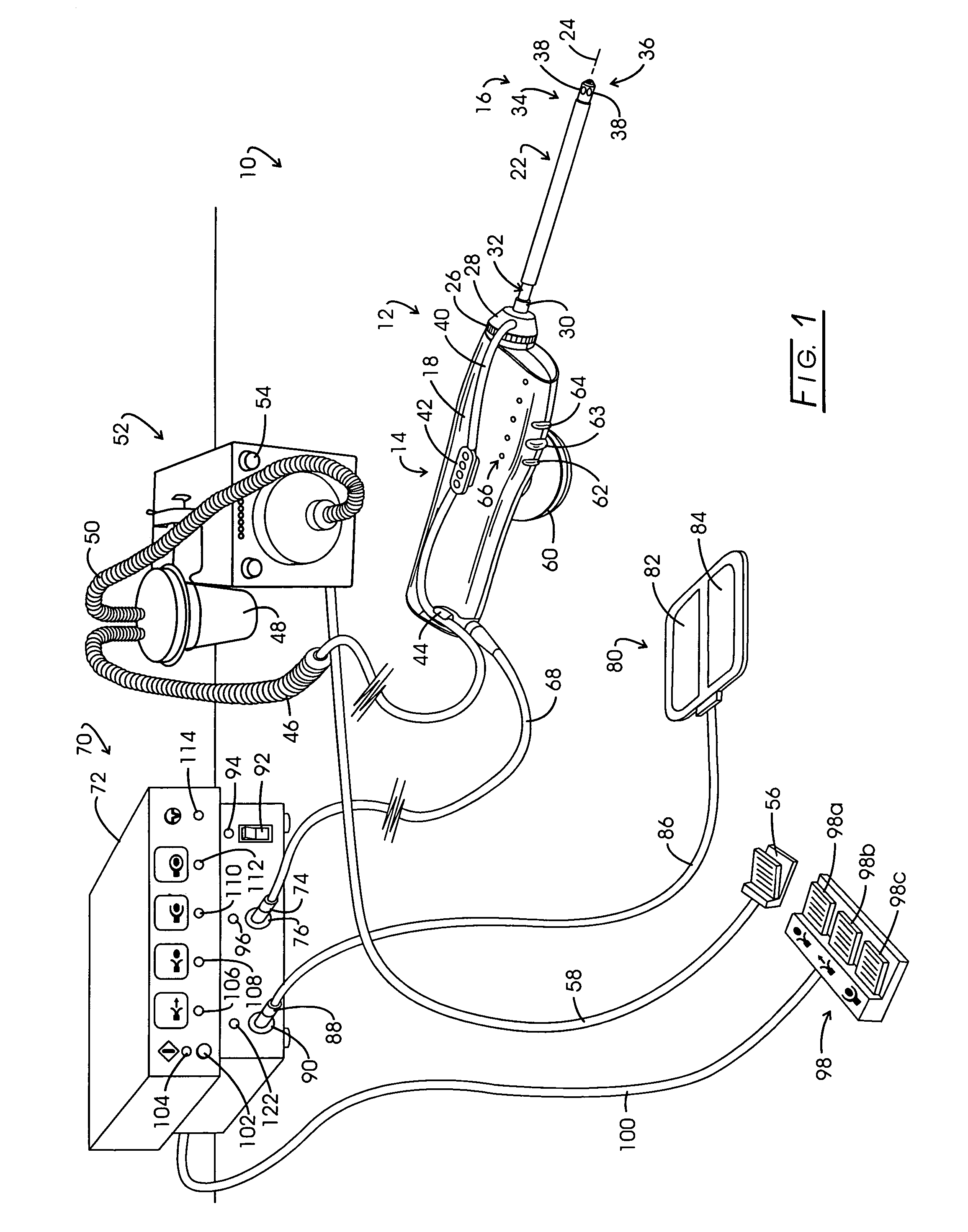
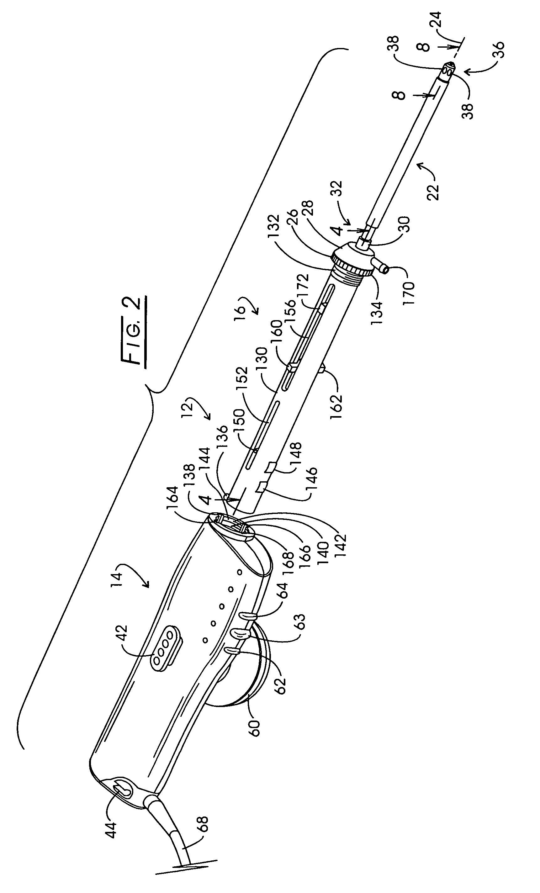
[0054]Referring to FIG. 1, the system for isolating and retrieving a target tissue volume or biopsy sample is illustrated in general at 10. System 10 comprises a tissue retrieval instrument represented generally at 12 which includes a reusable component represented generally at 14, sometimes referred to as a “handle”. Instrument 12 additionally includes a disposable component represented generally at 16, the rearward portion of which is removably mounted within the polymeric housing 18 of reusable component 14. The disposable component 16 is sometimes referred to as a “probe”.

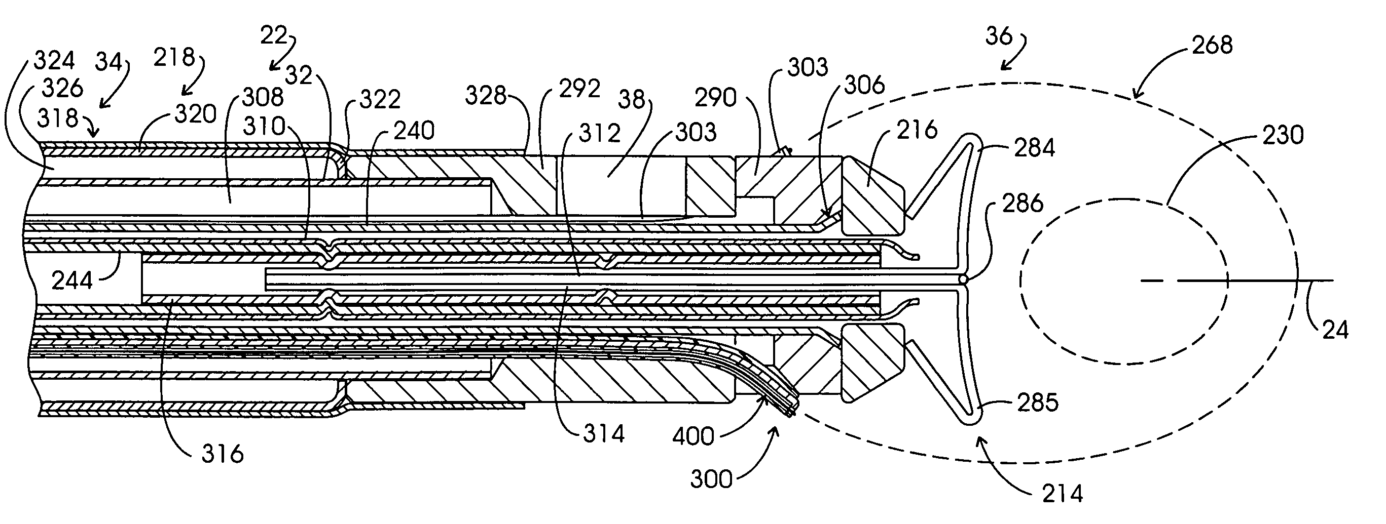
[0055]Disposable component 16 includes an elongate cannula or support assembly repre...


PUM


Abstract

Electrosurgical tissue specimen recovery apparatus and system employing a multi-leaf capture component configured with pursing cables which are electrosurgically excited to define a cutting leading edge. To complete a capture, the cables are loaded in tension to purse and thus converge the tips of the capture component leafs together. The forward regions of the leafs are configured with a combination of a thin flat stainless steel region over which a polymeric cable guide is positioned. The polymeric cable guide and stainless steel leaf driving combination improves frictional aspects as well as the resulting aspect ratio of a recovered tissue specimen.

Description

CROSS-REFERENCE TO RELATED APPLICATIONS[0001]This application is a continuation-in-part of prior application Ser. No. 10 / 630,336, filed Jul. 30, 2003 now U.S. Pat. No. 6,955,653.STATEMENT REGARDING FEDERALLY SPONSORED RESEARCH[0002]Not applicable.BACKGROUND OF THE INVENTION[0003]The detection of tumorous lesions in the breast has progressed from early observation and palpation procedures to a variety of somewhat sophisticated imaging systems. A consequence of these advances in tumor detection is the identification of suspect tumor at an early stage in its development. Generally, at such early stages the suspect tumor may be somewhat small. Rather than resort immediately to an open surgical resection upon such early detection, practitioners generally carry out a preliminary, minimally invasive biopsy procedure. Such preliminary biopsy approaches are of importance, inasmuch as statistically, only about 20% of these small tumors will be found to be malignant. Tumors determined to be be...

Claims


Application Information

Patent Timeline
Patent Type & Authority Patents(United States)
IPC IPC(8): A61D5/00A61B10/00B65D81/00A61B18/04A61F2/02A61B18/18A61B10/02A61B18/14A61B19/00
CPCA61B10/0233A61B10/0266A61B10/02A61B18/148A61B19/38A61B2018/144A61B2019/5408A61B90/40A61B2090/3908
Inventor EGGERS, PHILIP E.EGGERS, ANDREW R.EGGERS, ERIC A.
Owner COVIDIEN AG
Why Eureka
Try Eureka