System and method for joint resurface repair

a joint surface and joint technology, applied in the field of devices and methods for the repair of defects, can solve the problems of deterioration of joint function, less durability, and cell not being typically replaced, and achieve the effect of increasing the resistance of the implant to rotational forces and enhancing the load bearing or load transfer properties of the implan

Inactive Publication Date: 2009-03-31
ARTHROSURFACE
View PDF259 Cites 105 Cited by
  • Summary
  • Abstract
  • Description
  • Claims
  • Application Information

AI Technical Summary

Benefits of technology

[0024]In order to secure the implant to the fixation screw, a precision taper (or other component of a mating feature) is machined into a protrusion (or other component of a mating feature) on the back of the device. The implant may be constructed of cobalt chromium, or other materials. The implant may also include a slight outward taper or protrusion along the diametrical surface to enhance load bearing or load transfer properties of the implant to surrounding bone. Additionally, a series of radial cuts may create surfaces that increase resistance of the implant to rotational forces. These features may be located around the outer diameter of the implant.
[0029]Following fabrication of the implant, a second surgical procedure is performed. The radiused cover is removed exposing a precision taper (or, alternatively, the cover may be removed during the first procedure). A pin with a distally mounted element is placed through the central lumen of the fixation screw so that the distally mounted element is secured into the screw. This element carries one or more suture strands that now trail from the fixation screw. The sutures are then threaded through the implant and a knot or bead may be created proximal to the implant. By continuing to manipulate and tension the suture strands, the implant can be brought coaxial to the fixation screw. Once coaxial, the implant is aligned via engagement of the keyed elements and driven into place with a plastic driving rod and mallet. Finally, through the guide aperture on the surface of the implant, bone cement may be injected to enhance the contact surface between the implant and the subchondral bone.

Problems solved by technology

However, when injured, hyaline cartilage cells are not typically replaced by new hyaline cartilage cells.
While similar, fibrocartilage does not possess the same unique aspects of native hyaline cartilage and tends to be far less durable.
Such cartilage disease or deterioration can compromise the articular surface causing pain and further deterioration of joint function.
Although this procedure has been widely used over the past two decades and can provide good short term results, (1-3 years), the resulting fibrocartilage surface is seldom able to support long-term weight bearing, particularly in high-activity patients, and is prone to wear.
This procedure is technically difficult, as all grafts must be taken with the axis of the harvesting coring drill being kept perpendicular to the articular surface at the point of harvest.
If these plugs are not properly placed in relation to the surrounding articular surface, the procedure can have a very detrimental effect on the mating articular surface.
If the plugs are placed too far below the level of the surrounding articular surface, no benefit from the procedure will be gained.
Further, based on the requirement of perpendicularity on all harvesting and placement sites, the procedure requires many access and approach angles that typically require an open field surgical procedure.
Finally, this procedure requires a lengthy post-operative non-weight bearing course.
The disadvantages of this procedure are its enormous expense, technical complexity, and the need for an open knee surgery.
Further, this technique is still considered somewhat experimental and long-term results are unknown.
Some early studies have concluded that this approach offers no significant improvement in outcomes over traditional abrasion and microfracture techniques.
Although these patients are also managed with anti-inflammatory medications, eventual erosion of the remaining articular cartilage results in effusion, pain, and loss of mobility and / or activity for the patient.
Problems encountered after implanting such prostheses are usually caused by the eventual loosening of the prosthetic due to osteolysis, wear, or deterioration of the cements used to attach the device to the host bones.
Further, some prostheses used are actually much larger than the degenerated tissue that needs to be replaced, so that extensive portions of healthy bone are typically removed to accommodate the prostheses.
Patients who undergo TKR often face a long and difficult rehabilitation period, and the life span of the TKR is accepted to be approximately 20 years.

Method used


Image

  • System and method for joint resurface repair
  • System and method for joint resurface repair
  • System and method for joint resurface repair

Examples


Embodiment Construction

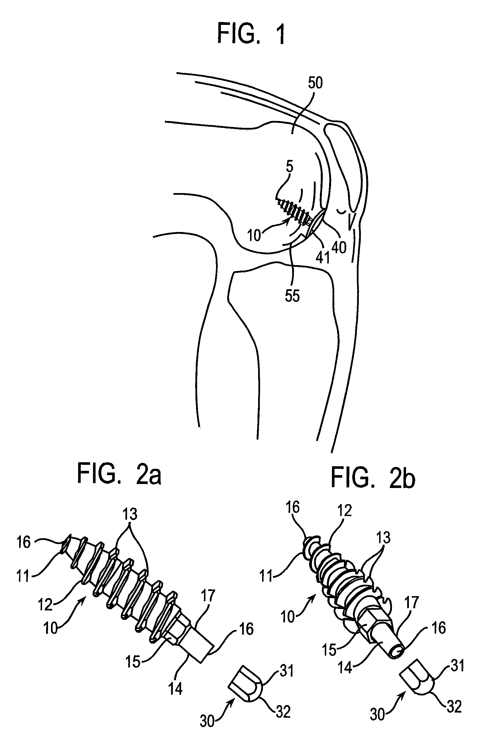
[0151]As an overview, FIG. 1 shows a surgically implanted articular joint surface repair system consistent with the present invention. As shown, the assembled fixation device includes fixation screw 10, implant 40, and anchoring pin 5, implanted in the defect in the medial femoral chondral surface 55 of knee 50. Implant 40 is configured so that bearing or bottom surface 41 of the implant reproduces the anatomic contours of the surrounding articular surface of the knee 50.

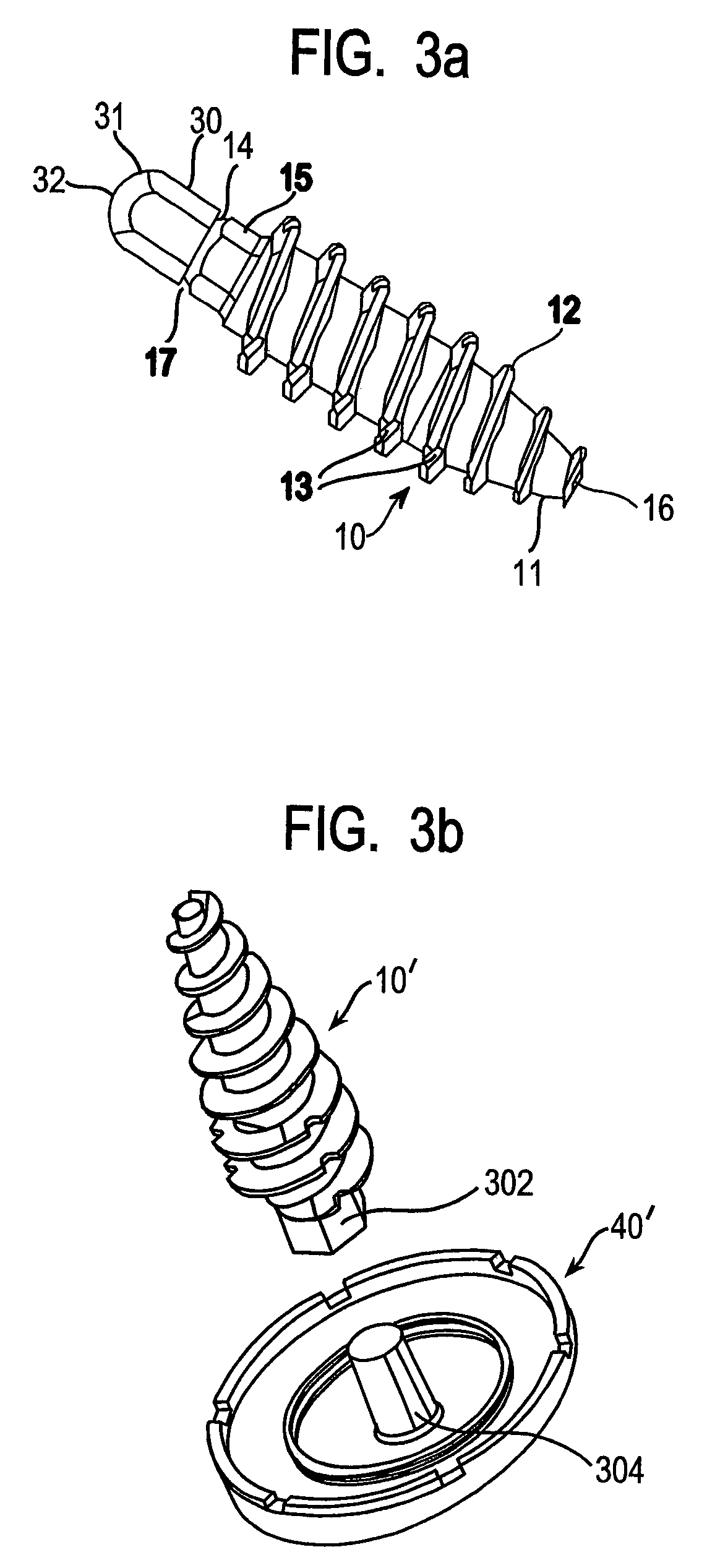
[0152]As illustrated in FIGS. 2a, 2b and 3a, fixation screw 10 comprises threads 12 running the length of the screw from tapered distal tip 11 to hex-shaped drive 15. In the embodiment shown, the screw includes a tapered distal end 11, and aggressive distal threads 12, so that, as screw 10 is driven into the subchondral bone 100 (as shown in FIG. 7a) the screw dilates open and radially compress the subchondral bone, increasing its local density and thereby increasing the fixation strength of the screw. The screw 10 ...


PUM

PropertyMeasurementUnit
transparentaaaaaaaaaa
dimensionsaaaaaaaaaa
perimeteraaaaaaaaaa

Abstract

A generic bone implant, or set of standardized implants, is created based on using a guide device to develop an axis normal to an articular surface of bone and collecting only one or two data points. A generic cutting tool is used to cut the bone to a point where a generic implant can be used. Several improved tools relating to the procedure for using such an implant, as well as methods for using implants consistent with the invention are further described, including: single-axis and biaxial drill guide tools and methods, generic single-axis implant methods and devices, generic biaxial implant methods and devices, tools and methods for holding or delivering an implant, removal or revision tools and methods, digital measuring systems and methods, and set of measuring gauges for determining the appropriate implant dimensions.

Description

CROSS-REFERENCE TO RELATED APPLICATIONS[0001]This application is a divisional of U.S. patent application Ser. No. 10 / 024,077, filed Dec. 17, 2001 now U.S. Pat. No. 6,610,067, which is a continuation-in-part of U.S. patent application Ser. No. 09 / 846,657, filed May 1, 2001 now U.S. Pat. No. 6,520,964, which claims benefit of U.S. provisional application Ser. No. 60 / 201,049, filed May 1, 2000.FIELD OF THE INVENTION[0002]This invention relates to devices and methods for the repair of defects that occur in articular cartilage on the surface of bones, particularly the knee.BACKGROUND OF THE INVENTION[0003]Articular cartilage, found at the ends of articulating bone in the body, is typically composed of hyaline cartilage, which has many unique properties that allow it to function effectively as a smooth and lubricious load-bearing surface. However, when injured, hyaline cartilage cells are not typically replaced by new hyaline cartilage cells. Healing is dependent upon the occurrence of bl...

Claims


Application Information

Patent Timeline
Patent Type & Authority Patents(United States)
IPC IPC(8): A61B17/58A61B17/56A61B1/00A61B17/00A61B17/04A61B17/06A61B17/16A61B17/17A61B17/86A61B17/88A61B19/00A61F2/00A61F2/30A61F2/38A61F2/46
CPCA61B5/4528A61B17/0401A61B17/1615A61B17/1675A61B17/1714A61B17/1764A61B17/8615A61B17/888A61B19/201A61B19/46A61F2/30756A61F2/30942A61F2/4618A61F2/4657A61B5/103A61B17/0487A61B17/06166A61B17/1635A61B17/1637A61B17/7098A61B17/8625A61B17/863A61B17/8695A61B17/8805A61B17/8827A61B17/8894A61B2017/00238A61B2017/00464A61B2017/0404A61B2017/0409A61B2017/044A61B2017/0445A61B2017/0448A61B2017/045A61B2019/461A61B2019/508A61F2/30767A61F2/38A61F2/3859A61F2002/30065A61F2002/30069A61F2002/30113A61F2002/30143A61F2002/30225A61F2002/30299A61F2002/30324A61F2002/30332A61F2002/30354A61F2002/30443A61F2002/30448A61F2002/30451A61F2002/30604A61F2002/30759A61F2002/30772A61F2002/30831A61F2002/30858A61F2002/30871A61F2002/30874A61F2002/30878A61F2002/30879A61F2002/30892A61F2002/30896A61F2002/30943A61F2002/30945A61F2002/30948A61F2002/30952A61F2002/30957A61F2002/4623A61F2002/4635A61F2002/4658A61F2002/4661A61F2002/4663A61F2002/4681A61F2210/0071A61F2220/0033A61F2220/0041A61F2220/005A61F2220/0058A61F2230/0006A61F2230/0017A61F2230/0069A61F2230/0093A61F2250/0036A61F2310/00017A61F2310/00023A61F2310/00029A61F2310/00059A61F2310/00089A61F2310/00095A61F2310/00203A61F2310/00239A61F2310/00796A61F2/4603A61F2002/30433A61F2002/30873A61B90/06A61B90/11A61B2034/108A61B2090/061
Inventor TALLARIDA, STEVEN J.EK, STEVEN W.
Owner ARTHROSURFACE
Why Eureka
Try Eureka