Detection of features in images

Inactive Publication Date: 2009-04-14
SIEMENS MEDICAL SOLUTIONS USA INC
View PDF46 Cites 66 Cited by
  • Summary
  • Abstract
  • Description
  • Claims
  • Application Information

AI Technical Summary

Benefits of technology

[0007]The present invention provides a method for detecting features of interest in an image based on the shape of the intensity profile of the feature, rather than its intensity. This is achieved by an intensity independent comparison of the inte

Problems solved by technology

For example, ultrasound images include a considerable amount of speckle which makes images difficult to interpret.
However, automating the process of detecting features of interest is made difficult by the presence of noise and imaging artefacts.
Similar problems arise in other imaging techniques, such as magnetic resonance imaging.
However, techniques based on thresholding are not entirely satisfactory because of the difficulty of setting a suitable global threshold.
For

Method used


Image

  • Detection of features in images
  • Detection of features in images
  • Detection of features in images

Examples


Embodiment Construction

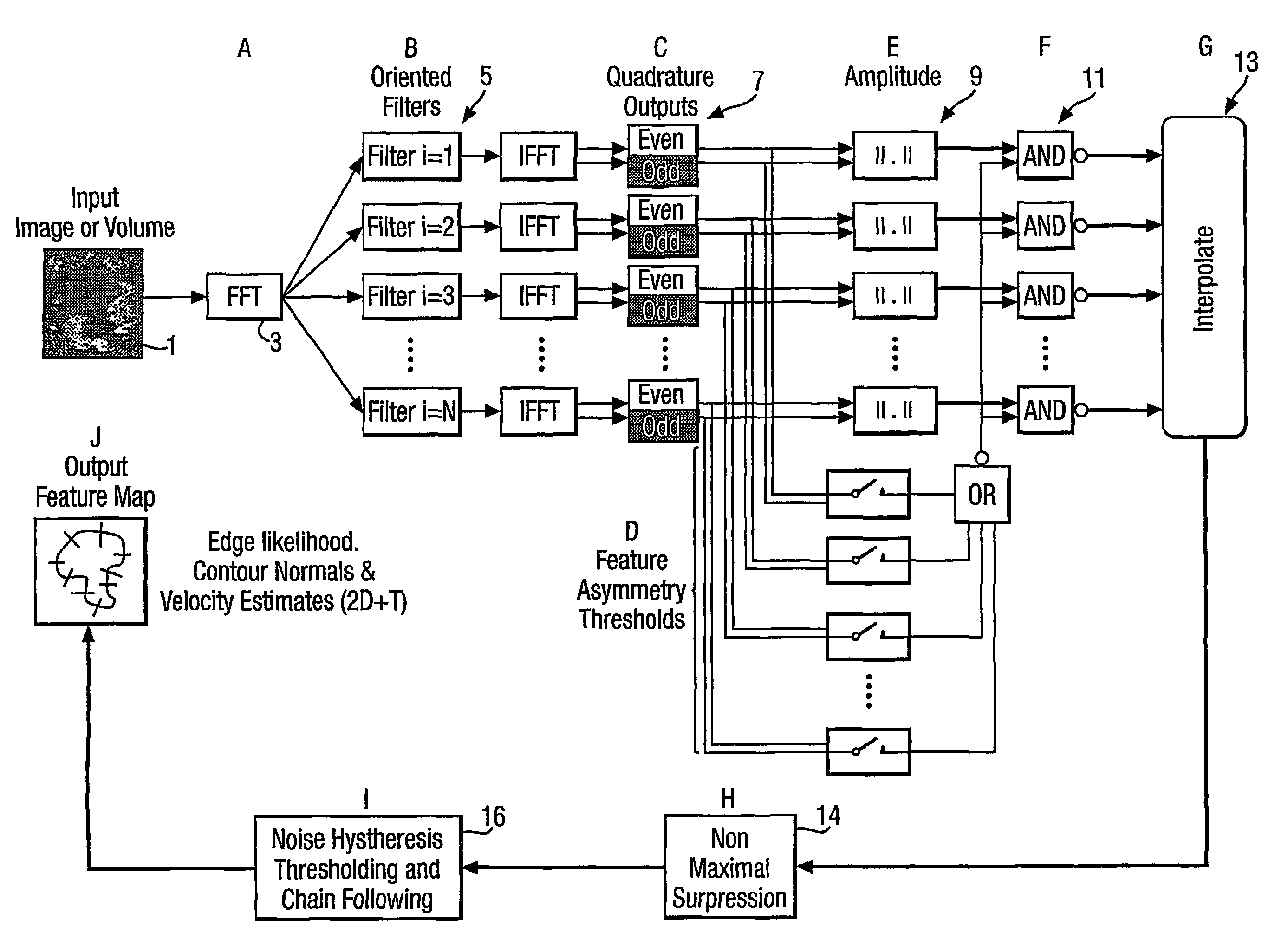
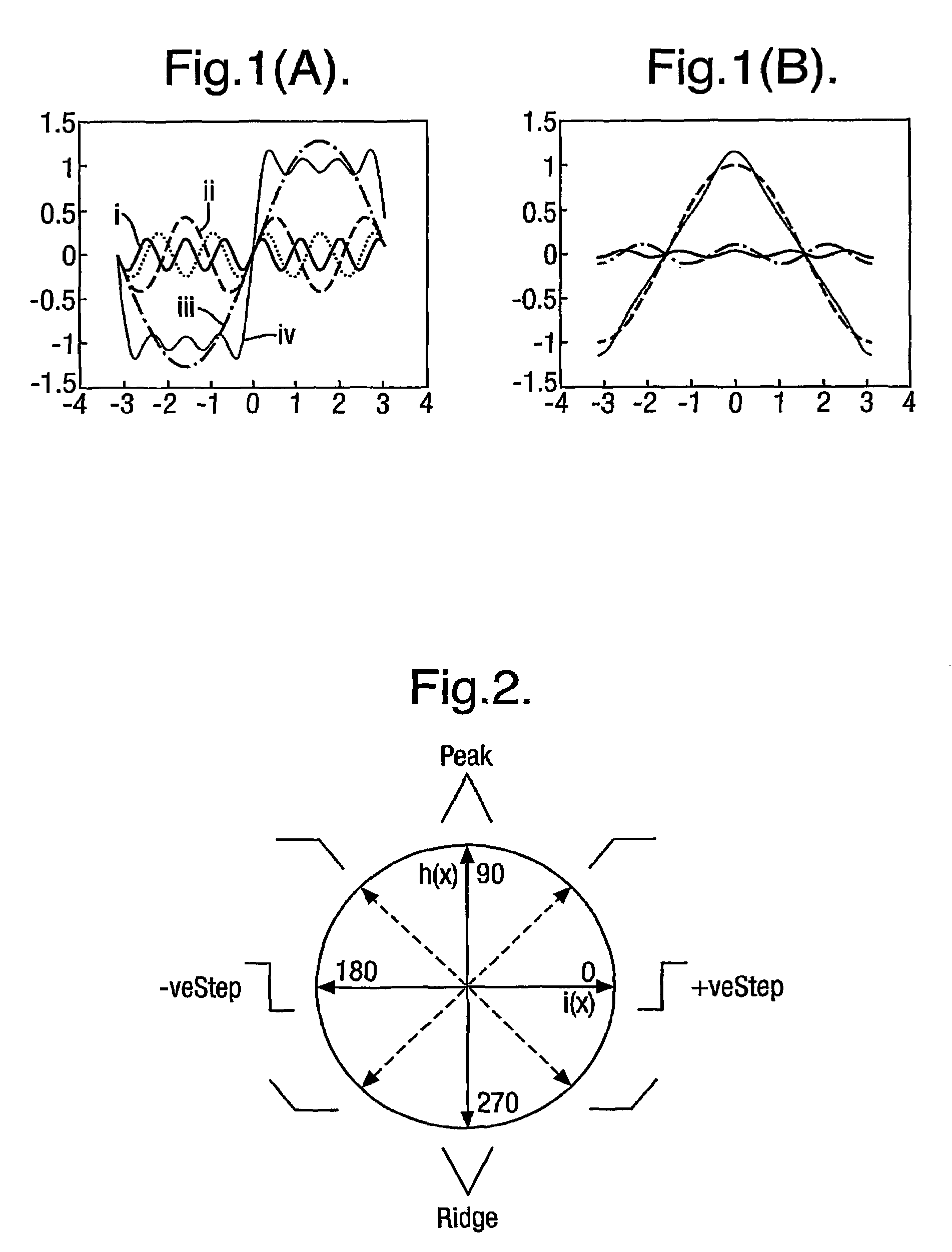
[0035]While traditional methods for detection of a step edge in an intensity profile rely on examining the derivatives of the intensity profile (i.e. its slope), to detect the sudden changes in intensity which mark the visible edge of features in the image, this embodiment of the invention examines a decomposition of the intensity profile in the spatial or spatio temporal frequency domains. For instance, FIG. 1 illustrates Fourier decompositions of a step edge (FIG. 1(a)) and a triangular function (FIG. 1(b)). In FIG. 1(a) it can be seen that three sinusoidal components i, ii and iii sum to make an approximation to a step edge iv. Further it will be noted that the phase of all three components is the same, zero, at the positive-going step edge. Further the phase value of all the components at the negative step edge is 180 degrees. Reference to FIG. 1(b) illustrates that for a triangular peak the phase value of all of the Fourier components is 90 degrees at the peak of the triangle.

[...


PUM


Abstract

An image processing technique which identifies pixels in images which are associated with features having a selected shape, such as but not exclusively step edge, roof, ridge or valley. The shape of the intensity profile in the image is compared in an intensity independent way with a shape model to select those pixels which satisfy the shape model and are thus associated with the feature of interest. This comparison is achieved by examining the phase and amplitude of a spectral decomposition of parts of the image profile in the spatial or spatio temporal frequency domain. This decomposition can be achieved using quadrature wavelet pairs such as log-Gabor wavelets. The difference between the odd and even components, known as the feature asymmetry, gives an indication of the shape of the feature. The analysis may be extended to the time domain by looking at the shape of the image profile across a time sequence of images, which gives an indication of the velocity of a moving feature. Pixels identified as belonging to a feature of the right shape are labelled with the value of feature asymmetry, the local amplitude, feature orientation and feature velocity, and this information can be used to improve the tracking of detected features through a sequence of images.

Description

[0001]This application is the U.S. national phase of international application PCT / GB01 / 05094 filed 19 Nov. 2001, which designated the U.S.BACKGROUND AND SUMMARY[0002]The present invention relates to a method and apparatus for processing images to detect particular features in the image. It is particularly concerned with the detection of features in noisy 2D, 2D+T, 3D and 3D+T images, and is thus particularly useful in medical imaging, for instance using ultrasound.[0003]There are a variety of techniques for processing images to enhance them, particularly to reduce the amount of noise and / or to detect features of interest in the image. Such techniques are particularly useful in areas where images are noisy, for instance in medical imaging. For example, ultrasound images include a considerable amount of speckle which makes images difficult to interpret. Skilled operators are able to interpret such images based on their experience and knowledge of the features of interest likely to be...

Claims


Application Information

Patent Timeline
IPC IPC(8): G06K9/00A61B5/05G06K9/40G06K9/46A61B8/00G06T5/00
CPCG06T7/0012G06T7/0083G06T7/0095G06T2207/10132G06T2207/30048G06T7/12G06T7/168
Inventor MULET-PARADA, MIGUELNOBLE, JULIA ALISON
Owner SIEMENS MEDICAL SOLUTIONS USA INC
Why Eureka
Try Eureka