
Cardiac resynchronization via left ventricular pacing

a technology of left ventricular pacing and cardiac resynchronization, which is applied in the field of medical devices, can solve the problems that the implantable medical device employing this technique may consume less power, and achieve the effect of less power

Active Publication Date: 2009-07-07
MEDTRONIC INC
View PDF31 Cites 22 Cited by
  • Summary
  • Abstract
  • Description
  • Claims
  • Application Information

AI Technical Summary

Benefits of technology

[0005]The invention is directed to techniques for cardiac resynchronization. In particular, the invention is directed to techniques for synchronizing delivery of pacing pulses to the left ventricle with intrinsic right ventricular depolarizations. One exemplary situation in which the invention may be applied is the provision of cardiac resynchronization therapy to patients with left bundle branch block (LBBB) who have adequate atrial-right ventricular conduction. Implantable medical devices employing these techniques may provide a more physiological interval between atrial and ventricular contractions, in the sense that the interval between the atrial and ventricular contractions is a function of an intrinsic, rather than paced, depolarization of the right ventricle. Further, implantable medical device employing this technique may consume less power than conventional devices that provide cardiac resynchronization therapy by delivering pacing pulses to both the right and left ventricles.
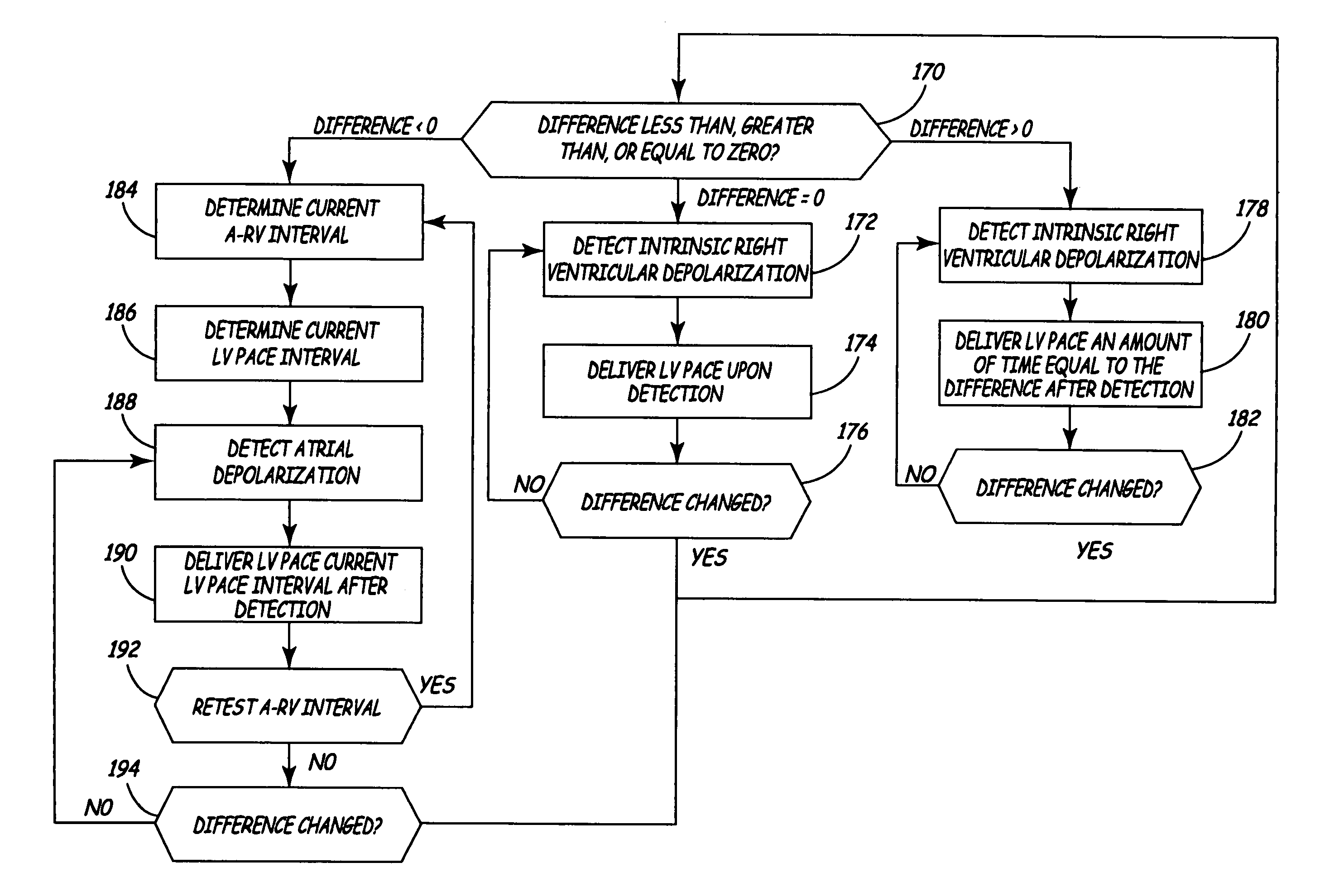
[0009]Where the pacing interval is greater than the measured interval, i.e., the left ventricular pace should be delivered after the intrinsic right ventricular depolarization, the implantable medical device may pace the left ventricle based on a determined difference between pacing interval and the measured interval. In particular, the implantable medical device paces the left ventricle upon expiration of an interval that is initiated upon detection of subsequent intrinsic right ventricular depolarizations. The interval is equal to the determined difference between the selected pacing interval and the measured interval. Pacing the left ventricle based on the determined difference may allow an implantable medical device to maintain ventricular synchrony despite beat-to-beat changes in the interval between atrial depolarizations and intrinsic right ventricular depolarizations due to changes in patient activity level, medication, or the like.

Problems solved by technology

Further, implantable medical device employing this technique may consume less power than conventional devices that provide cardiac resynchronization therapy by delivering pacing pulses to both the right and left ventricles.

Method used


Image

  • Cardiac resynchronization via left ventricular pacing
  • Cardiac resynchronization via left ventricular pacing
  • Cardiac resynchronization via left ventricular pacing

Examples


Embodiment Construction

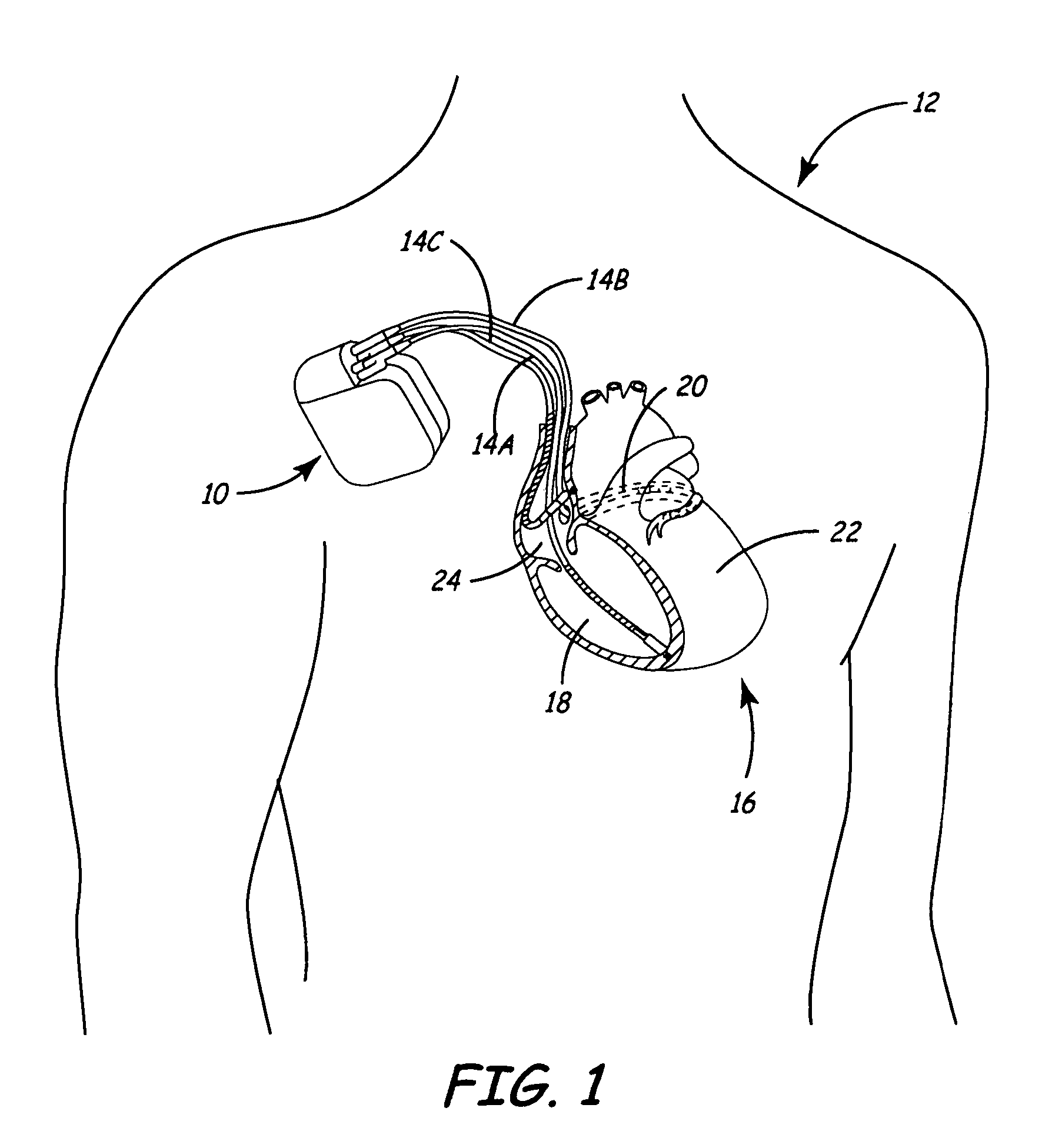
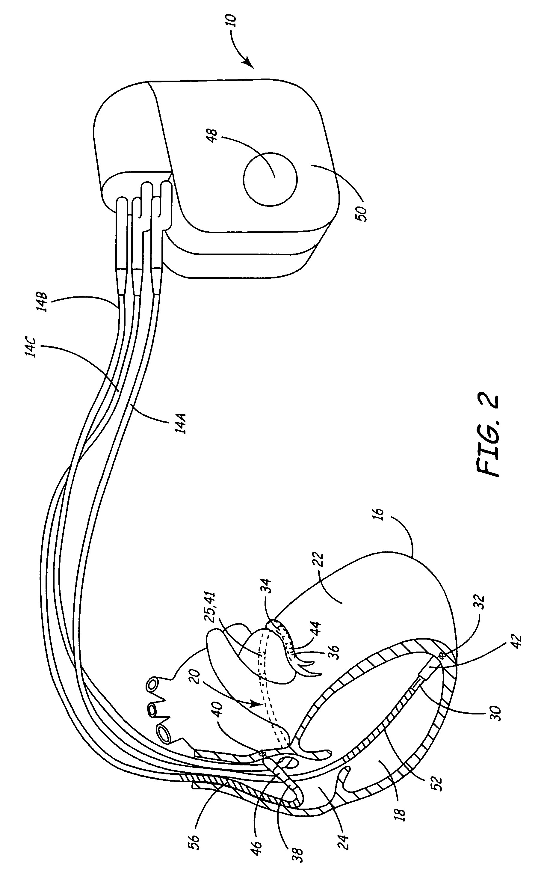
[0024]FIG. 1 is a conceptual diagram illustrating an exemplary implantable medical device (IMD) 10 implanted in a patient 12. IMD 10 may, as shown in FIG. 1, take the form of a multi-chamber cardiac pacemaker. In the exemplary embodiment illustrated in FIG. 1, IMD 10 is coupled to leads 14A, 14B and 14C (collectively “leads 14”) that extend into the heart 16 of patient 12

[0025]More particularly, right ventricular (RV) lead 14A may extend through one or more veins (not shown), the superior vena cava (not shown), and right atrium 24, and into right ventricle 18. Left ventricular (LV) coronary sinus lead 14B may extend through the veins, the vena cava, right atrium 24, and into the coronary sinus 20 to a point adjacent to the free wall of left ventricle 22 of heart 16. Right atrial (RA) lead 14C extends through the veins and vena cava, and into the right atrium 24 of heart 16.

[0026]Each of leads 14 includes electrodes (not shown), which IMD 10 may use to sense electrical signals attend...


PUM


Abstract

The invention is directed to techniques for providing cardiac resynchronization therapy by synchronizing delivery of pacing pulses to the left ventricle with intrinsic right ventricular depolarizations. An implantable medical device measures an interval between an atrial depolarization and an intrinsic ventricular depolarization is measured. In various embodiments, the intrinsic ventricular depolarization may be an intrinsic right or left ventricular depolarization. The implantable medical device delivers pacing pulses to the left ventricle to test a plurality of pacing intervals. The pacing intervals tested may be within a range around the measured interval between the atrial depolarization and the intrinsic ventricular depolarization. One of the pacing intervals is selected based on a measured characteristic of an electrogram that indicates ventricular synchrony. For example, the pacing interval may be selected based on measured QRS complex widths and / or Q-T intervals. The implantable medical device paces the left ventricle based on the selected pacing interval.

Description

FIELD OF THE INVENTION[0001]The invention relates to medical devices and, more particularly, to implantable medical devices used for cardiac pacing.BACKGROUND OF THE INVENTION[0002]Many patients that suffer from congestive heart failure (CHF) develop a wide QRS complex resulting from a delayed activation of one of the ventricles in the heart, and inter- and / or intraventricular electrical-mechanical dysynchrony. This ventricular “dysynchrony” may be caused by dilation of the heart, which disrupts the conductive pathways and interferes with depolarization sequences. Ventricular dysynchrony may worsen heart failure symptoms.[0003]In a classic case of ventricular dysynchrony, the right ventricle of the heart activates first, and the left ventricle activates at a later time. Delayed activation of the left ventricle may be caused by a particular disruption of the conductive pathways of the heart, referred to as a left bundle branch block (LBBB). A patient who has LBBB often experiences a ...

Claims


Application Information

Patent Timeline
Patent Type & Authority Patents(United States)
IPC IPC(8): A61N1/36A61N1/368
CPCA61N1/368A61B5/4836A61B5/686A61N1/3627A61N1/3682A61B5/366
Inventor CORBUCCI, GIORGIO
Owner MEDTRONIC INC
Learn More