
Method of attaching soft tissue to bone

a technology of soft tissue and bone, applied in the field of improved methods of attaching soft tissue to bone, can solve the problems of reconstructed ligaments or tendons being more susceptible to damage, affecting the healing effect of joints or tendons, and reducing the risk of re-injury, so as to avoid transmission loss

Active Publication Date: 2009-09-29
SOFTWAVE TISSUE REGENERATION TECH LLC
View PDF26 Cites 19 Cited by
  • Summary
  • Abstract
  • Description
  • Claims
  • Application Information

AI Technical Summary

Benefits of technology

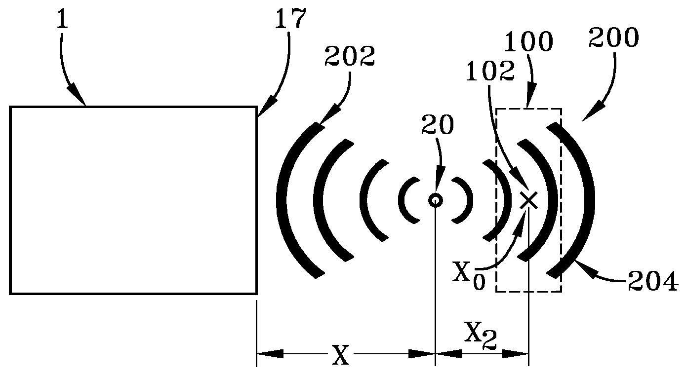
[0012]A method of attaching or reattaching a ligament, tendon, cartilage or other soft tissue to a bone mass has the steps of: positioning or placing the ligament, tendon, cartilage or other soft tissue adjacent or internal to the bone mass; anchoring or otherwise holding or fastening the ligament, tendon, cartilage or soft tissue to the bone mass; and transmitting shock waves to the ligament, tendon, cartilage or other soft tissue and the bone mass. Preferably the ligament, tendon, cartilage or other soft tissue is positioned in the path of the emitted shock waves and away from geometric focal volume or point of the emitted shock waves. The shock waves may be transmitted during the surgical procedure or post operatively in one or more treatment dosages or both. In so treating the ligament, tendon, cartilage or other soft tissue should be positioned at a distance away from any geometric focal point to minimize hemorrhaging. The soft tissue may include cartilage or muscle tissue. In the case of cartilage, the tissue can be inserted into a bone mass prepared cavity and optionally anchored there by a covering bone plug.
[0013]The transmitted shock waves are emitted using an applicator device which may be used by placing it inside a disposable sterile sleeve or cover. In such a case the applicator can be simply cleaned with a disinfecting agent prior to use as it is not directly exposed to the tissue. Alternatively the applicator without a sleeve or cover can be used wherein the applicator should be sterilized prior to use. In either use the sleeve or cover or the applicator without a cover should be coupled acoustically to the treated tissue or organ by a sterile coupling fluid or viscous gel like ultrasound gels or even NaCl solution to avoid transmission loss. Additionally a sterile cover or foil may be placed surrounding the treatment site.DEFINITIONS

Problems solved by technology

Often the ligaments or tendons are so damaged that they must be replaced using cadaver ligaments or tendons or other sources of such tissue.
Accordingly the period of time immediately after surgery and during rehabilitation are critical in that the reconstructed ligaments or tendons are more susceptible to damage until regrowth and reattachment to the bone mass occurs.
With young healthy patients the time to achieve regrowth is usually quicker, but the natural high activity of these patients' means that the risk of re-injury is greater during the time after surgery and prior to full attachment.
Conversely more elderly patients require a longer time to achieve reattachment, but are less active generally and therefore the risk of re-injury is over a longer time, but at a lower presumed level.

Method used


Image

  • Method of attaching soft tissue to bone
  • Method of attaching soft tissue to bone
  • Method of attaching soft tissue to bone

Examples


Embodiment Construction

[0046]When soft tissue tears away from bone, reattachment becomes necessary. Various devices including sutures alone, screws, staples, wedges and plugs have been used to secure soft tissue to the bone. Recently various types of threaded suture anchors have been employed for this purpose. Suture anchors are fasteners that are screwed into predrilled holes or otherwise self-tapping into a bone mass such that the suture anchor can be embedded in the bone mass wherein a suture can be placed through an opening in the anchor which can therefore be used to tie the ligaments, tendons or other soft tissue to the bone structure. This means to anchor the soft tissue around the bone insures that the ligament or tendon stays in a position that is most suitable for repair in that during the healing process the ligament or tendon can reattach itself to the underlying bone structure without being displaced or otherwise remain unattached.

[0047]As shown in FIG. 13, a representative suture anchor 70 i...


PUM


Abstract

A method of attaching or reattaching a ligament, tendon, cartilage or other soft tissue to a bone mass has the steps of: positioning or placing the ligament, tendon, cartilage or other soft tissue adjacent to the bone mass; anchoring or otherwise fastening the ligament, tendon, cartilage or soft tissue to the bone mass; and transmitting shock waves to the ligament, tendon or other soft tissue and the bone mass. Preferably the ligament, tendon, cartilage or other soft tissue is positioned in the path of the emitted shock waves and away from geometric focal volume or point of the emitted shock waves. The shock waves may be transmitted during the surgical procedure or post operatively in one or more treatment dosages or both. In so treating the ligament, tendon, cartilage or other soft tissue should be positioned at a distance away from any geometric focal point to minimize hemorrhaging. The soft tissue may include cartilage or muscle tissue. In the case of cartilage, the tissue can be inserted into a bone mass prepared cavity and optionally anchored there by a covering bone plug.

Description

RELATED APPLICATIONS[0001]This application claims benefit of priority to U.S. Provisional Patent Application Ser. No. 60 / 818,993 filed Jul. 6, 2006 entitled Method of Attaching Soft Tissue to Bone, the disclosure of which is incorporated herein by reference in its entirety.FIELD OF THE INVENTION[0002]The invention relates to an improved method of attaching soft tissue to bone, more particularly one of the methods relates to the use of shock waves combined with mechanical fasteners to accelerate healing and increase attachment adhesion, another deals with enhancing cartilage transplant healing.BACKGROUND OF THE INVENTION[0003]In severe injuries ligaments, tendons and muscle tissue surrounding the bones can be torn or otherwise detached from the skeletal structure. Often the ligaments or tendons are so damaged that they must be replaced using cadaver ligaments or tendons or other sources of such tissue. In other cases tissue such as cartilage is worn or degenerated and the repair incl...

Claims


Application Information

Patent Timeline
Patent Type & Authority Patents(United States)
IPC IPC(8): A61F2/08A61H1/00A61H1/02A61H5/00
CPCA61B17/0401A61N7/00A61B2017/0409A61B2017/044A61B2017/0414
Inventor WARLICK, JOHNSCHULTHEISS, REINERSCHADEN, WOLFGANG
Owner SOFTWAVE TISSUE REGENERATION TECH LLC
Learn More