

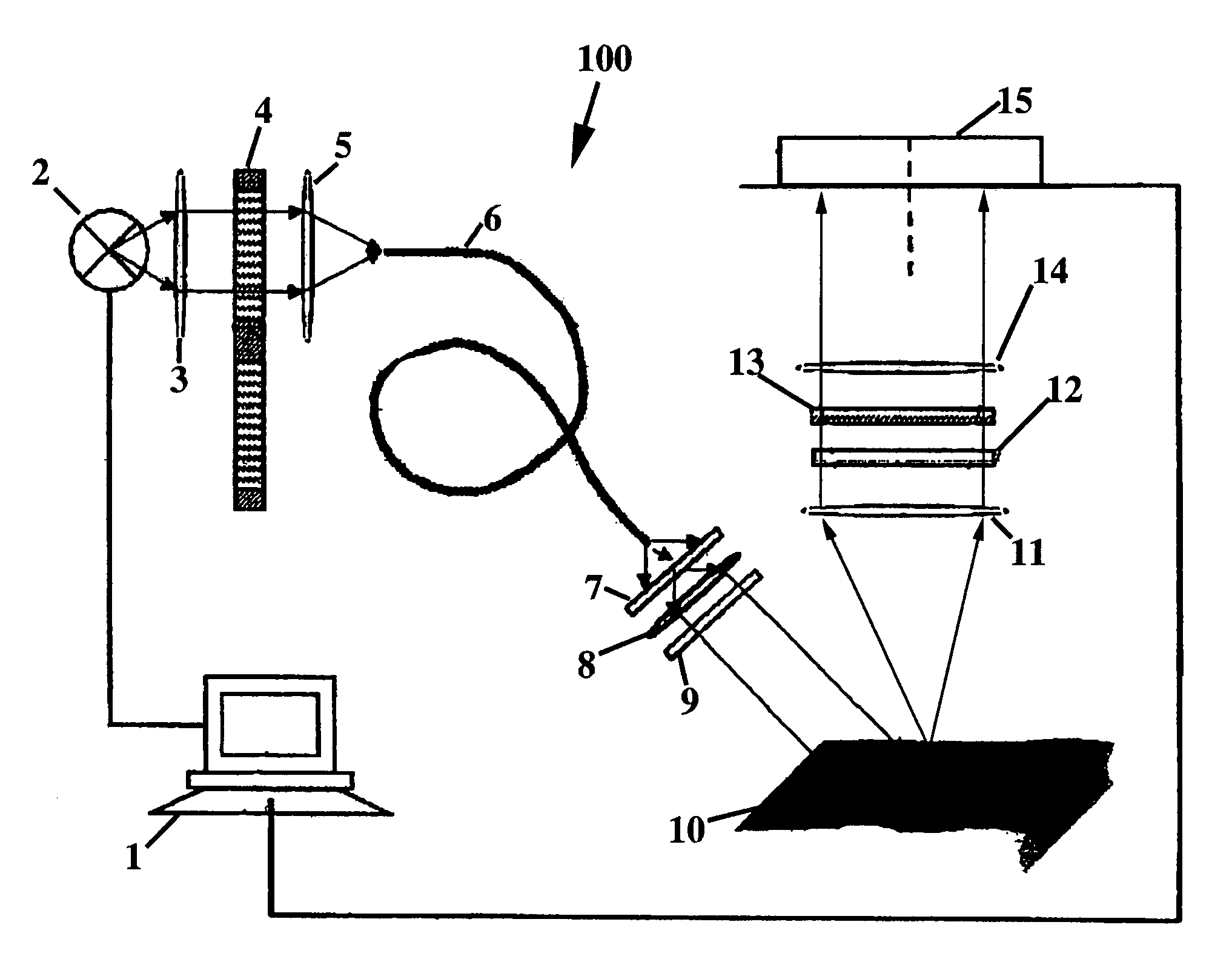
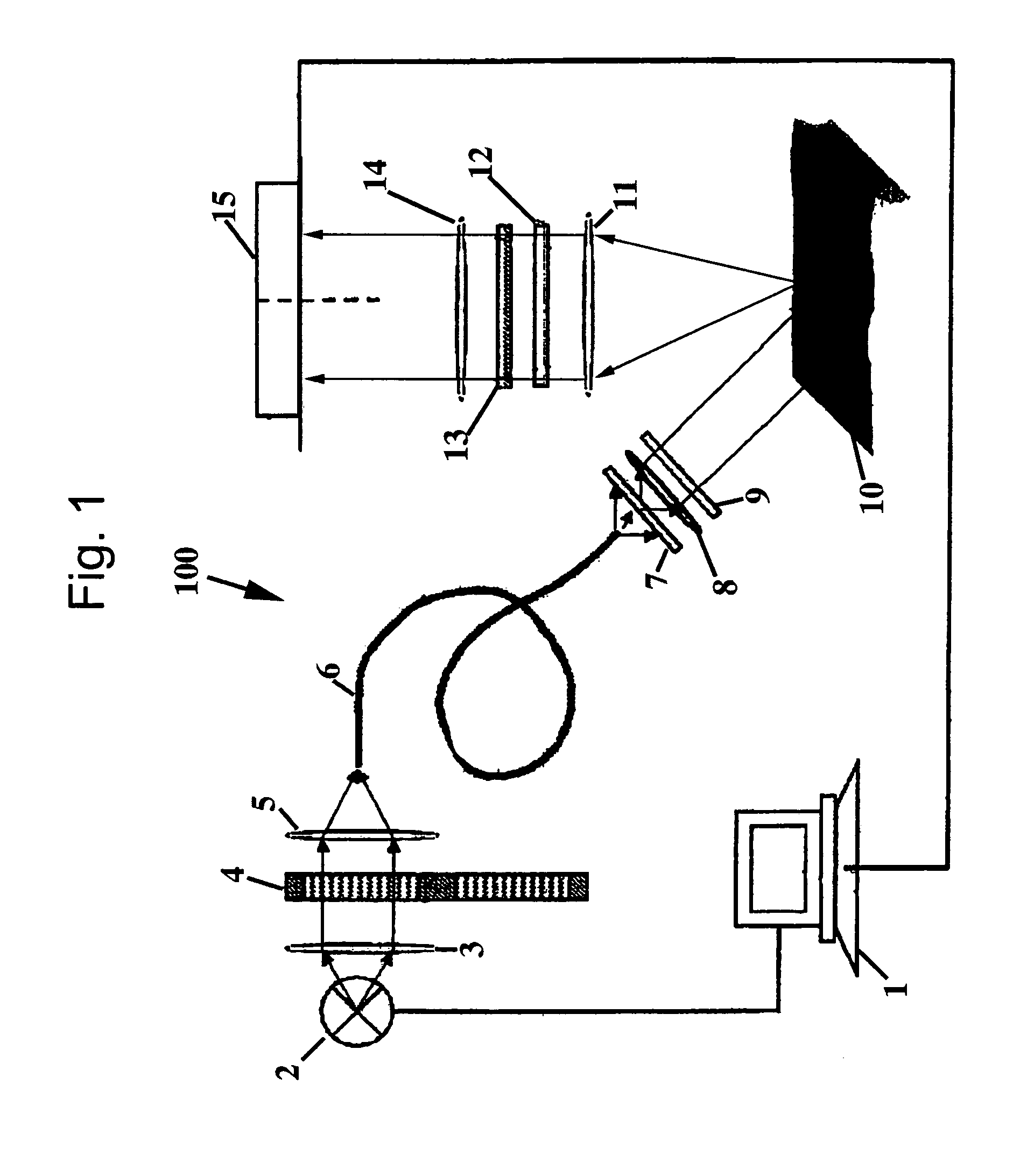
Polarized light imaging devices and methods
a polarized light and imaging device technology, applied in the field of real-time methods for imaging organic tissue, can solve the problems of inability to achieve the effect of real-time imaging, lack of one or more elements necessary for clinical use, and inability to achieve the effect of achieving the effect of superior spatial resolution and limited field of view
- Summary
- Abstract
- Description
- Claims
- Application Information
AI Technical Summary
Benefits of technology
Problems solved by technology
Method used
Image

Examples
examples
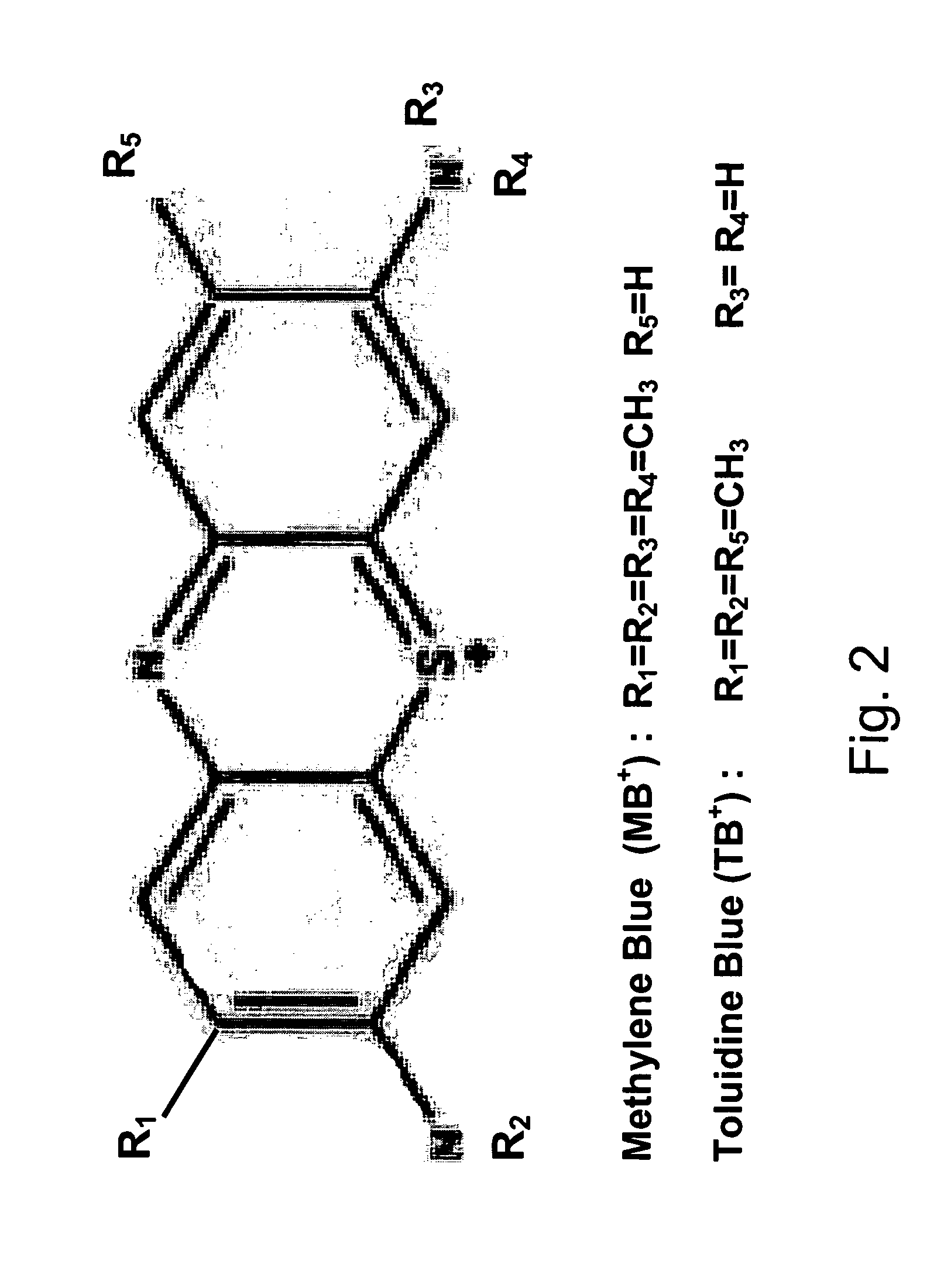
[0047]The present invention will be further described by referring to FIGS. 5-10, which depict images of tissues acquired by the PLI method. As will be readily apparent, applying contrast agents, such as MB and TB, greatly improves the contrast of the tumor within the image.
[0048]Example images of infiltrative morpheaform BCC obtained at the wavelengths of 410 nm and 610 nm before and after staining are presented in FIGS. 5a-5c and 6a-6d. The images acquired at the wavelength of 410 nm before and after contrast agent application are presented in FIGS. 5a and 5b, respectively. In FIGS. 5a and 5b, the areas contaminated with blood appear dark in the image. On the upper right and lower left boundaries of the sample, the Mohs stain can be seen. Due to high blood content in the sample and enhanced hemoglobin absorption at 410 nm, it is difficult to locate the tumor in the conventional images, presented in FIGS. 5a and b. FIG. 5a (specimen before staining) does not differ significantly fr...
PUM
| Property | Measurement | Unit |
|---|---|---|
| wavelength | aaaaa | aaaaa |
| wavelength | aaaaa | aaaaa |
| wavelengths | aaaaa | aaaaa |
Abstract
Description
Claims
Application Information