

Anti-HIV antibody
- Summary
- Abstract
- Description
- Claims
- Application Information
AI Technical Summary
Benefits of technology
Problems solved by technology
Method used
Image

Examples
example 1
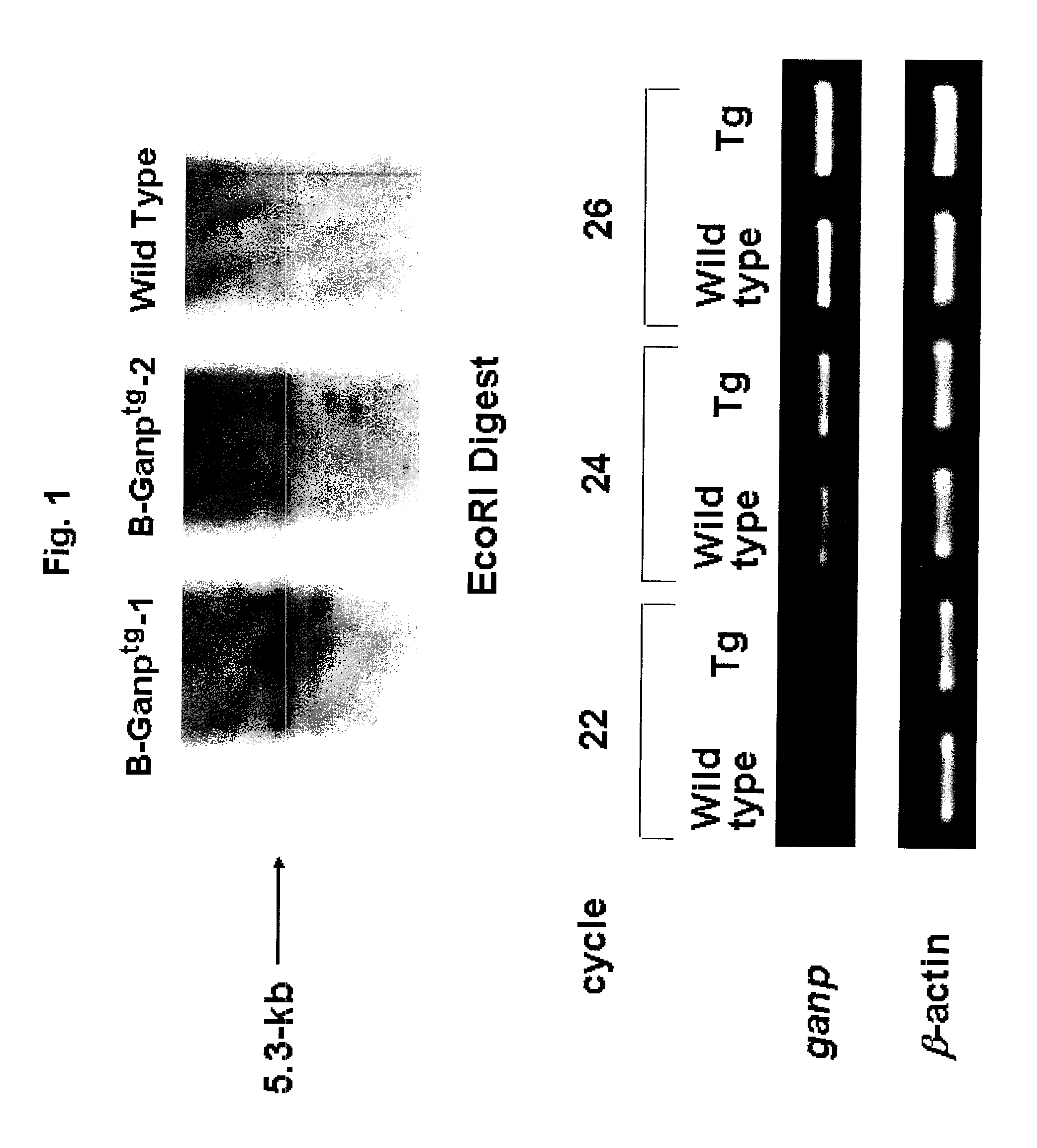
Preparation of GNAP Transgenic (Tg) Mouse
[0137]A transgene to be introduced into mice was prepared by inserting a 5.3 kb mouse GANP gene into the EcoRI site of pLG vector. This vector having a human immunoglobulin intron enhancer domain (2 kb EcoRi fragment) is a specific vector that directs strong expression in B cells. This gene was linearized and transferred into mice. Briefly, a linearized pLG vector (Koike, M. et al., Int. Immunol. 7, 21-30 (1995)) comprising the full-length mouse GANP cDNA was micro-injected into fertilized eggs of C57BL / 6 mice. The presence of the transferred gene was screened using the genomic DNA obtained from mouse tail, the following primers and the reaction solution (upper panel, FIG. 1). In the upper panel of FIG. 1, the band appearing at around 5.3 kb represents the GANP gene.
[0138]
1-5′ primer:5′-TCCCGCCTTCCAGCT GTGAC-3′(SEQ ID NO: 7)1-3′ primer:5′-GTGCTGCTGTGTTATGTCCT-3′(SEQ ID NO: 8)
Composition of the Reaction Solution
[0139]
DNA (50 ng / μl) 1 μl10× bu...
example 2
Preparation of Antibodies
(1) Materials
[0145](a) Animals: Balb / c mouse, wild type (WT) mouse and GANP transgenic (Tg) mouse[0146](b) Immunizing antigen: HIV24NL43 peptide / KLH conjugation[0147](c) ELISA antigen: HIV24NL43 peptide (sequence: CNNTRKSIRI QRGPGRAFVT IGKI (SEQ ID NO: 9))[0148](d) Myeloma cell: P3-X63.Ag8.U1[0149](e) Secondary antibody: HRP-labeled anti-mouse antibodies IgC, IgA and IgM
(2) Methods
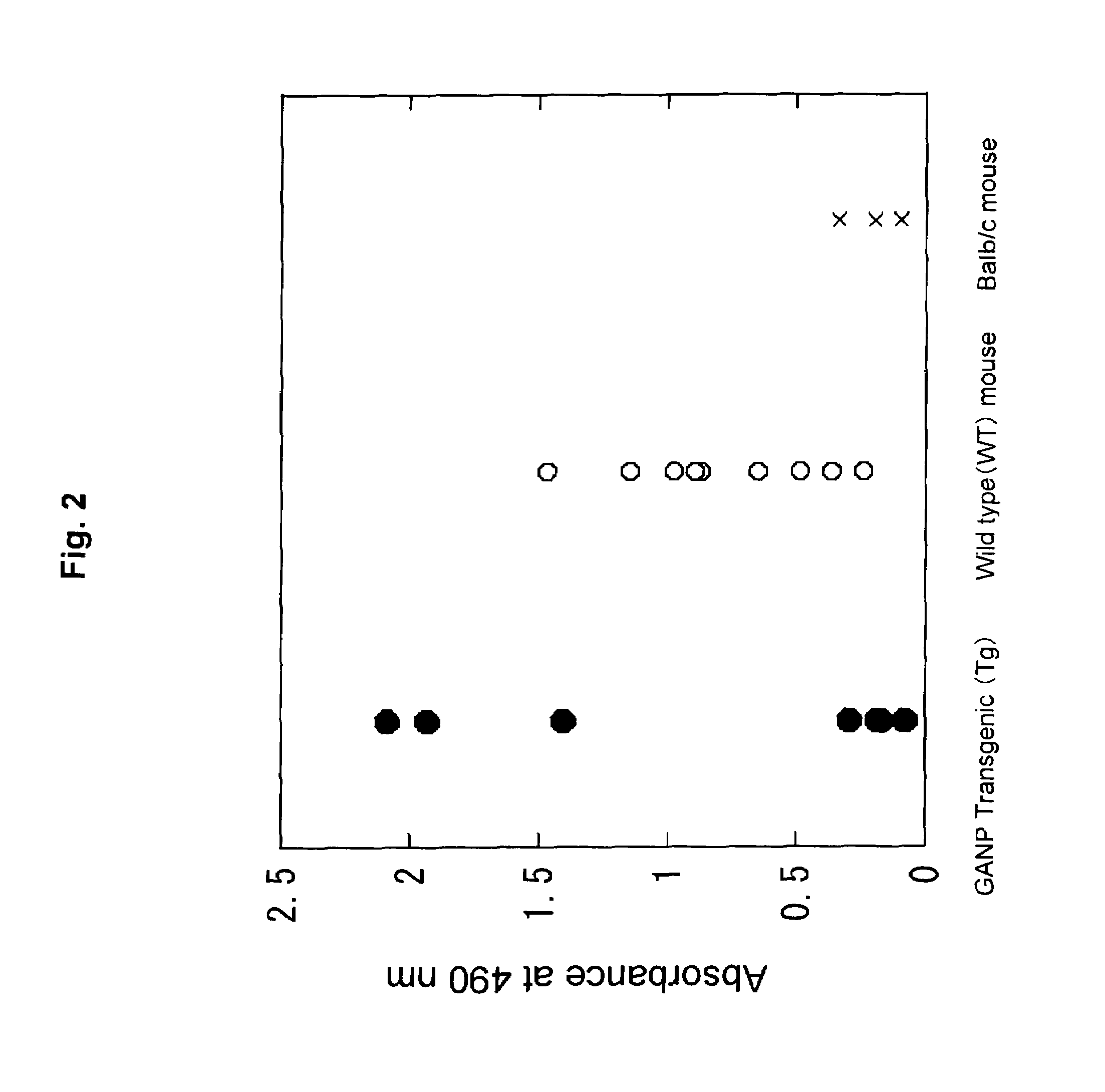
[0150]Five individuals each of the above-described three types of mice were immunized with NL43 peptide (carrier protein: KLH) three times at intervals of two weeks. After the third immunization, antisera were collected and subjected to ELISA to measure antibody titers.
[0151]Two individuals showing high antibody titers were selected from each type of mice used, and antibody-producing cells (splenic cells) were collected therefrom and fused to P3U1 myeloma cell. Cells were plated to give a density of 0.2×10⁵ GANP-Tg mouse splenic cells / well. Balb / c mouse-derived 5448 clones, wild-ty...
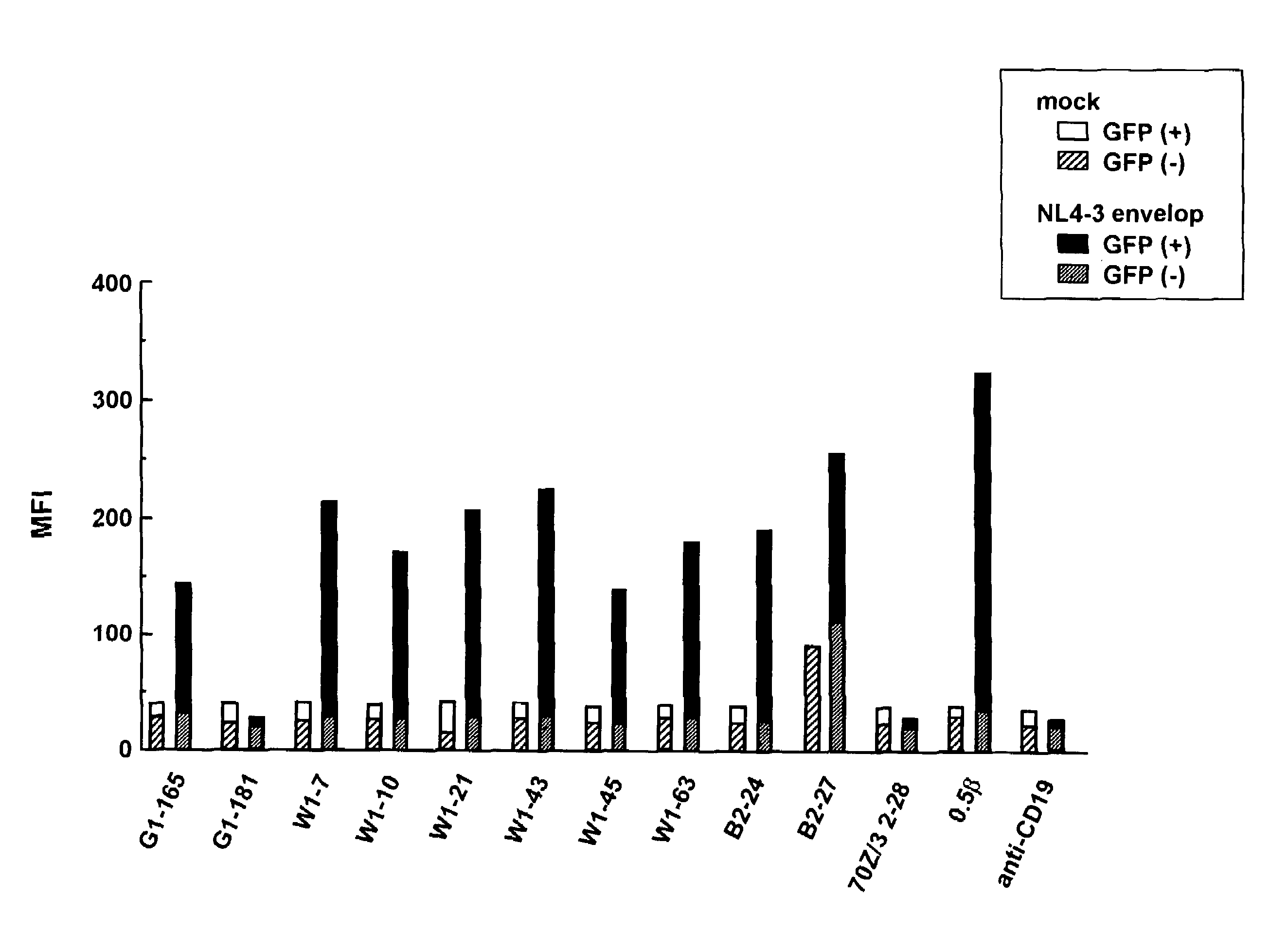
example 3
Measurement of Affinity
[0156]Using the monoclonal antibodies prepared in Example 2 above, the following evaluation and examination were carried out.
[0157]In order to evaluate the affinity of each antibody, analyses by ELISA and with a Biacore system were conducted.
[0158]First, in ELISA, HIV24NL43 peptide (1 μg / ml) was used as immobilized antigen and immobilized at room temperature for one hour. The antigen-immobilized plate was washed with PBSTween 20 and blocked with 2.0% skim milk. After further washing with PBSTween 20, the antigen was reacted with the anti-peptide monoclonal antibodies (0.457-1 μg / ml) to be evaluated, at room temperature for one hour. Then, the resultant samples were washed with PBSTween 20 and reacted with HRP-labeled anti-mouse IgG, IgA or IgM at room temperature for one hour. After washing with PBSTween 20, color was developed with ortho-phenylene diamine (OPD) for 5 minutes, followed by temlination of the reaction with 2N sulfuric acid.
[0159]Absorbance was m...
PUM
| Property | Measurement | Unit |
|---|---|---|
| Molar density | aaaaa | aaaaa |
| Affinity | aaaaa | aaaaa |
Abstract
Description
Claims
Application Information