

Use of islet 1 as a marker for isolating or generating stem cells
a stem cell and marker technology, applied in the field of in vitro expansion and propagation of undifferentiated progenitor cells, can solve the problems of encumbrance in research as well as therapeutic potential, less study, and inability to study the origin of cells with characteristics of a second tissue or the clonal origin of differentiated cells. to achieve the effect of enhancing expression of islet1
- Summary
- Abstract
- Description
- Claims
- Application Information
AI Technical Summary
Benefits of technology
Problems solved by technology
Method used
Image

Examples
example 1
Experimental Procedures
Mouse Mutants
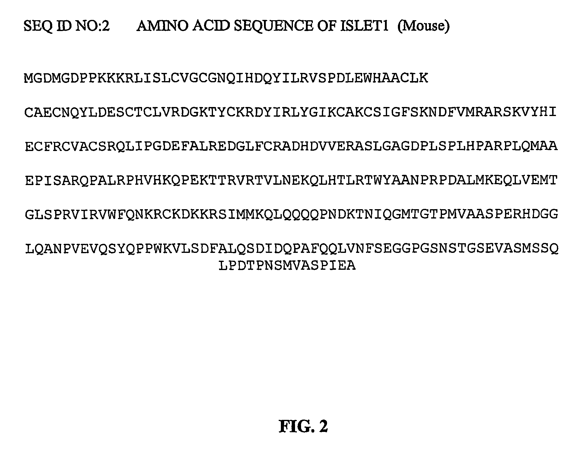
[0096]The generation of islet null mutants has been previously described (Pfaff et al, 1996). The knockout construct deleted the exon encoding the second LIM domain of Islet 1. Islet1-cre mice were generously provided by Thomas M. Jessell, and have been previously described (Srinivas et al, 2001). An IRES-cre cassette was inserted into the exon encoding the second LIM domain of Islet1, disrupting islet gene expression.
Whole Mount RNA In Situ Hybridization
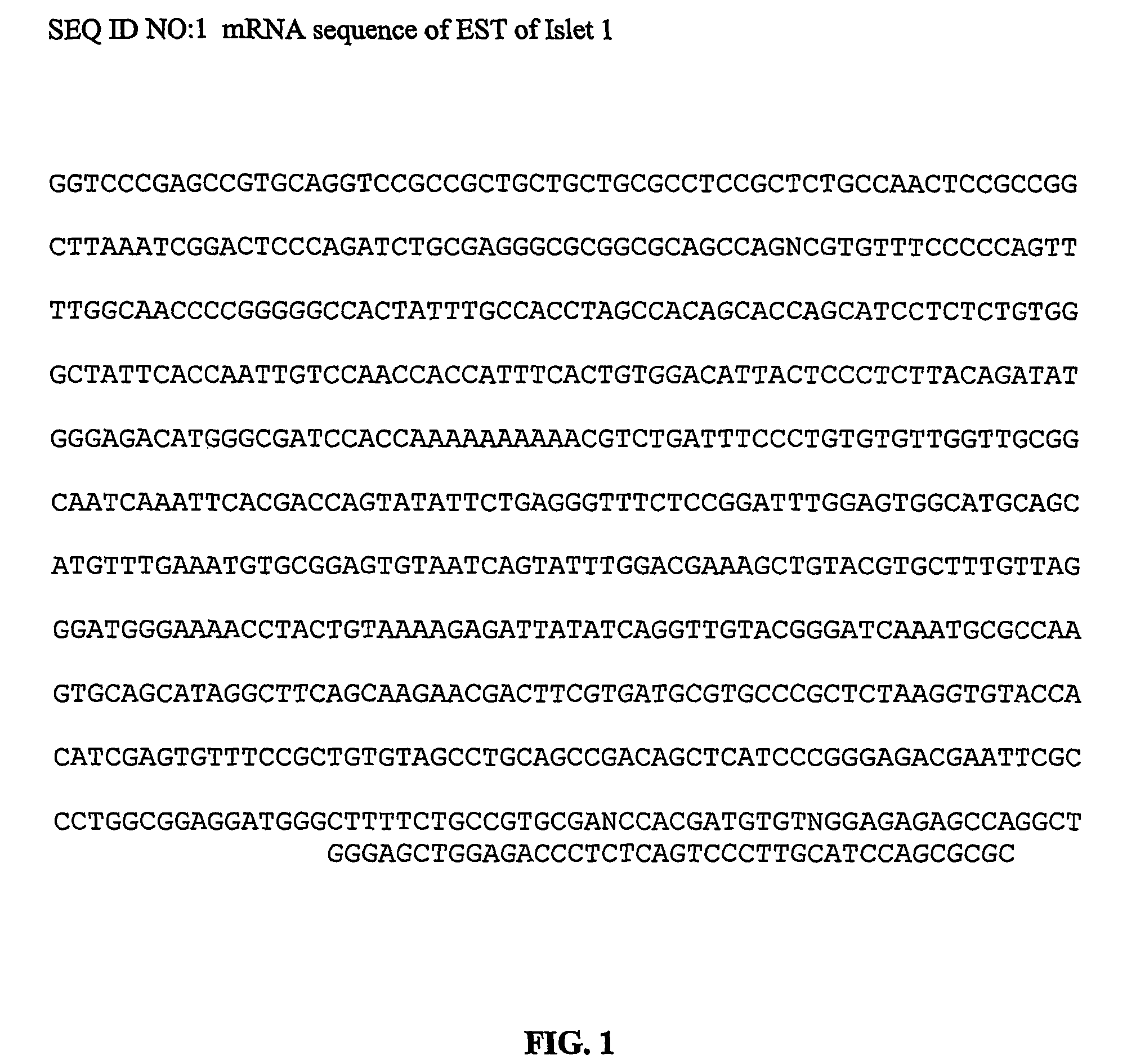
[0097]Whole mount RNA in situ hybridization was carried out as previously described (Wilkinson, 1999). References for specific RNA probes which were used are as follows: MLC2a (Kubalak et al., 1994); MLC2v (O'Brien et al., 1993); tbx5 (Bruneau et al., 1999); tbx20 (unpublished results); BMP2, BMP6, BMP7 BMP4, BMP5 (Kim et al., 2001; Lyons et al., 1995); FGF4, FGF8 and FGF10 (Feldman et al., 1995; Sun et al., 1999); EHand (Cross et al., 1995); islet1 (EST, GenBank Accession No.: AA198791)(SEQ ID NO...
example 2
Culture of Undifferentiated I-Cells
[0100]To isolate cardiac progenitors from murine hearts, the methods used were similar to those used for cardiomyocyte isolations from the adult organ. In a trypsin-digested state
[0101]i-cells and cardiac fibroblasts share a similar cell diameter of around 35 μm and copurify in the same fractions on Percoll gradients. I-cell cultures were developed by testing multiple conditions, including cultures on fibronectin, collagen-type-IV or laminin. Media conditions tested included several concentrations of fetal calf serum (FCS), epidermal growth factor (EGF), platelet derived growth factor (PDGF-BB), acidic fibroblast growth factor (aFGF), basic fibroblast growth factor (bFGF), bone morphogenetic protein (BMP) 2+4, insulin-like growth factor (IGF) 1, sonic hedgehoc (Shh) and dexamethasone, as set forth below.
[0102]Approximately 5-10% of 96-well plates seeded with 10 isl 1 positive cells yielded continuous growing cultures, indicating that only around 5-...
example 3
In vitro Differentiation of Single I-Cells
[0104]Next in vitro differentiation capacity of i-cells obtained from mouse and rat hearts was tested by adding cytokines chosen on the basis of what has been reported for ES cell differentiation to neuroectoderm and mesoderm. Differentiation required that i-cells had to be replated without a feeder layer at a density around 1-2×10⁴ cells cm−2 in medium containing no serum, but lineage-specific cytokines. Neuroprogenitors can be expanded with PDGF-BB and induced to differentiate by addition of bFGF (Palmer, et al., 1999).
[0105]Under the bFGF treatment around 45% of the i-cells acquired morphologic and phenotypic characteristics of astrocytes with immunohistochemical positivity for glial acidic fibrillary protein (GFAP) and neurons which stained positive for neurofilament 200 (NF-200). Myocytic differentiation acquired cells that showed positive signals for α-sarcomeric actin and α-actinin in immunohistochemical experiments.
PUM
| Property | Measurement | Unit |
|---|---|---|
| pH | aaaaa | aaaaa |
| pH | aaaaa | aaaaa |
| cell diameter | aaaaa | aaaaa |
Abstract
Description
Claims
Application Information