
Biomarkers of tuberculosis that distinguish disease categories: use as serodiagnostic antigens

a tuberculosis biomarker and disease category technology, applied in the field of microbiology, immunology and medicine, can solve the problems of difficult to complete the serological analysis of patients' serological reactivity to a large proportion of the i>mycobacterium tuberculosis /i>(mtb) proteome, difficult to achieve high-throughput testing, and easy to use methods

Inactive Publication Date: 2010-08-17
NEW YORK UNIV +1
View PDF6 Cites 8 Cited by
  • Summary
  • Abstract
  • Description
  • Claims
  • Application Information

AI Technical Summary

Benefits of technology

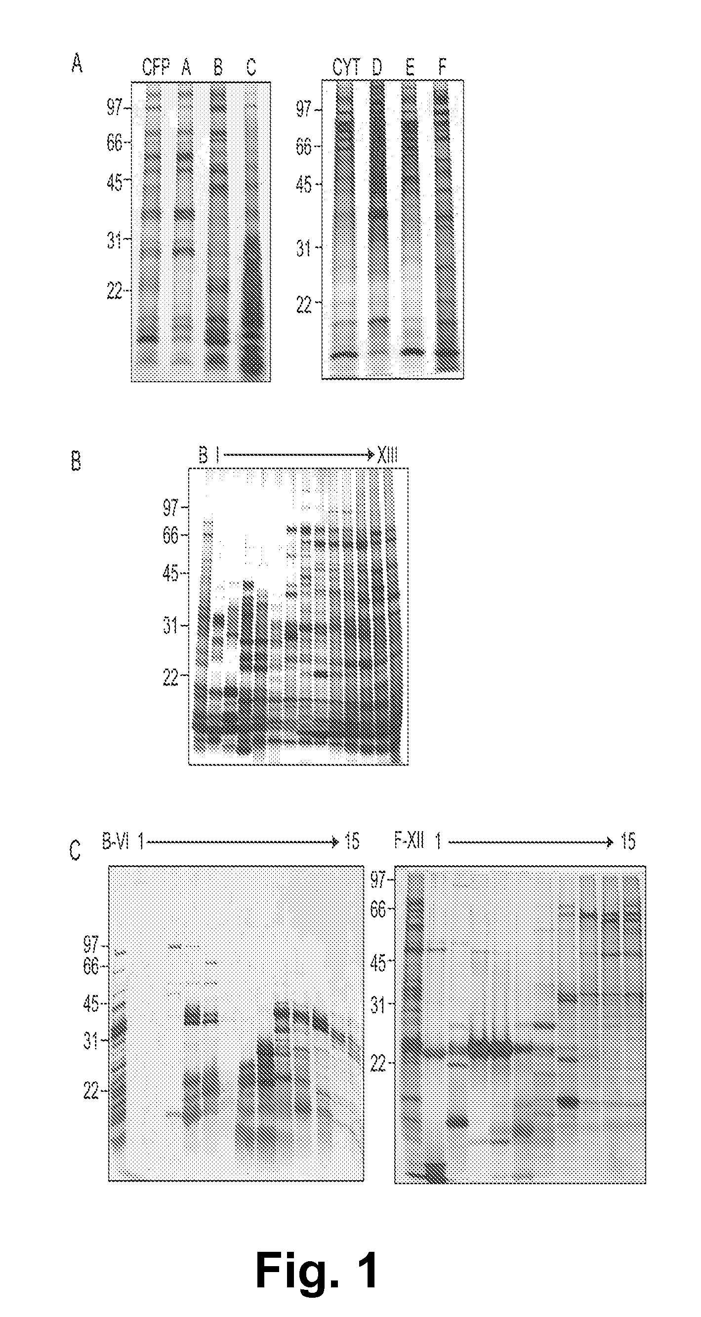
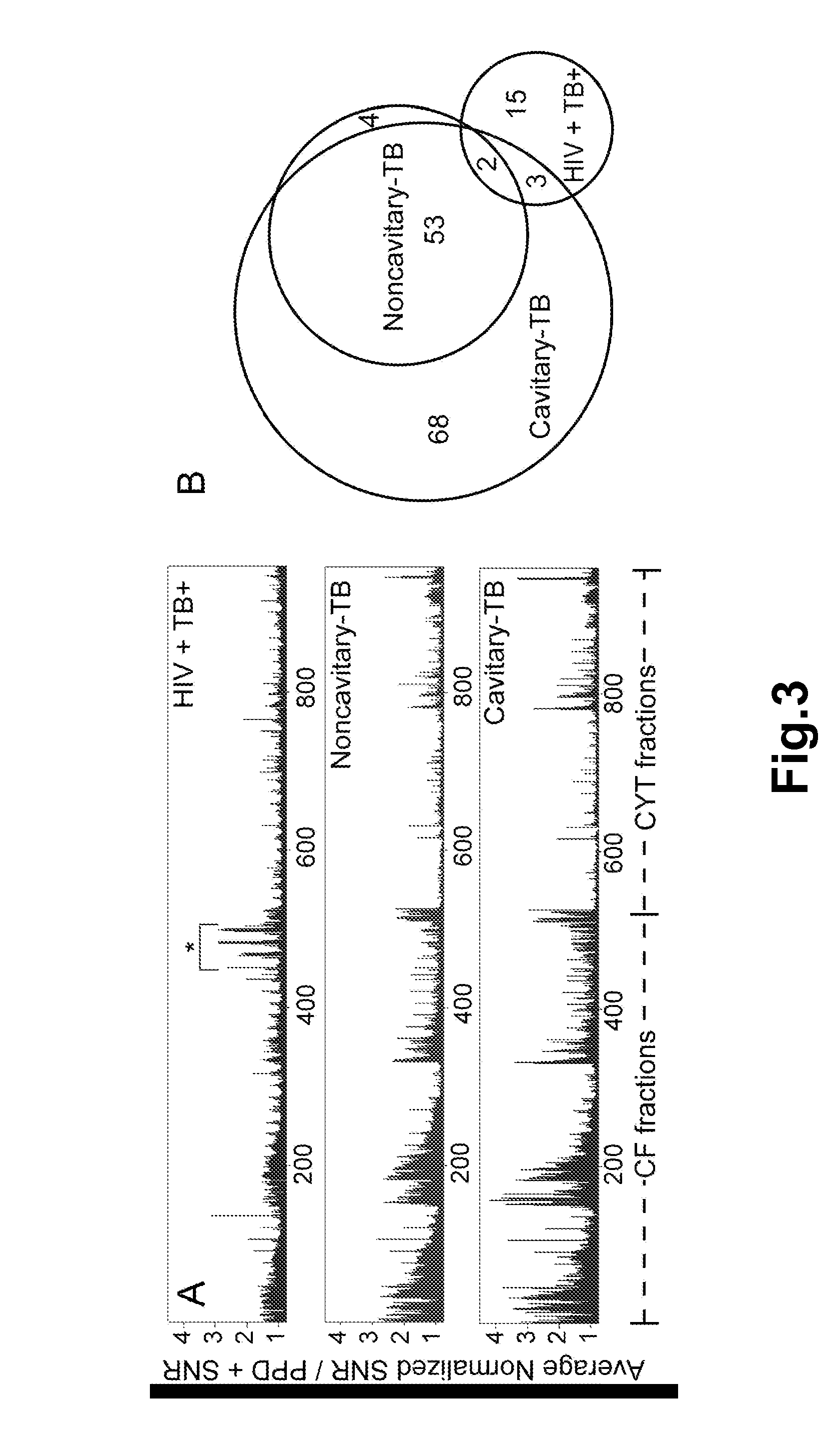
The patent text describes a method for identifying proteins in the tuberculosis bacteria, Mycobacterium tuberculosis, using a combination of two-dimensional separation and mass spectrometry. The technique was used to separate native M. tuberculosis cytosol proteins and culture filtrate proteins, resulting in 960 unique protein fractions. The antibodies from patients with tuberculosis and healthy individuals were tested for recognition of the proteins, and a total of 21 antigens were identified. The invention also includes isolated M. tuberculosis proteins that are recognized by antibodies from patients with tuberculosis, including LppZ, SodC, BfrB, and TrxC. The peptides that represent the antibody recognition sites on these proteins are also provided. The technique can be used to develop protein microarrays for the diagnosis and treatment of tuberculosis.

Problems solved by technology

The development of such a test has proven to be one of the greatest challenges in TB research.
However, a complete analysis of patients' serological reactivity to a large proportion of the Mycobacterium tuberculosis (Mtb) proteome is hindered by the inability to evaluate the reactivity of the nearly 4,000 predicted proteins of Mtb in a high-throughput fashion.
Moreover, this methodology is facile and allows reproducible screening of sera from a large number of individual patients.
However, this methodology is not suitable for high-throughput testing.

Method used


Image

  • Biomarkers of tuberculosis that distinguish disease categories: use as serodiagnostic antigens
  • Biomarkers of tuberculosis that distinguish disease categories: use as serodiagnostic antigens
  • Biomarkers of tuberculosis that distinguish disease categories: use as serodiagnostic antigens

Examples


example i

Experimental Procedures

Preparation of Mtb Subcellular Fractions

[0133]Mtb strain H37Rv was expanded from a 1-ml frozen stock (approximately 108 colony-forming units per ml) to 24 L of late log culture in glycerol-alanine salts medium (17). The culture supernatant was separated from the cells and processed to generate the CFPs of Mtb as previously described (18). The Mtb H37Rv cells (88.9 g wet weight) were washed 3 times with PBS (pH 7.4), frozen at −70° C. and inactivated with 24 kGy of γ-irradiation. Lysis of these cells was achieved by suspending in 44 ml of TSE buffer [10 mM Tris-HCl (pH 7.4), 150 mM NaCl, 1 mM EDTA] containing 0.06% DNase, 0.06% RNase, 0.07% pepstatin, 0.05% leupeptin and 20 μM PMSF and passing through a French Press five times at 1,500 psi. The resulting lysate was diluted with 1 vol of TSE buffer and centrifuged at 2,000×g for 5 min to remove unbroken cells. The cytosol was obtained as the final supernatant of sequential centrifugations at 27,000×g and 100,000...

example ii

Multi-Dimensional Protein Fractionation Reduced the Complexity of Mycobacterial Protein Pools

[0147]Recent advances in proteomics have enabled the separation and identification of individual proteins or peptides from complex biological samples. Specifically, multi-dimensional chromatography of peptides, derived from tryptic digests of crude biological samples, followed by MS / MS analysis (“MudPIT”) has provided experimental validation of substantial portions of theoretical proteomes (25). Recently, this approach was applied to Mtb and resulted in more than a threefold increase in the number of proteins that had previously been identified by 2-D gel electrophoresis methods (26).

[0148]Applying a similar strategy, the present study employed a multi-dimensional fractionation scheme (FIG. 6) for efficient separation of complex pools of intact Mtb proteins into relatively simple and enriched fractions that could be evaluated for serological reactivity in a high-throughput fashion.

[0149]Aliq...

example iii

Validation of the Protein Microarray Format

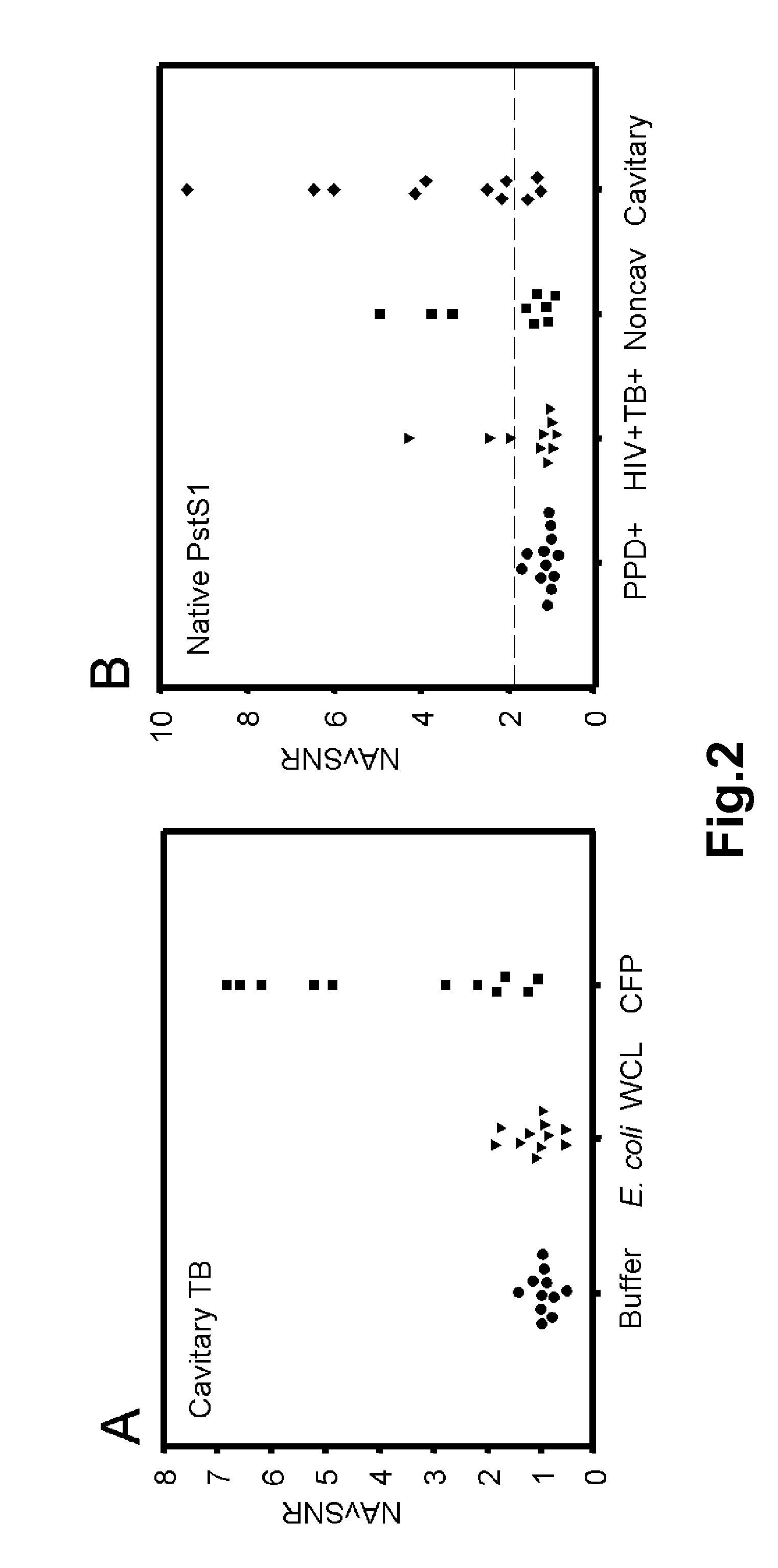
[0150]Each multidimensional chromatography fraction, as well as intermediate fractions and recombinant proteins were printed to nitrocellulose microarray slides. Serum samples from PPD+ healthy subjects (n=12), noncavitary-TB subjects (n=9), cavitary-TB subjects (n=11), and HIV+TB+ (n=10) subjects were probed against the microarrays in triplicate. For each slide the NAvSNR (see Example I) was calculated for each protein or protein fraction. The integrity of the protein microarray and validation of this platform was determined by assessing the reactivity of TB patients' sera with selected protein fractions and with purified proteins that were spotted on the microarray slides as controls, and comparing these results to published results obtained by plate ELISA (5, 6).

[0151]To demonstrate specificity, the reactivity of TB patients' serum samples towards unfractionated CFP was compared to reactivity towards either E. coli WCL control or FAST® p...


PUM

PropertyMeasurementUnit
concentrationaaaaaaaaaa
molecular weightaaaaaaaaaa

Abstract

Mycobacterial proteins from culture filtrate or cytosol are disclosed as being useful B cell antigens for early diagnosis of mycobacterial disease, particularly in humans. These proteins include four that had not previously been recognized as B cell antigens (LppZ protein encoded by Mtb gene Rv3006; SodC protein encoded by Mtb gene Rv0432; BfrB protein encoded by Mtb gene Rv3841 and TrxC protein encoded by Mtb gene Rv3914). Antigenic compositions include these proteins and / or peptide fragments thereof, in various combinations with each other or with one or more of a set of 10 additional Mtb proteins known to be antigens (in particular early antigens. Methods and kits for using these antigenic composition for early diagnosis of mycobacterial infection and disease are also disclosed.

Description

[0001]This application claims priority from U.S. provisional application No. 60 / 836,653, filed Aug. 10, 2006, and incorporates by reference all the contents thereof.STATEMENT OF RIGHTS TO INVENTIONS MADE UNDER FEDERALLY SPONSORED RESEARCH[0002]This invention was funded in part by grants and contracts from the National Institute of Health, specifically, NIAID grant RO1 AI-056257 and Contract NO1 AI-75320, which provides to the United States government certain rights in this invention.BACKGROUND OF THE INVENTION[0003]1. Field of the Invention[0004]The invention in the field of microbiology, immunology and medicine relates to novel proteins, peptides and antigenic compositions for early diagnosis of mycobacterial disease, kits comprising such compositions, and serodiagnostic methods using such compositions.[0005]2. Description of the Background Art[0006]Recent World Health Organization data show that the global incidence of tuberculosis (TB) is increasing at 1% per year and that there ...

Claims


Application Information

Patent Timeline
Patent Type & Authority Patents(United States)
IPC IPC(8): A61K39/04A61K39/00A61K39/02
CPCC07K14/35G01N33/5695
Inventor BELISLE, JOHN T.SARTAIN, MARK J.LAAL, SUMAN
Owner NEW YORK UNIV
Learn More