Detection of tuberculosis and infection by Mycobacterium tuberculosis using HBHA

a technology of mycobacterium tuberculosis and detection field, which is applied in the field of detection of tuberculosis and infection by i > myco, can solve the problems of limiting the utility of culturing as a diagnostic test of the first intention, and affecting the detection accuracy

Inactive Publication Date: 2010-11-16
INSTITUT PASTEUR DE LILLE +3
View PDF2 Cites 21 Cited by
  • Summary
  • Abstract
  • Description
  • Claims
  • Application Information

AI Technical Summary

Problems solved by technology

In 2004, tuberculosis still represented a major public health problem as it is responsible for more than two million deaths per year worldwide and a third of the worldwide population is infected by the responsible agent, Mycobacterium tuberculosis, routinely known as Koch's bacillus or KB.
However, the results of mycobacterial cultures can only be interpreted after 6 to 8 weeks, and developing countries do not always have access to the infrastructure required for this technique, which greatly limits the utility of culturing as a diagnostic test of the first intention (3).
The determination of acido-resistant bacillae in sputum thus remains the most useful test for rapid diagnosis of pulmonary tuberculosis, but identification is unfortunately only positive in 50% to 60% of cases of pulmonary tuberculosis, partly due to the fact that 5000 to 10000 bacillae per μl of sputum are required for the test to be positive (3).
Diagnosing pulmonary tuberculosis is even more difficult in a child who, when young, rarely expectorates, and from whom a gastric aspiration is usually collected.
However, direct examination of such samples is positive in less than 20% of children with proven tuberculosis, which is much lower than the results obtained in the adults (4).
Finally, a diagnosis of extra pulmonary tuberculosis, which is more frequent in children than in adults, is still difficult to make.
This last test cannot, however, readily differentiate individuals infected with M. tuberculosis from those vaccinated with BCG, and cross reactions with environmental mycobacteria render its specificity poor.
That test, which is based on the demonstration of a cellular immune response, also has poor sensitivity in immunodeficient individuals.
Finally, though it enables to identify persons infected with M. tuberculosis in a population of unvaccinated individuals, it unfortunately cannot differentiate individuals presenting a latent tuberculosis (symptom-free) from those with an active disease.
The practical use of that test, then, is extremely limited.
However, the technique is expensive, limiting its application.
An analysis of IFN-γ secretion induced by tuberculin, a complex mixture of mycobacterial antigens, however, has the same limitations as its use for skin tests.
However, discrimination between these two groups of individuals on the basis of these preliminary results was insufficient for conducting a diagnostic test.

Method used


Image

  • Detection of tuberculosis and infection by <i>Mycobacterium tuberculosis </i>using HBHA
  • Detection of tuberculosis and infection by <i>Mycobacterium tuberculosis </i>using HBHA
  • Detection of tuberculosis and infection by <i>Mycobacterium tuberculosis </i>using HBHA

Examples


example 1

Origin of Blood Samples

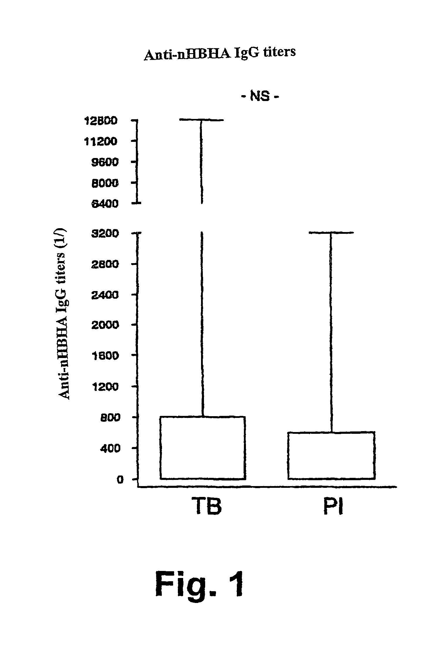
[0129]Blood samples were obtained from tuberculous patients and from patients with latent tuberculous after having obtained their consent. The tuberculous patients were selected on the basis of a positive direct examination and / or a positive culture for Mycobacterium tuberculosis. All subjects were enrolled before the end of the first three weeks of treatment. The subjects having a latent form of tuberculosis were selected on the basis of a positive delayed hypersensitivity test to tuberculin (diameter of induration over 18 mm at the time of diagnosis). An active form of tuberculosis was excluded on the basis of a normal thorax radiograph. All of the patients were seronegative for HIV and none had received immunosuppressor treatment. All of the subjects were living in Europe at the time of recruitment

example 2

Other Samples

[0130]Broncho-alveolar lavage fluid (BAL) was removed, by fibroscopy, after injecting about 200 ml of physiological water. The volume removed was then centrifuged (about 10 ml).

[0131]Pleural, articular, peritoneal, cephalorachidian and other fluids were collected in sterile syringes and were centrifuged as soon as they arrived at the laboratory.

[0132]For ganglia or other suspect masses, surgical exeresis was carried out. Upon its arrival at the laboratory, the anatomical fragment collected was morcellated and incubated in a culture medium to allow progressive release of cells from the tissue.

example 3

Antigens

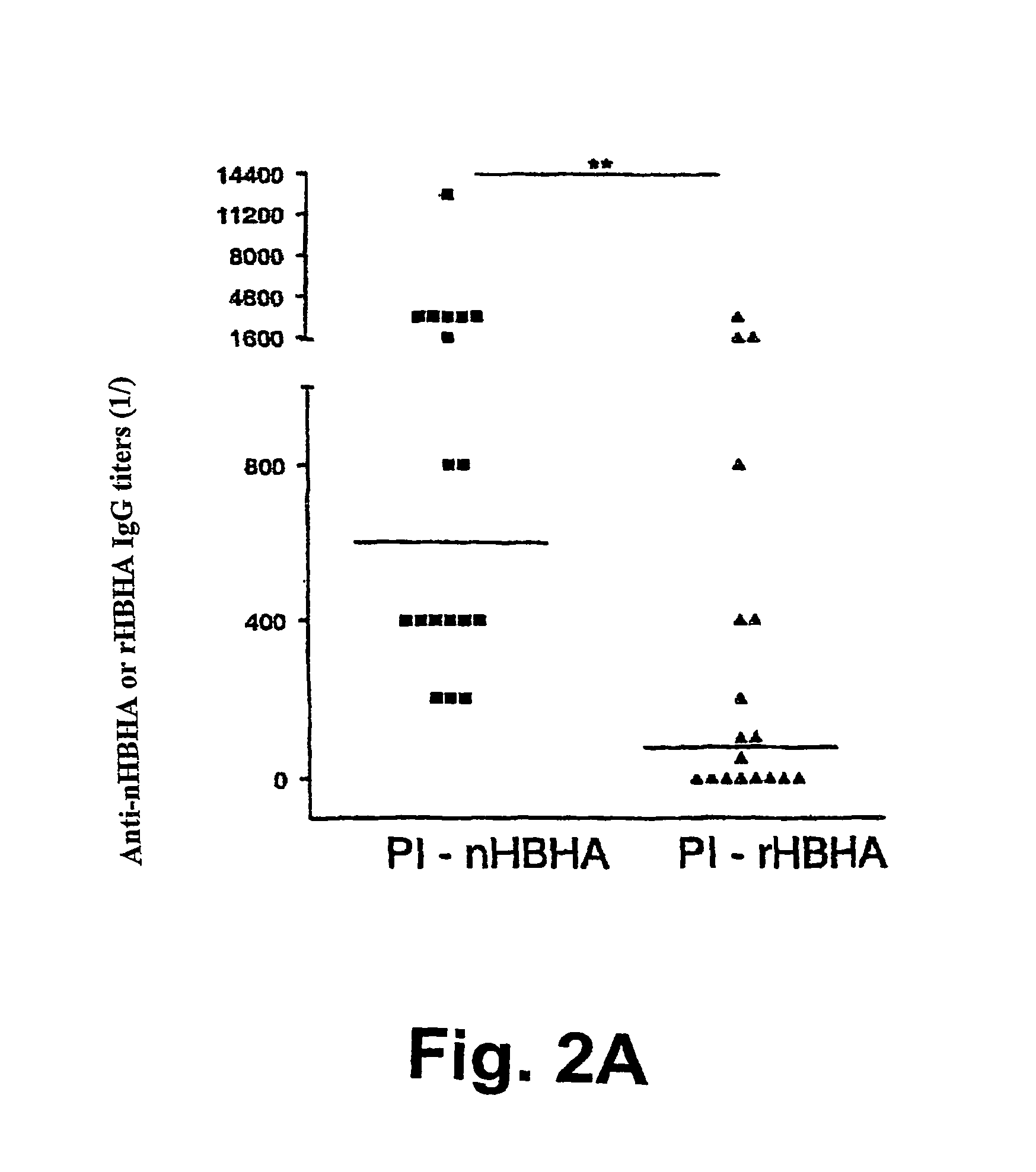
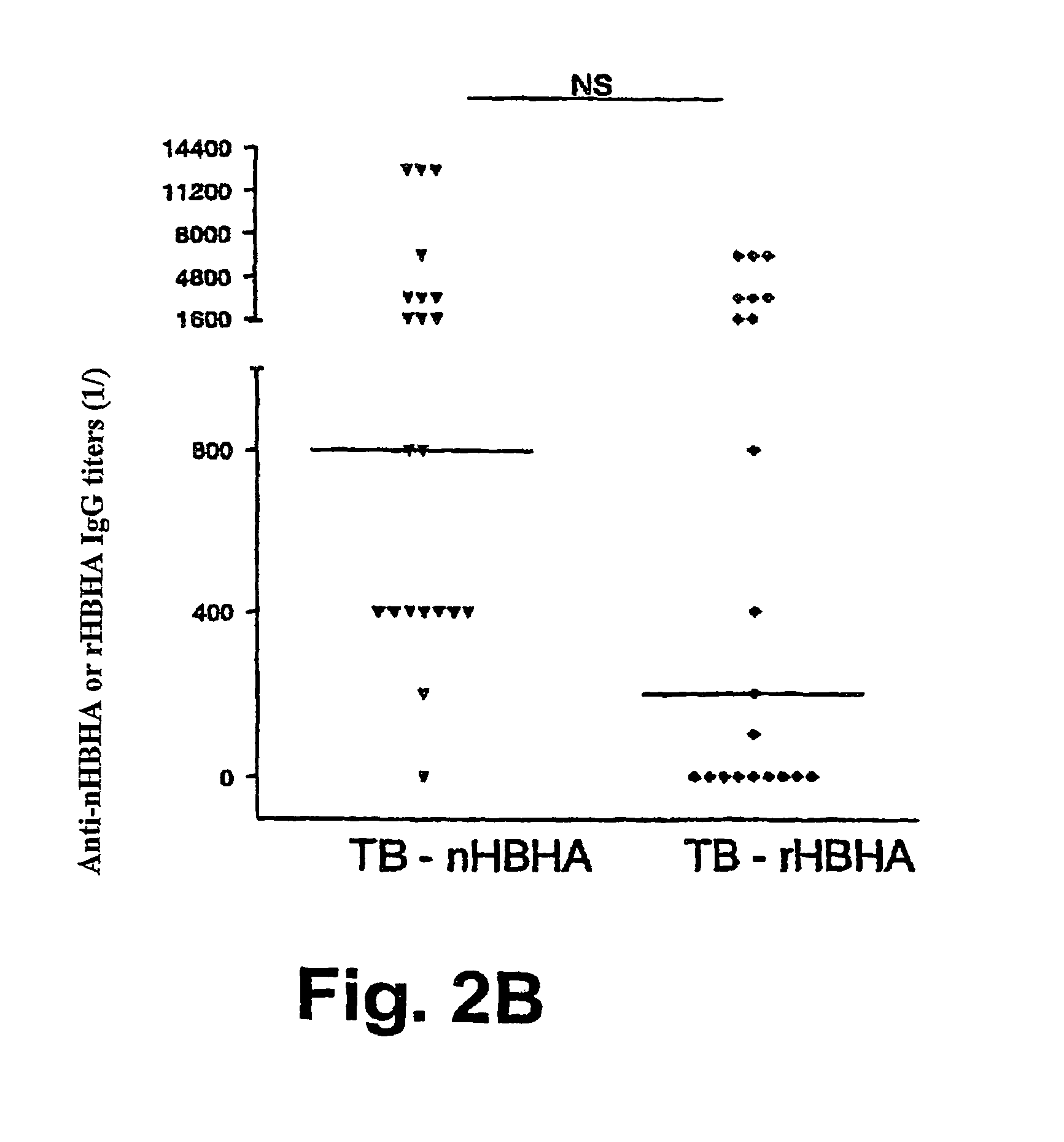
[0133]The native form of HBHA (nHBHA) was purified from M. bovis BCG by heparin-sepharose chromatography followed by high pressure fluid chromatography (HPLC) as described elsewhere (14). An HPLC chromatogram and SDS-PAGE analysis after staining with Coomassie Blue proved that the preparation had no protein contamination. No traces of glycolipids were found using gas chromatography. The limulus test showed that the lipopolysaccharide concentration was less than 10 pg / ml.

[0134]The non methylated recombinant form (rHBHA) was purified from the E. coli strain (BL21(DE3)(pET-HBHA). The degree of methylation of the recombinant protein compared with that of the native protein was described in French patent FR-A-01 / 14953.

[0135]The truncated recombinant form of the C terminal portion (rHBHAΔC) was purified from the E. coli strain (BL21(DE3)(pET-rHBHAΔC). This truncated form rHBHAΔC was described in Pethe et al (2000 Journal of Biological Chemistry 275(19): 14273-14280, incorporated i...


PUM

PropertyMeasurementUnit
temperatureaaaaaaaaaa
concentrationaaaaaaaaaa
pHaaaaaaaaaa

Abstract

The present invention concerns methods for in vitro detection of an infection by Mycobacterium tuberculosis in mammals, and methods for in vitro distinction between mammals infected with Mycobacterium tuberculosis in which the disease is declared (active form) and mammals which are infected but asymptomatic for tuberculosis (latent form), and a method for in vitro distinction between mammals presenting an active form of tuberculosis and mammals not infected by M. tuberculosis or presenting a latent form of tuberculosis. The present invention also pertains to kits for detection and distinction between infected mammals presenting tuberculosis symptoms and infected mammals with no disease development, and a kit for distinguishing between mammals presenting an active form of tuberculosis and mammals not infected by M. tuberculosis or presenting a latent form of tuberculosis.

Description

[0001]The present invention relates to methods for in vitro detection of infection with Mycobacterium tuberculosis in mammals and to methods for in vitro distinction between mammals infected with Mycobacterium tuberculosis for which the disease is declared (active form) and mammals infected but asymptomatic for tuberculosis (latent form), and to a method for in vitro distinction between mammals presenting an active form of tuberculosis and mammals not infected by M. tuberculosis or presenting a latent form of tuberculosis. The present invention also relates to kits for detecting and distinguishing between infected mammals presenting the symptoms of tuberculosis and infected mammals not having developed the disease, and to a kit for distinguishing between mammals presenting an active form of tuberculosis and mammals not infected by M. tuberculosis or presenting a latent form of tuberculosis.[0002]Tuberculosis is a bacterial disease which primarily affects the lungs (pulmonary tubercu...

Claims


Application Information

Patent Timeline
Patent Type & Authority Patents(United States)
IPC IPC(8): C12Q1/00A61K39/38A61K39/04G01N33/569
CPCG01N33/5695
Inventor LOCHT, CAMILLEMASCART, FRANCOISETEMMERMAN, STEPHANEHOUGARDY, JEAN-MICHELPLACE, SAMMYSERGHERAERT, CHRISTIAN
Owner INSTITUT PASTEUR DE LILLE
Why Eureka
Try Eureka