Method for determining the spatial distribution of magnetic resonance signals with use of local spatially encoding magnetic fields

a magnetic resonance signal and spatial distribution technology, applied in the field of determining the spatial distribution of magnetic resonance signals with use of local spatially encoded magnetic fields, can solve the problems of large mechanical stress on the nuclear resonance apparatus, excessive noise production, and required gradient strengths, and achieve simplified representation, reduced excitation pulse length, and high spatial resolution

Inactive Publication Date: 2010-11-30
BRUKER BIOSPIN MRI
View PDF18 Cites 17 Cited by
  • Summary
  • Abstract
  • Description
  • Claims
  • Application Information

AI Technical Summary

Benefits of technology

[0010]The multidimensional selective excitation by means of multidimensional RF pulses [10, 11, 12], in which the excitation volume is limited in more than one direction or the excitation is modulated in more than one direction, has also given rise to numerous applications. Noteworthy examples in this case include the excitation of a small three-dimensional volume or multiple volumes simultaneously within a much larger object under examination for localized spectroscopy, the imaging of a selectively excited region of interest (ROI) with a reduced field of view (FOV) for the purpose of reducing the measurement time, the excitation of special volumes adapted to the structures of the object under examination, and echo-planar imaging with reduced echo train lengths. The amplitude and phase modulation in the excitation can also be used to compensate for disadvantageous effects of an inhomogeneous B1 field of the RF antennas used for transmission. This is an application that has become immensely more important because of the large increase in high-field MRI systems [12, 13].
[0011]MRI and MRS methods are also known in which nuclear spins are simultaneously selectively excited within one or more spatially separated regions under examination by means of multidimensional RF excitation, and phase encoding is superposed on the magnetic resonance signals during this excitation by means of a suitable encoding scheme. In simultaneous acquisition of the magnetic resonance signals of all regions under examination, this phase encoding permits separation of the signals based on their region of origin and / or determination of their spatial distribution within these regions [14, 15, 16].
[0012]For the practical deployment of multidimensional RF pulses, a further aspect of technical progress over the past few years has proven advantageous and is described in detail in [12]. In the past, spatially selective excitation has usually been performed using a single RF transmitter antenna, having an essentially homogeneous transmission field (B1 field), in conjunction with the gradient system. Inspired by the success of parallel imaging, in which signal acquisition is performed with a configuration of multiple RF receiver antennas, also termed antenna array in the specialist literature, one has passed on to now use such arrays for transmission in selective excitation as well. This makes it possible to partially replace the spatial encoding of the excitation locations, which is implemented in selective excitation by variation of gradient fields by analogy with acquisition, by so-called sensitivity encoding and thereby to reduce the length of the excitation pulses. This means that information is used that is contained in the different spatial variations of the transmission fields of the individual array elements, hereinafter also referred to as transmission profiles [17, 18].
[0013]One of the basic issues in the use of spatially selective excitation is determination of the RF pulses that have to be played out by the transmitter antenna equipment to generate the desired excitation pattern in conjunction with the k-space trajectory produced with the gradients. In [10], Pauly et al. describe a method for one-channel spatially selective excitation in which, because of a mathematical analogy of selective excitation with Fourier imaging, the sought pulse shape B1(t) can be essentially calculated by Fourier transformation of the desired excitation pattern and sampling of the Fourier transformation along the defined k-space trajectory. Katscher et al. extended this calculation method to an antenna array having multiple independent transmission channels [17].
[0014]In addition to these methods of selective excitation that are characterized by the fact that, during excitation of the nuclear spins by RF pulses, gradient pulses with a spatially encoding effect are simultaneously applied, techniques have also been developed in which, without the additional effect of gradient fields, spatial amplitude and / or phase modulation of the transverse magnetization is achieved by pure superposition of accordingly designed RF pulses that are irradiated simultaneously using at least two transmitter antenna elements [19, 20, 21].
[0015]In magnetic resonance imaging and in spatially resolved magnetic resonance spectroscopy, gradient fields that rise or fall monotonically in one spatial direction in the entire volume under examination of the nuclear resonance apparatus are usually used for spatial encoding of the magnetic resonance signals. Because of this property of covering the entire volume under examination, these gradient fields are termed global gradients and the generating system component is termed a global gradient system. Moreover, to simplify representation in the following description, it is assumed that the basic field is oriented in the z-direction of a magnet-bound coordinate system and that the gradient fields can be switched in three variants Ggx, Ggy, and Ggz, whose z-components essentially increase linearly in mutually orthogonal directions with a settable strength [7, 8].

Problems solved by technology

One of the basic issues in the use of spatially selective excitation is determination of the RF pulses that have to be played out by the transmitter antenna equipment to generate the desired excitation pattern in conjunction with the k-space trajectory produced with the gradients.
It is a disadvantage in the deployment of global gradients that the gradient strengths required for typical applications correspond to considerable magnetic field differences between the edges of the imaging region.
Moreover, quickly varying Lorentz forces occur during fast switching of these magnetic fields that can disadvantageously result in very large mechanical stresses on the nuclear resonance apparatus and excessive noise production.
A further limitation are neural stimulations in living objects under examination by rapidly varying large magnetic field strengths so that in many cases the image quality that could be technically achieved in principle cannot be implemented in practice because of physiological restrictions re acoustical strain or nerve stimulation.
If magnetic resonance signals from the entire volume under examination are spatially encoded using such a local gradient system, globally unique spatial assignment based on this spatial encoding is generally not possible in the event of non-spatially selective signal generation or acquisition.
In the case of a single MSEM region, it cannot be ascertained which portion of the signal originates from that region and which portion from outside of the region; in the case of multiple MSEM regions, it is furthermore generally not possible to distinguish which signal components originate from which of these MSEM regions.
The disadvantage of this prior art is firstly the necessity to use RF coils or coil arrays with limited sensitivity profiles, given that with the number of MSEM regions the number of receiver elements and channels increases accordingly, making the apparatus much more complex.
Moreover, the image reconstruction method is very complex and can result in image artifacts under non-ideal measuring conditions.
Because the coils used have especially high sensitivity for object regions near to the surface and much reduced sensitivity for low-lying regions, the methods of prior art are unsuitable for selection of MSEM regions that are removed from the surface.
Using these methods in conjunction with techniques of parallel imaging [7] results in the disadvantage that the potential of the multi-element receiver coils to shorten the measurement time can only be exploited to a limited degree because part of the additional measurement information first has to be used to identify the MSEM region producing the signal.
This disadvantage can only be compensated for by a corresponding increase in the number of receiver elements and channels, that is, by more complex apparatus.

Method used


Image

  • Method for determining the spatial distribution of magnetic resonance signals with use of local spatially encoding magnetic fields
  • Method for determining the spatial distribution of magnetic resonance signals with use of local spatially encoding magnetic fields
  • Method for determining the spatial distribution of magnetic resonance signals with use of local spatially encoding magnetic fields

Examples


Embodiment Construction

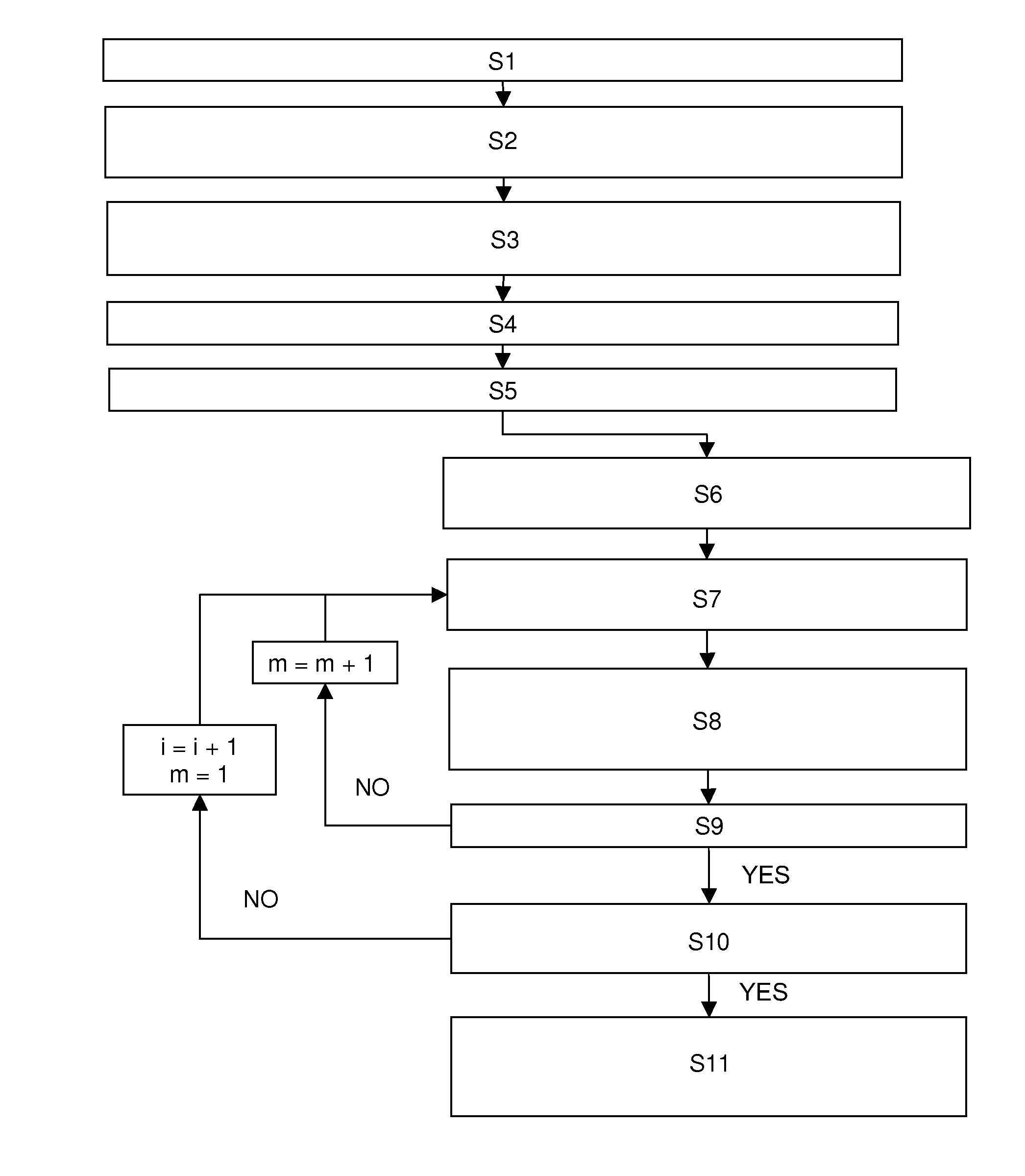
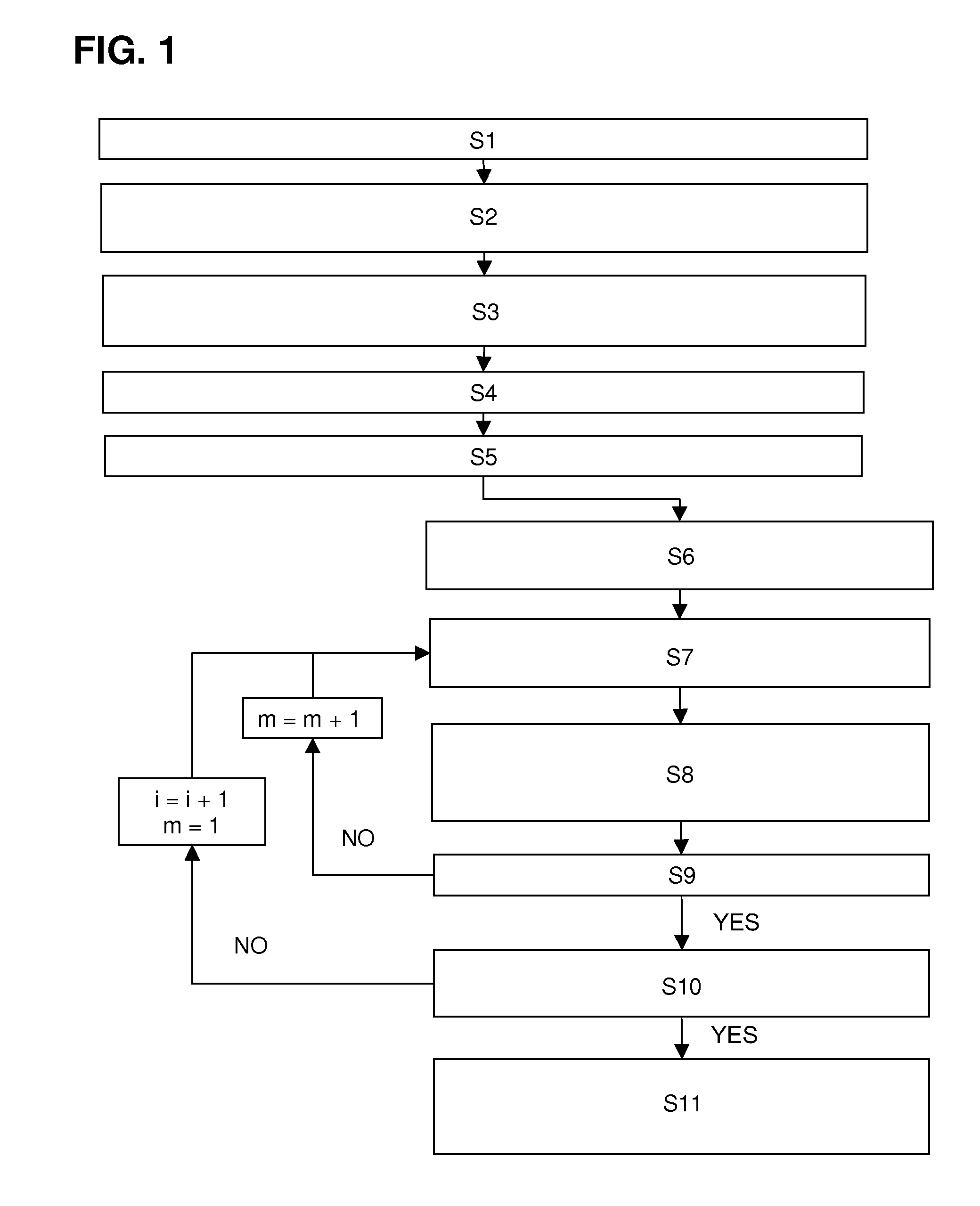
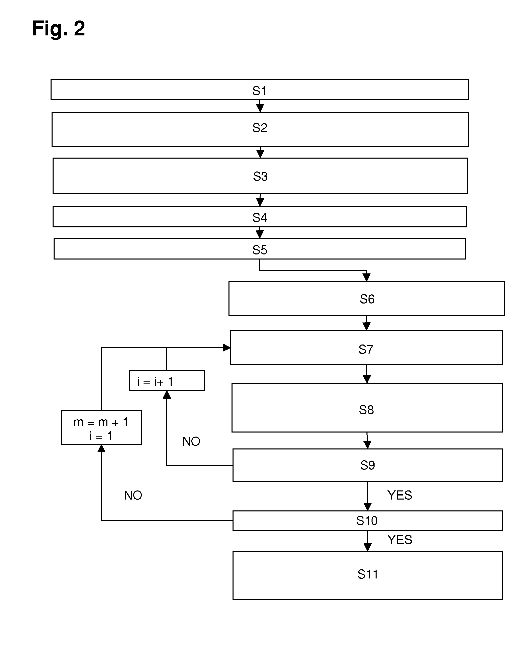
[0064]FIG. 1 shows the inventive method steps, which are described in more detail below. In particular, FIG. 1 illustrates the following method steps:[0065]S1=Selection of the imaging region[0066]S2=Definition of the encoding scheme P for the spatial encoding by local and / or global gradients after excitation[0067]S3=Definition of the phase encoding scheme A for the spatial encoding of the MSEM regions during excitation[0068]S4=Definition of an excitation pattern for each phase encoding step of A[0069]S5=Calculation of the RF pulses for each phase encoding step of A[0070]S6=Initialization and start of a spatially resolving MR measurement with phase encoding step m=1, i=1[0071]S7=Excitation of the object with RF pulse for phase encoding step i[0072]S8=Evolution of the spin system in the object, encoding according to step m of P and acquisition of the magnetic resonance signals[0073]S9=All encoding steps of P traversed?[0074]S10=All excitation phase encoding steps of A traversed?[0075]...


PUM


Abstract

A method for determining the spatial distribution of magnetic resonance (MR) signals from an imaging region has a preparatory step in which an encoding scheme with I phase encoding steps is defined, for each phase encoding step according to the phase encoding scheme, an excitation pattern of the transverse magnetization is defined and RF pulses to be irradiated to implement this pattern are calculated, wherein the same phase is defined at all spatial locations of the imaging region within an MSEM region and, in the execution step, according to the spatial encoding scheme each encoding step is performed I times according to the phase encoding scheme, wherein selection of the imaging region, amplitude modulation, and phase encoding are performed with the calculated RF pulses during excitation of the nuclear spin. This results in unique determination of the spatial distribution of the magnetic resonance signals with a simple RF receiver configuration using local gradient systems.

Description

BACKGROUND OF THE INVENTION[0001]The invention relates to a method for determining the spatial distribution of magnetic resonance signals from a predetermined imaging region that is completely covered by N MSEM regions (MSEM=monotonic spatially encoding magnetic field) within a volume under examination of a magnetic resonance apparatus, with N≧1, wherein, in a preparatory step, a spatial encoding scheme P with M encoding steps, M≧1, for spatial encoding in L spatial dimensions within the imaging region is defined; wherein, in an execution step, for each encoding step of the spatial encoding scheme P, nuclear spins are excited in the volume under examination by at least one RF pulse by means of RF transmitter antenna equipment having at least one transmitter element; after this RF excitation, spatial encoding is performed according to the spatial encoding scheme P by means of additional magnetic fields, variable over time and space, of a global and / or local gradient system, wherein t...

Claims


Application Information

Patent Timeline
Patent Type & Authority Patents(United States)
IPC IPC(8): G01V3/00
CPCG01R33/3415G01R33/4836G01R33/561G01R33/4824G01R33/4833G01R33/5611
Inventor RUHM, WOLFGANG
Owner BRUKER BIOSPIN MRI
Why Eureka
Try Eureka