Human fetal bladder-derived epithelial cells

a human fetal bladder and epithelial cell technology, applied in the field of development biology and cell biology, can solve the problems of occupational exposure, arylamine exposure, and rare surgery to palliate symptoms in patients

Inactive Publication Date: 2010-12-07
RAVEN BIOTECHNOLOGIES INC +1
View PDF12 Cites 10 Cited by
  • Summary
  • Abstract
  • Description
  • Claims
  • Application Information

AI Technical Summary

Benefits of technology

[0025]In still another aspect of this invention, the invention relates to methods of generating a human prostate tissue model using a substantially pure population of human fetal urinary bladder-derived epithelial cells and administering the human fetal urinary bladder-derived epithelial cells into a non-human, mammalian recipient at a location within said recipient able to support growth of said human fetal urinary bladder-derived epithelial cells, wherein the human fetal urinary bladder-derived epithelial cells have been recombined ex vivo with mesenchymal tissue able to effect further differentiation of said human fatal urinary bladder-derived epithelial cells

Problems solved by technology

However, because about 50% of patients with high-stage, high-grade tumors eventually relapse following cystectomy, surgery is seldom performed to palliate symptoms i

Method used


Image

  • Human fetal bladder-derived epithelial cells
  • Human fetal bladder-derived epithelial cells
  • Human fetal bladder-derived epithelial cells

Examples


example 1

Isolation of Fetal Urinary Bladder-Derived Epithelial Cells

[0083]Human fetal bladder tissue, gestational age from 14-21 weeks, was obtained from Advanced Bioscience Research at Alameda, Calif. As soon as the tissues arrived, they were rinsed three times with 20 ml of cool PBS. The bladders were cleaned of excess connective tissue, rinsed an additional two times with cool PBS, then cut into small segments (approximately 1 mm mince) with a razor blade or a pair of scissors under dissecting a microscope.

[0084]The minced tissue was suspended in 10 ml of OPTI-MEM® (Invitrogen Life Technologies), then transferred into a 15 ml centrifuge tube using a plastic pipette which had been pre-coated with bovine serum albumin to minimize tissue binding to the walls of the pipet. The minced tissue was centrifuged at 1000 rpm for 5 minutes in Heraeus Megafuge 2.0 (Cat#75003485) to pellet the minced tissue. The supernatant was aspirated and the pellet resuspended in 6 ml of fresh OPTI-MEM®.

[0085]The t...

example 2

Immunohistochemical Characterization

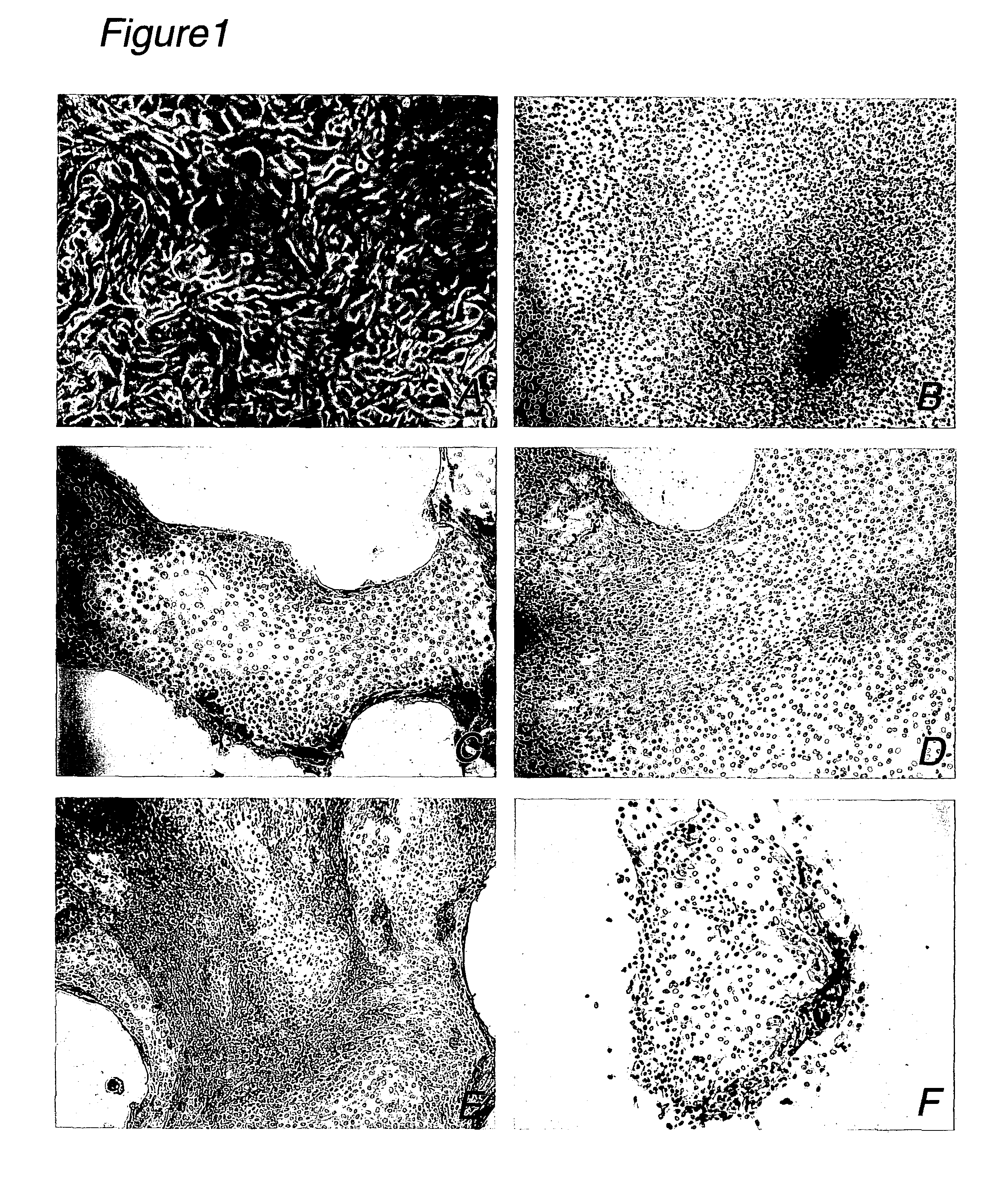
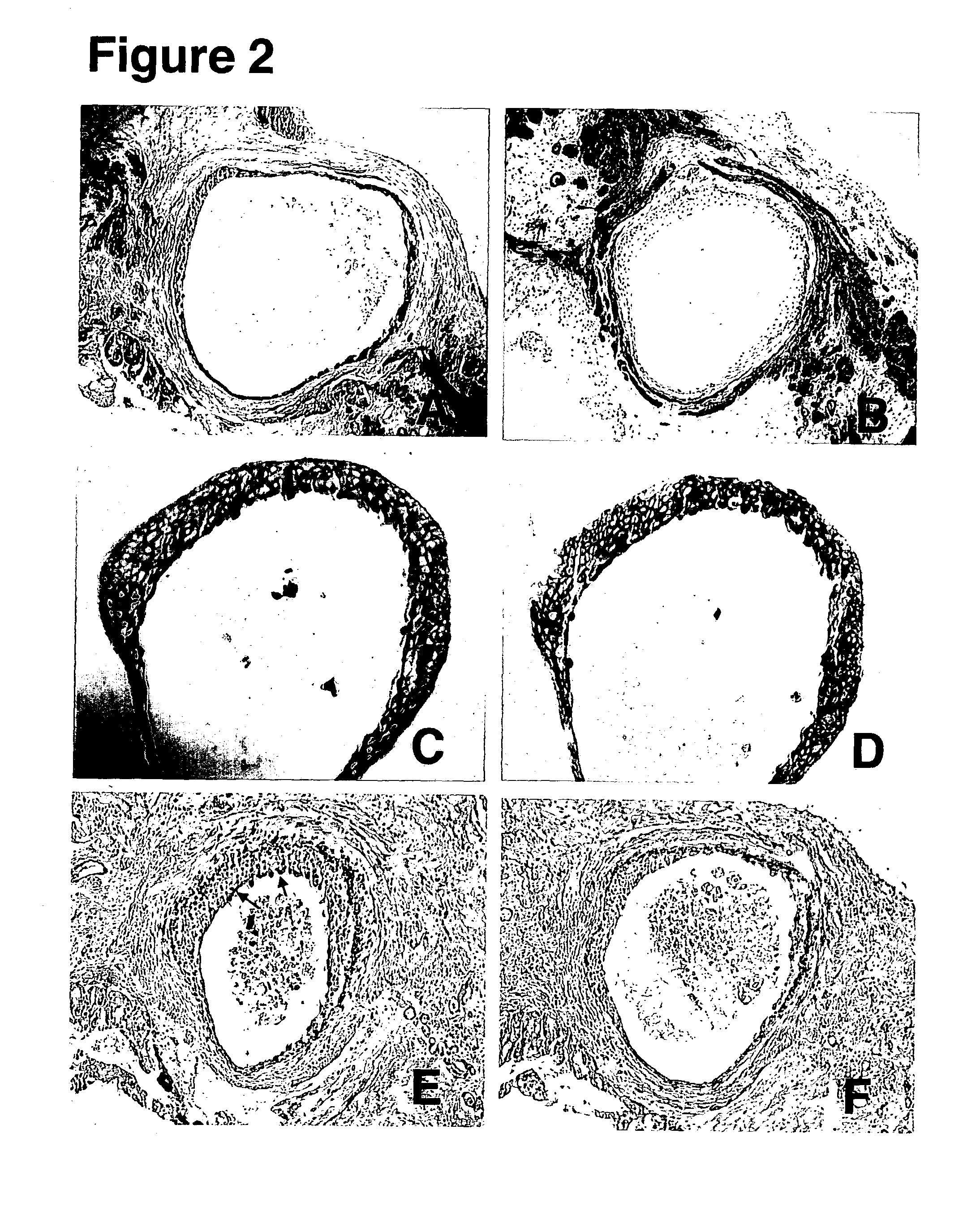
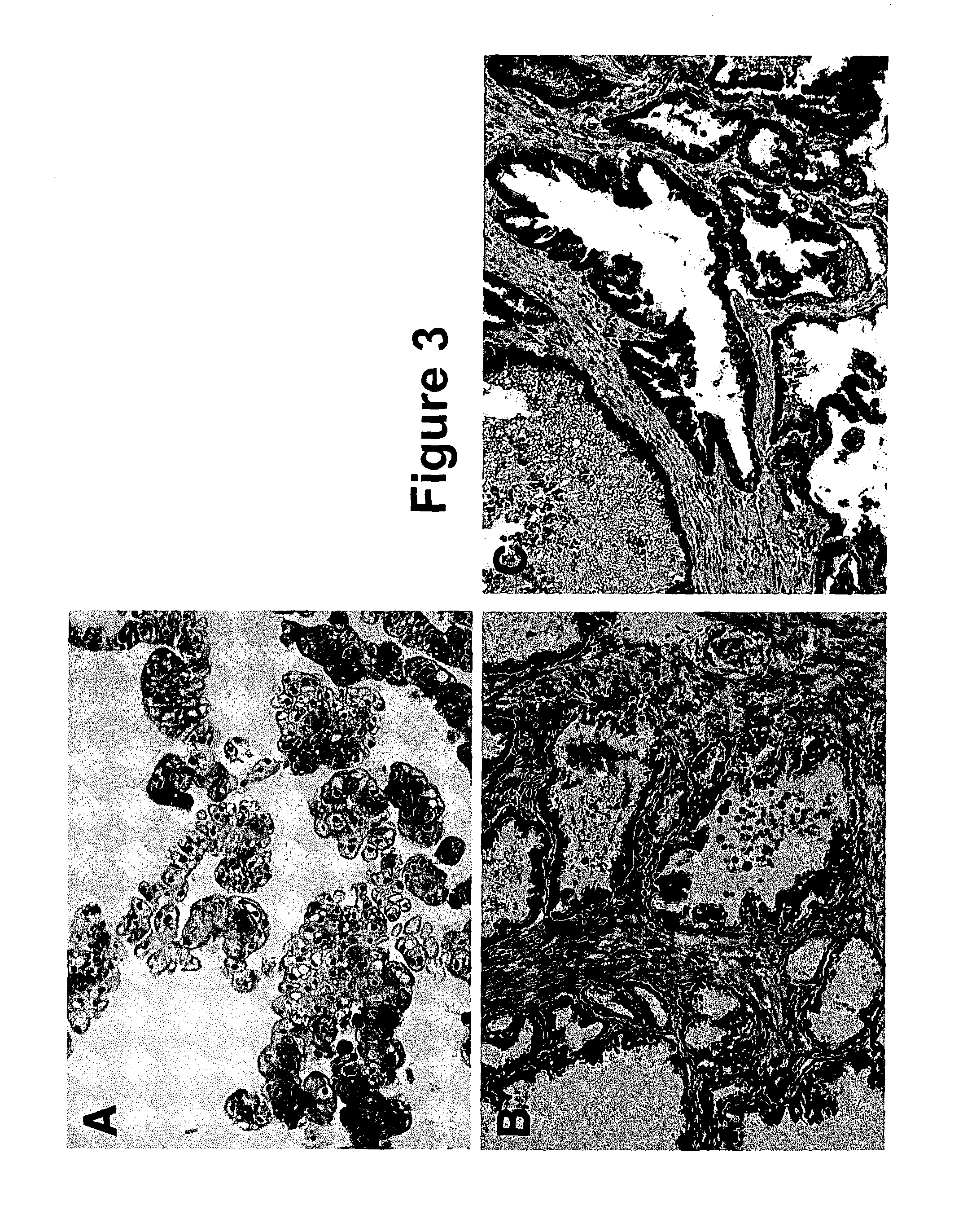
[0088]Monolayers of fetal urinary bladder-derived epithelial cells cultured in four-well chamber slides were washed three times with cool PBS and fixed with 100% reagent alcohol at −20° C. for 5 minutes. Then, the fixed slides were air dried overnight. The cells were incubated sequentially in blocking buffer (5% goat serum in PBS) for about 1 hour, in primary antibodies for overnight, and in peroxidase-conjugated affinipure F(ab)2 fragment of goat anti-mouse IgG+IgM(H+L) for about 1 hour. The cells were washed three times with PBS for 5 minutes per wash between those steps. The primary antibodies used were cytokeratins 7 and 19, uroplakin III, and alpha-actin at the dilution recommended by the supplier. To visualize staining of cells by the antibodies, the fetal urinary bladder-derived epithelial cells were incubated in peroxidase substrate DAB / H2O2 prepared from Sigma.

[0089]Some cells were positive for uroplakin III and cytokeratins 7 and 19. Tho...

example 3

Detection of Fetal Bladder Cell Molecular Marker H19 Imprinted Gene mRNA

[0090]H19 imprinted gene mRNA has been characterized as a marker specifically expressed in fetal human bladder cells but not expressed in normal human adult bladder cells (Elkin et al., 1995, FEBS Lett. 374(1):57-61; Cooper et al., 1996, J. Urol. 155(6):2120-27). To detect H19 mRNA, replicate fetal bladder-derived epithelial cell cultures of passage 2, approximately 105 cells each, were extracted for total RNA with RNeasy kit (Cat#74104) purchased from Qiagen according to the protocol provided by the supplier. The final total RNA was eluted in 50 μl water for each sample. The total RNA, 9.5 μl for each sample, was reverse transcribed into first strand cDNA in a 20 μl reaction with a cDNA synthesizing kit (Promega Corporation Cat A3500) with random primer according to the protocol provided by the supplier. PCR reactions were set up as following with high fidelity PCR master mix manufactured by Roche Biochemical (...


PUM

PropertyMeasurementUnit
Timeaaaaaaaaaa
Volumeaaaaaaaaaa

Abstract

This invention discloses a substantially pure population of human urinary bladder-derived epithelial cells and methods of isolating and culturing the urinary bladder-derived epithelial cells. In addition, several uses of human urinary bladder-derived epithelial cells and cells differentiating therefrom are disclosed herein.

Description

REFERENCE TO RELATED APPLICATIONS[0001]The application claims priority under 35 U.S.C. §119(e) to U.S. Provisional Patent Application Ser. No. 60 / 357,035, filed Feb. 12, 2002, the entirety of which is incorporated herein by reference.TECHNICAL FIELD[0002]The invention relates to the field of developmental biology and cell biology, and more particularly to purified fetal human bladder-derived epithelial cells and fetal human bladder epithelium cell lines.BACKGROUND ART[0003]The human urinary bladder is essentially a two-layered organ, consisting of a muscular outer wall and a highly specialized epithelium lining the lumen. Differentiated urinary bladder epithelium is characterized by the presence of urothelial plaques on the lumenal surface of urinary bladder superficial or umbrella cells. These plaques are characterized by a highly unusual membrane structure, i.e., the asymmetric unit membrane (AUM), with a lumenal leaflet twice as thick as the cytoplasmic leaflet. The thickening of...

Claims


Application Information

Patent Timeline
IPC IPC(8): C12N5/00C12N5/02C12M3/00A61K39/00C12N5/071C12Q1/02C12Q1/68
CPCC12N5/0683C12N5/0685A61K39/00A61K2039/515C12N2500/25C12N2500/38C12N2500/99C12N2501/117C12N2501/195C12N2501/392C12N2501/70C12N2502/246C12N2503/02C12N2506/25C12N2500/90
Inventor LI, RONGHAOPAN, ZHUANGYU
Owner RAVEN BIOTECHNOLOGIES INC
Why Eureka
Try Eureka