
Solution phase electrophoresis device, components, and methods

a technology of electrophoresis and solution phase, applied in the direction of fluid pressure measurement, liquid/fluent solid measurement, peptide, etc., can solve the problem of complex eukaryotic proteome, the total number of distinct protein species present concurrently in a eukaryotic cell, and the resolving capacity of current analytical techniques, so as to achieve the effect of convenient disassembly

Active Publication Date: 2010-12-14
LIFE TECH CORP
View PDF44 Cites 3 Cited by
  • Summary
  • Abstract
  • Description
  • Claims
  • Application Information

AI Technical Summary

Benefits of technology

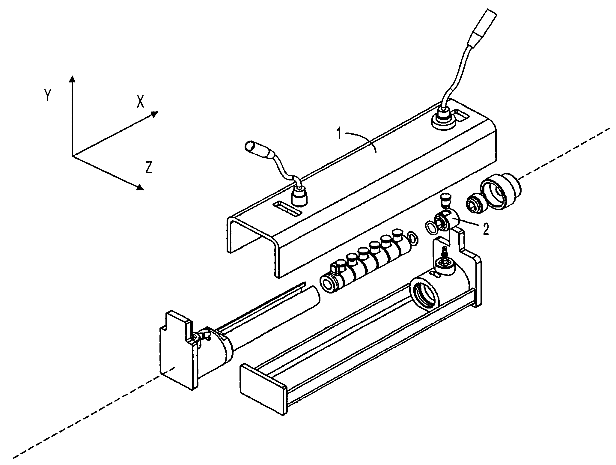
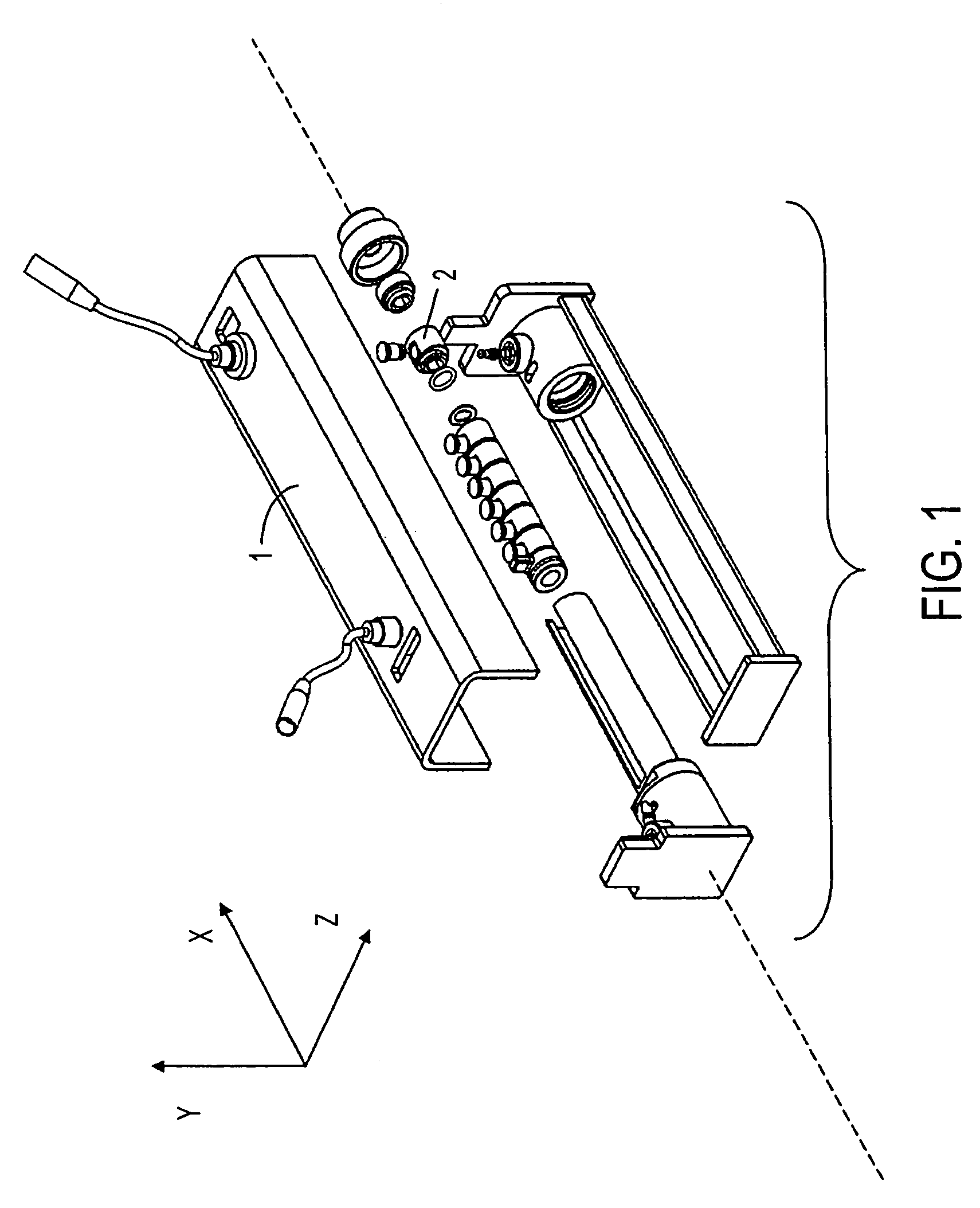
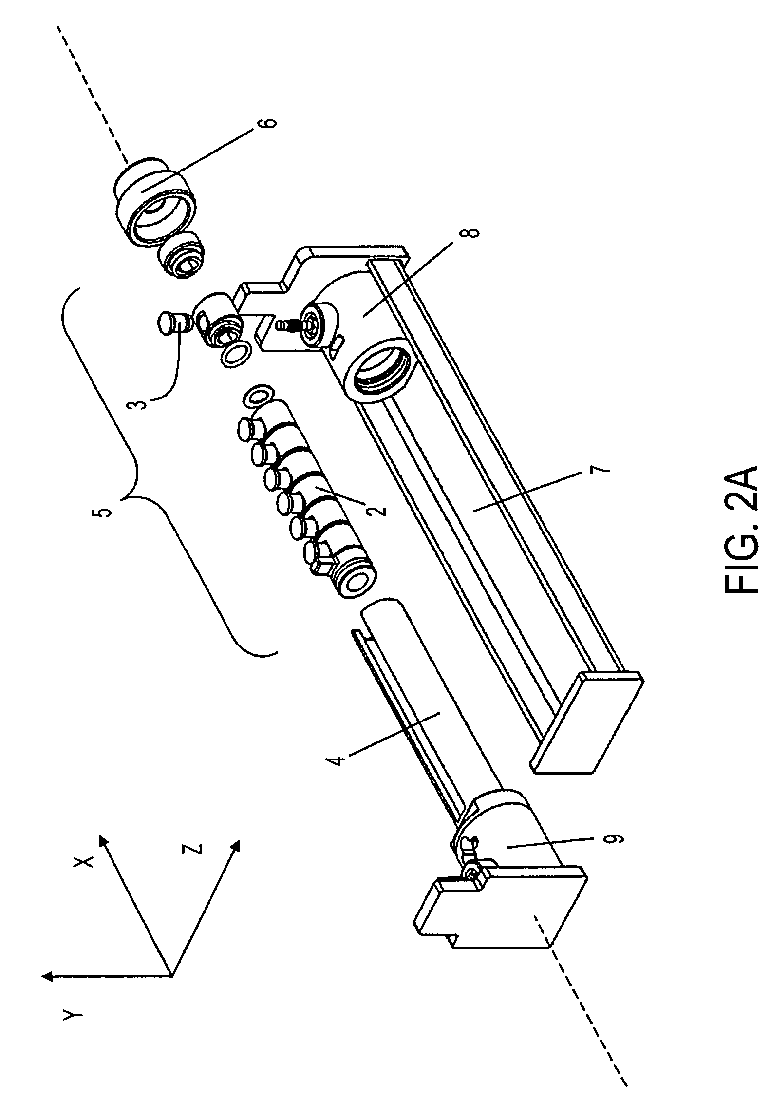
The present invention is a solution phase isoelectric focusing device that is easy to assemble and disassemble. The device uses fixed pH membrane disks and does not require means for sample recirculation or mixing during operation. The device can be positioned on a rocker platform or a rotary platform to effect sample agitation. The sample chambers are held tightly in position by a screw cap and sealed with O-rings to prevent leaks. The device also includes an anode end piece for easy removal of the sample chambers, electrode wires protected from damage, and a cap seal that prevents air bubbles during sealing. The invention also includes methods and designs for sealing sample chambers without trapping air bubbles and using immobilized buffer membranes. Kits for solution IEF are also provided. The invention can be used for separating, fractionating, and characterizing proteins and other biomolecules during electrophoresis.

Problems solved by technology

The complexity of eukaryotic proteomes—that is, the total number of distinct protein species present concurrently in a eukaryotic cell, including alternatively spliced isoforms and variants differing in post-translational modification—typically exceeds the resolving capacity of current analytical techniques.
The limited spatial resolution in turn constrains the dynamic detection range of the technique: efforts to observe low abundance species by increasing the initial protein load lead to increased obscuration by high abundance species.
None of the devices, however, provides particularly convenient solution phase IEF prefractionation of small volume protein samples with a simple device in a format that readily interfaces with subsequent analytical techniques such as 2D PAGE.

Method used


Image

  • Solution phase electrophoresis device, components, and methods
  • Solution phase electrophoresis device, components, and methods
  • Solution phase electrophoresis device, components, and methods

Examples


example 1

Fractionation of Rat Liver Lysate

[0289]Rat liver tissue is lysed by sonication at a final concentration of 5% (w / v) in 7M urea, 2M thiourea, 4% CHAPS (collectively, “UTC”) and protease inhibitors. After reduction, alkylation, centrifugation, and determination of the protein concentration of the supernatant fraction, samples are diluted to 0.6 mg / ml protein in UTC containing 1% ZOOM® ampholytes, pH 3-10 (Invitrogen Corp., Carlsbad, Calif., USA), 20 mM DTT, and a trace of bromophenol blue dye.

[0290]An aliquot of 3.35 ml is distributed equally into five central sample chambers of seven total chambers, each with capacity of about 670 μl, designed and assembled according to the device of the present invention. The five chambers are partitioned from one another by IBDs having pH 3.0, 4.6, 5.4, 6.2, 7.0, and 10.0.

[0291]After fractionation for 3 hours, the resulting fractions are collected and a 155 μl aliquot of each fraction is loaded onto a separate ZOOM® IPG strips (Invitrogen Corp., Ca...

example 2

Improvement in Detection of Low Abundance Proteins

[0297]Rat liver lysate is prepared and fractionated in a device of the present invention, essentially according to Example 1.

[0298]Separate 155 μl aliquots of the pH 4.6-5.4 fraction are loaded respectively on a pH 4.5-5.5 narrow range ZOOM® IPG strip (Invitrogen Corp., Carlsbad, Calif., USA) and a pH 4-7 ZOOM® IPG strip (Invitrogen Corp., Carlsbad, Calif., USA) and allowed to rehydrate overnight. The applied proteins are then focused using the ZOOM® IPGRunner™ System (Invitrogen Corp., Carlsbad, Calif., USA). The focused ZOOM® Strips are separately applied to NuPAGE® Novex 4-12% Bis-Tris ZOOM gels. The resulting 2DE gels are stained with SimplyBlue™ SafeStain and scanned.

[0299]Unfractionated rat liver lysate is analogously applied to a pH 4.5-5.5 narrow range ZOOM® IPG strip (Invitrogen Corp., Carlsbad, Calif., USA) as a control.

[0300]FIG. 13A shows the solution phase fraction run on a pH 4-7 IPG strip, demonstrating that prefractio...


PUM

PropertyMeasurementUnit
volumeaaaaaaaaaa
volumeaaaaaaaaaa
volumeaaaaaaaaaa

Abstract

A device for fluid phase electrophoresis, particularly solution phase isoelectric focusing, components thereof, and methods for use are presented.

Description

FIELD OF THE INVENTION[0001]The present invention is in the field of devices, components, and methods for electrophoresis, particularly isoelectric focusing, more particularly solution phase isoelectric focusing (IEF).BACKGROUND OF THE INVENTION[0002]The complexity of eukaryotic proteomes—that is, the total number of distinct protein species present concurrently in a eukaryotic cell, including alternatively spliced isoforms and variants differing in post-translational modification—typically exceeds the resolving capacity of current analytical techniques.[0003]For example, the number of distinct protein species in eukaryotic cells typically far exceeds the spatial resolution of two-dimensional polyacrylamide gel electrophoresis (2D PAGE) gels, with large numbers of distinct protein species appearing to comigrate. The limited spatial resolution in turn constrains the dynamic detection range of the technique: efforts to observe low abundance species by increasing the initial protein lo...

Claims


Application Information

Patent Timeline
Patent Type & Authority Patents(United States)
IPC IPC(8): G01N27/00B01D61/00B01D61/58C07K1/26G01L1/20G01N27/447
CPCG01N27/44704G01N27/44795
Inventor AMSHEY, JOSEPH W.UPDYKE, TIMOTHY V.JACKSON, THOMAS R.THACKER, MICHAEL
Owner LIFE TECH CORP
Learn More