Apparatus and method for processing an ultrasound spectrum image

a spectrum image and ultrasound technology, applied in the field of ultrasound diagnostic systems, can solve the problems of inability to perform accurate contour tracing, inability to provide accurate peak tracing, and disadvantages of conventional ultrasound diagnostic systems, and achieve the effect of removing noise, accurately performing both contour tracing and peak tracing

Active Publication Date: 2011-01-04
MEDISON CO LTD
View PDF12 Cites 5 Cited by
  • Summary
  • Abstract
  • Description
  • Claims
  • Application Information

AI Technical Summary

Benefits of technology

[0006]It is, therefore, an object of the present invention to address and resolve the above-mentioned deficiencies of the prior art. In this regard, the object of the present invention is to provide an apparatus and method for processing an ultrasound spectrum image, which can accurately perform both contour tracing and peak tracing by removing the noise from a noise-containing spectrum image and removing aliasing from an aliasing-containing spectrum image through automatically shifting the baseline.

Problems solved by technology

However, even when the spectrum image has aliasing, the conventional ultrasound diagnostic system does not consider the magnitude and direction of the aliasing when performing contour tracing.
For this reason, the conventional ultrasound diagnostic system is disadvantageous since it cannot perform accurate contour tracing, as shown in FIG. 1.
Therefore, the conventional ultrasound diagnostic system cannot provide accurate peak tracing.
Further, as the pulse wave (PW) gain for a spectrum image increases, the noise also tends to increase.
Also, the noise varies for each spectrum image.
However, when the conventional ultrasound diagnostic system performs the contour tracing on a spectrum image with increased noise, it determines a threshold for removing the noise based on the PW gain without analyzing the noise.
Thus, since the noise varies depending on external environments (e.g., gel existence, probe type, etc.), the conventional ultrasound diagnostic system is disadvantageous in that it cannot perform accurate contour tracing, as shown in FIG. 2.
Therefore, the conventional ultrasound diagnostic system cannot provide accurate peak tracing.

Method used


Image

  • Apparatus and method for processing an ultrasound spectrum image
  • Apparatus and method for processing an ultrasound spectrum image
  • Apparatus and method for processing an ultrasound spectrum image

Examples


Embodiment Construction

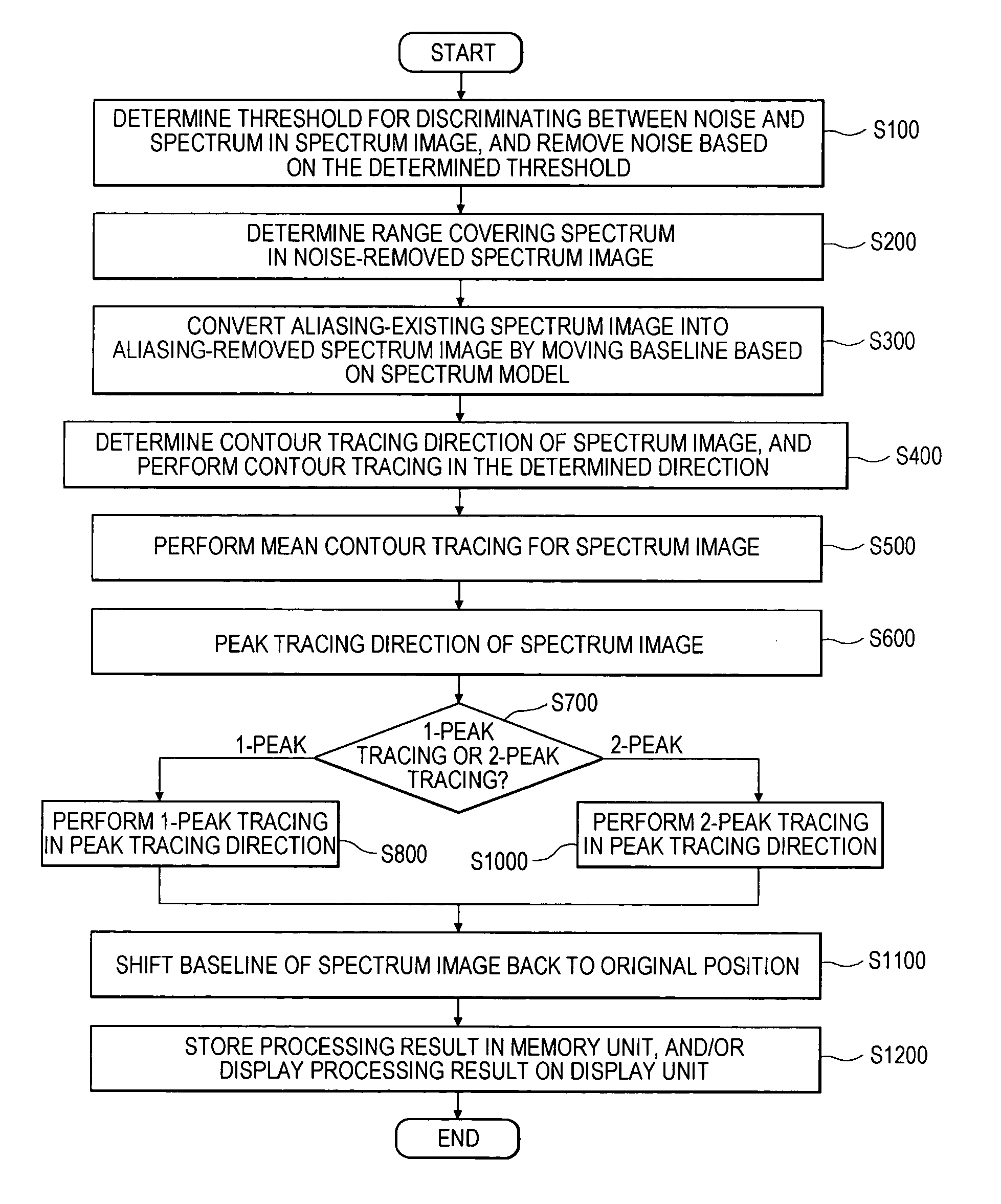
[0039]The preferred embodiments of the present invention will be described below with reference to FIGS. 3 to 22.

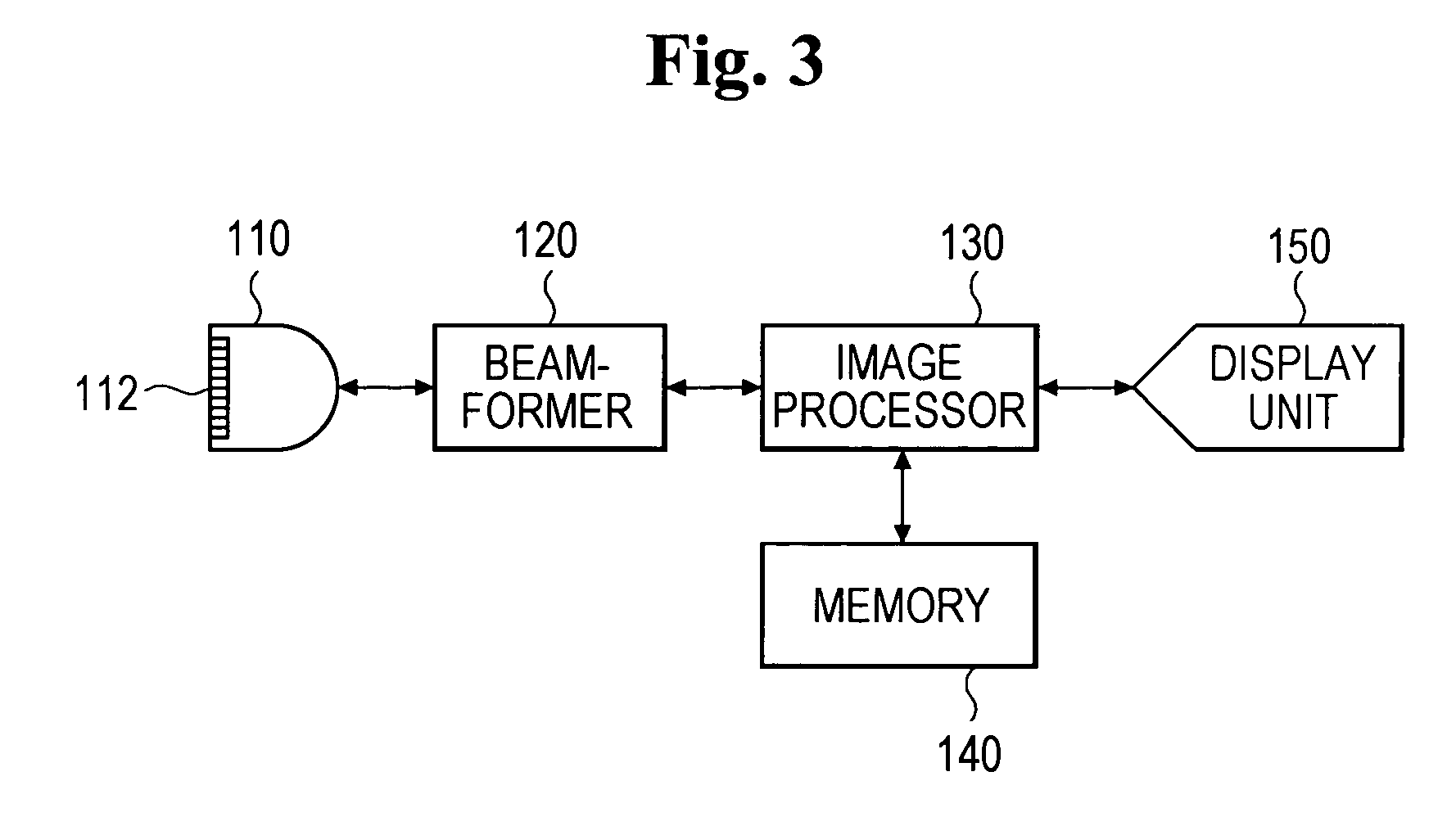
[0040]Referring now to FIG. 3, an ultrasound diagnostic system 100, which is constructed in accordance with the present invention, generally includes a probe 110, a beam-former 120, an image processor 130, a memory 140 and a display unit 150.

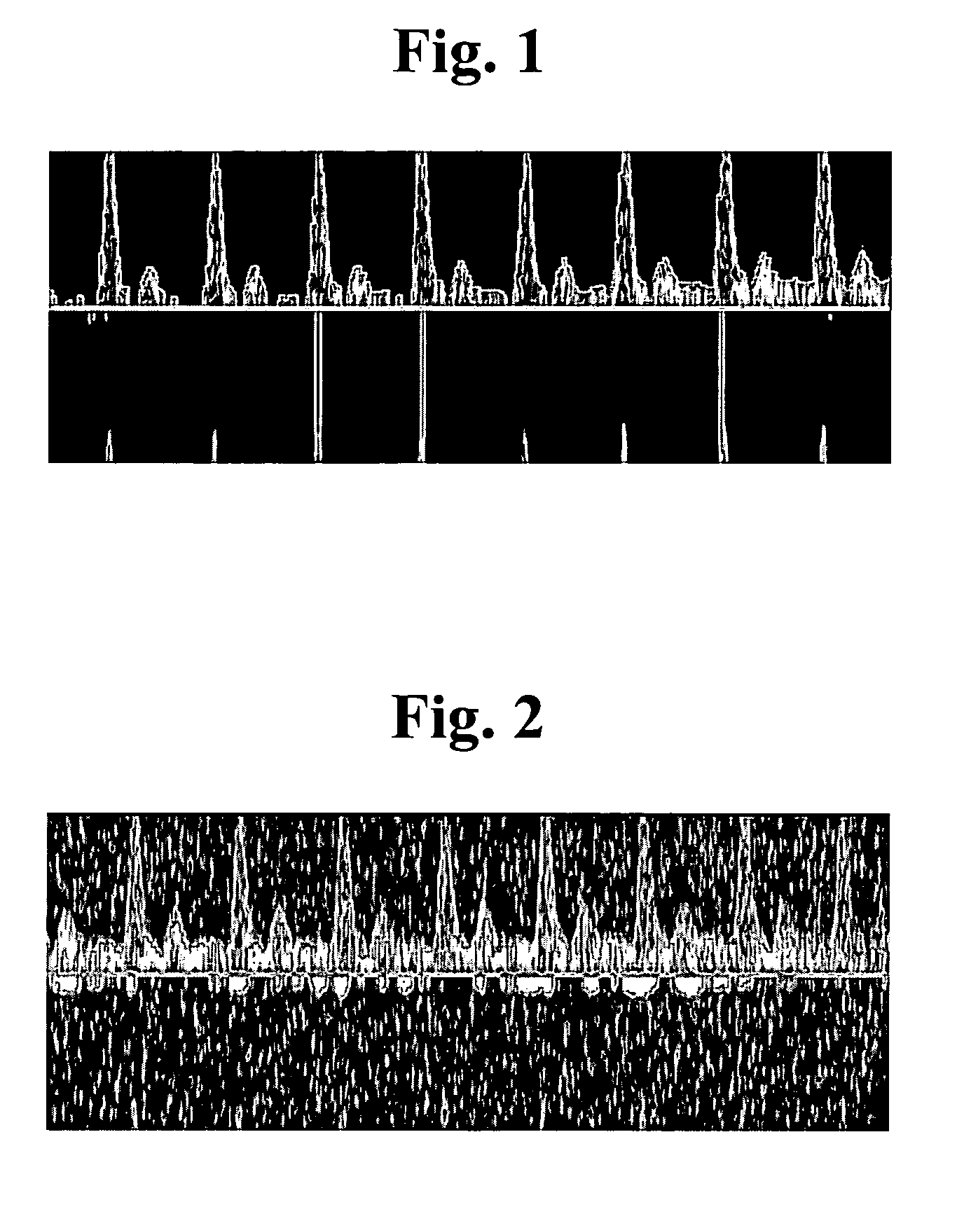
[0041]The probe 110 preferably includes a one-dimensional (1D) or two-dimensional (2D) array of transducers 112. The probe 110 is configured to transmit ultrasound signals to a target object and receive ultrasound echo signals. The beam-former 120 controls the transmission and reception of the probe 110. Further, in order to form a coherent beam of the echo signals from the target object, the beam-former 120 processes the received ultrasound echo signals. The image processor 130 produces spectrum signals based on the ultrasound echo signals and produces a noise-removed spectrum image based on the spectrum signals, wherein a frequency ...


PUM


Abstract

There is provided a method of processing an ultrasound spectrum image. According to such method, a spectrum image is formed based on ultrasound data and then the noise is removed from the spectrum image. The noise-removed spectrum image is matched with one or more spectrum models representing specific spectrum types. Then, whether or not the noise-removed spectrum image contains an aliasing is checked. If the noise-removed spectrum image contains the aliasing, then the aliasing is removed from the noise-removed spectrum image to provide a noise-removed spectrum image without the aliasing. Thereafter, contour tracing is performed on the noise-removed spectrum image without the aliasing to detect contour points. Further, peak tracing is performed on the noise-removed spectrum image without the aliasing to detect peaks.

Description

FIELD OF THE INVENTION[0001]The present invention generally relates to ultrasound diagnostic systems, and more particularly to an ultrasound diagnostic system for processing an ultrasound spectrum image and a method of implementing the same.BACKGROUND OF THE INVENTION[0002]An ultrasound diagnostic system is now widely used to inspect the internal condition of a human body. The ultrasound diagnostic system may obtain an image of the single layer or blood flow of a soft tissue without using an invasive needle. This is typically performed through the process of radiating an ultrasound signal from the body surface of a target object to be diagnosed to a desired portion in the body, receiving the reflected ultrasound signal, and processing the received ultrasound signal (ultrasound echo signal). Compared to other medical imaging systems (e.g., X-ray diagnostic system, X-ray Computerized Tomography (CT) scanner, Magnetic Resonance Imaging (MRI) system, nuclear medicine diagnostic system, ...

Claims


Application Information

Patent Timeline
Patent Type & Authority Patents(United States)
IPC IPC(8): G06K9/00
CPCA61B8/06G01S7/52026G01S7/52077G06K9/0055G01S15/584G01S15/8979G06F2218/16A61B8/00
Inventor KIM, HYE JUNGLEE, KI JONGKIM, SUNG HOYANG, EUN HOKIM, CHEOL AN
Owner MEDISON CO LTD
Why Eureka
Try Eureka