Rapid particle detection assay

a particle detection and particle technology, applied in the field of optical detection methods, immunology, cellular biology, can solve the problems of adding to the complexity and cost of instruments, and achieve the effect of simplicity and cos

Active Publication Date: 2011-04-19
BECTON DICKINSON & CO
View PDF34 Cites 30 Cited by
  • Summary
  • Abstract
  • Description
  • Claims
  • Application Information

AI Technical Summary

Benefits of technology

[0012]The present invention provides an optical instrument and method for the analysis of an analyte of interest in a fluid sample. The instrument enables measuring the total amount of analyte in the sample without the need to optically scan the sample. The assay is carried out in a “homogeneous” format in which the fluid sample is not removed from the sample chamber during the analysis. The invention provides significant advantages in simplicity and cost that will be particularly useful in applications such as the measurement of CD4+ T cells.
[0014]The sample chamber can be made from any suitable material, such as glass or plastic. In a preferred embodiment, the sample chamber is made from plastic, such as polystyrene, has a clear (transparent) top to enable optical detection, and has black sides and bottom to minimize ambient or scattered light from interfering with the detection.
[0016]The instrument further comprises excitation optics adapted to illuminate the detection region and detection optics adapted to measure light emitted from the detection region. The excitation and detection optics are positioned above the sample chamber, on the opposite side of the sample chamber from the focusing magnet. The positioning of the excitation and detection optics opposite from the focusing magnet enables uniform illumination of the magnetically concentrated analyte and detection of the emitted light from the analyte while the magnet remains in position and the analyte remains bound to the tip of the magnet, without interference from the magnet itself. In a preferred embodiment, a laser is used to provide excitation light, and a photodiode is used to measure the emitted light.
[0021]In some embodiments, a single species of capture reagent is used, e.g., antibodies for a particular cell-surface antigen. In other embodiments, multiple species of capture reagents, each specific for a different cell-surface antigen expressed by the cells of interest, can be used to increase the number of magnetic particles that are bound to each cell and to increase the cumulative magnetic force applied to the cells during the concentration step.
[0027]The combination of features of the instrument of the present invention provides several advantages. The assays of the present invention is carried out without washing the sample after magnetic concentration, or otherwise separating the concentrated analyte from the sample fluid. The use of the focusing magnet enables concentration of the analyte sufficiently such that the amount of analyte can be detected without optically scanning the sample. The placement of the excitation and detection optics on the opposite side of the sample chamber from the focusing magnet facilitates optical detection in microliter-sized samples without physical interference from the focusing magnet, while the design of the sample chamber minimizes the amount of sample fluid in the excitation and detection light paths, thus reducing background signal caused by the presence of sample fluid in the light path.

Problems solved by technology

The requirements of scanning the sample add to the complexity and cost of the instrument.

Method used


Image

  • Rapid particle detection assay
  • Rapid particle detection assay
  • Rapid particle detection assay

Examples


example 1

CD4+ T Cell Concentration Assay: Antibody Labeling

Instrument

[0087]Assays were carried out using an instrument essentially as shown in FIG. 11.

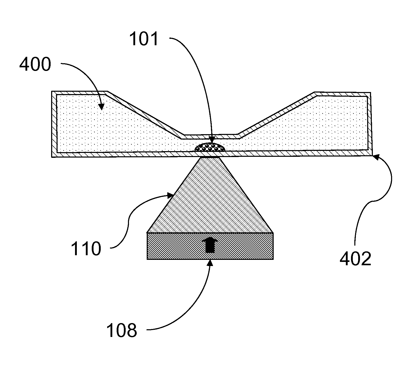
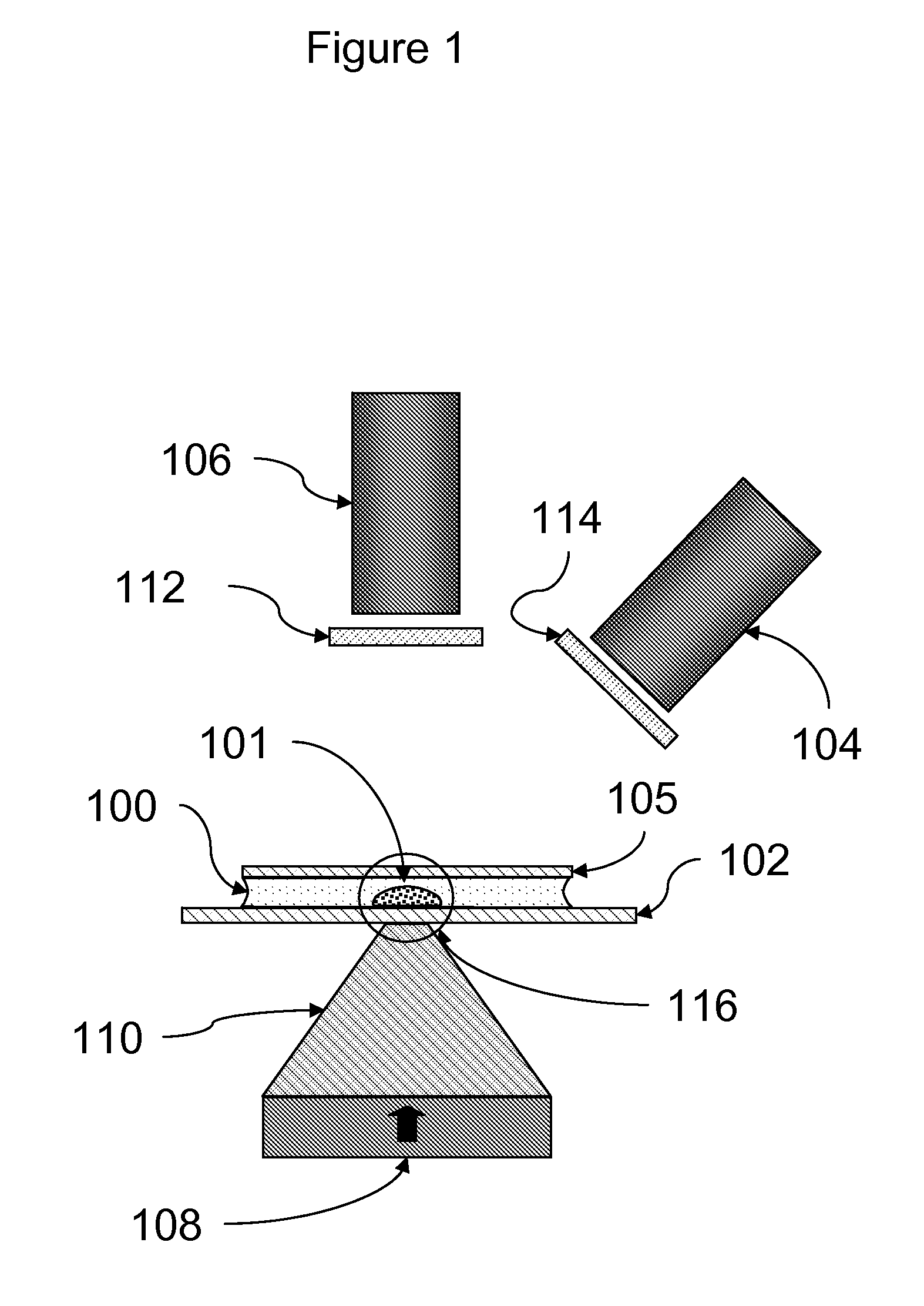
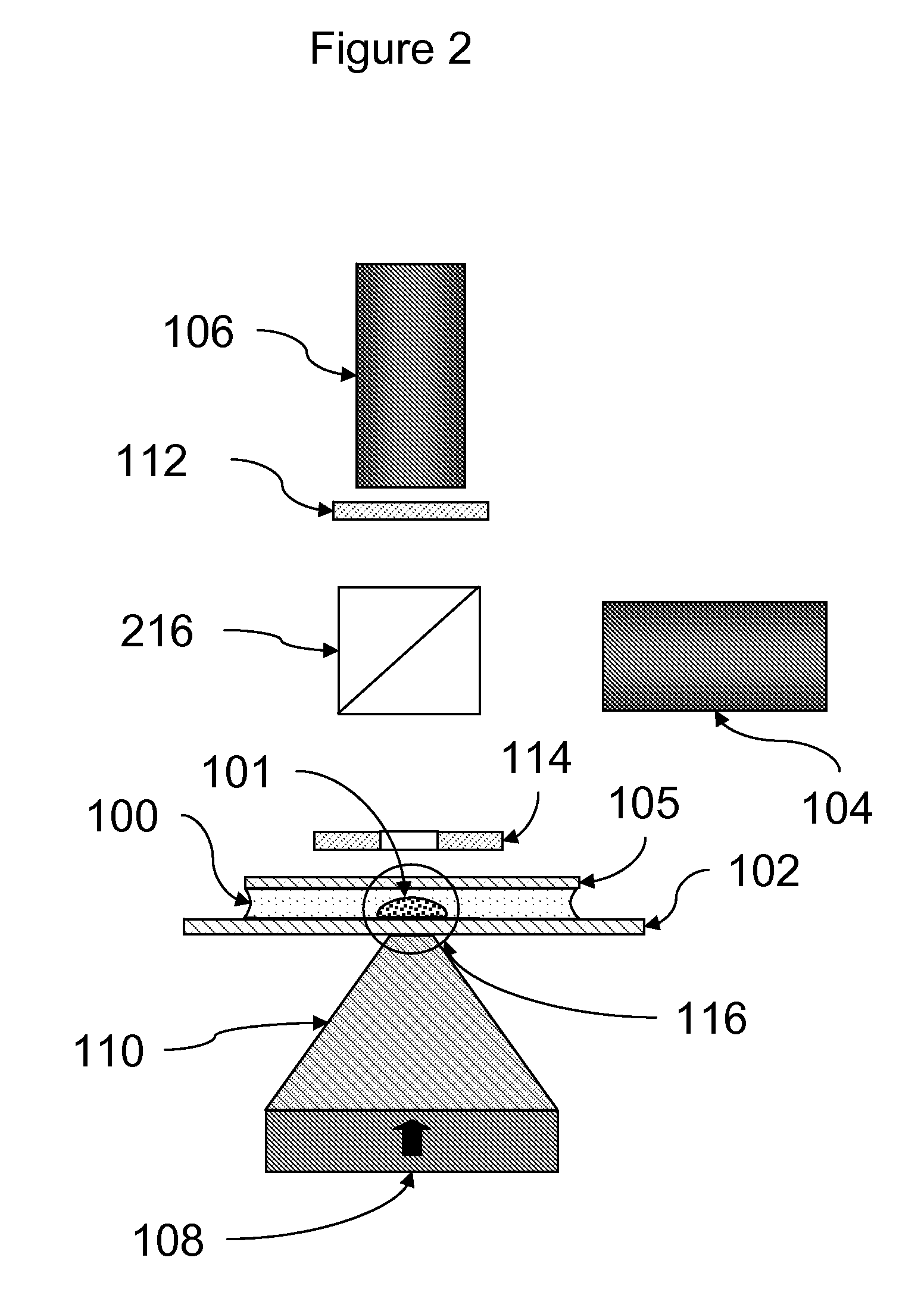
[0088]A rectangular sample holder 1102 containing a sample chamber essentially as shown in FIG. 4 was used. The diameter of the sample chamber was about 7 mm, and the total volume of the sample chamber was about 20 μl. The detection region was a region about 2 mm in diameter in the center of the sample chamber. The upper surface of the sample chamber was recessed about 700 μm below the upper surface over the perimeter of the sample chamber such that the depth of the sample chamber in the detection region was about 50 μm. The sample holder was carried in a sample holder carrier 1105, which facilitated inserting the sample chamber into the instrument and shielded the sample chamber from ambient light. Once inserted, the sample chamber was position directly over the focusing magnet 1109.

[0089]The focusing magnet consisted of a cylindrical (disc) ...

example 2

CD4+ T Cell Concentration Assay: DNA-Dye Labeling

[0106]This example describes the use of a nucleic acid stain to fluorescently label all cells in the sample. The instrument and assay methods were essentially as described in example 1, with the exception of the changes described, below.

Reagents

[0107]Commercially available CD4 antibody-coated magnetic particles (BD Imag™ beads, BD Biosciences, San Jose, Calif.) were used as magnetic capture reagents, as described in example 1.

[0108]A permeant DNA-binding dye, DRAQ5 (Biostatus Ltd., Leicestershire, England) was used as a detection reagent. Use of this dye results in the labeling of all nucleated cells in the blood sample.

Assay

[0109]Sample of whole blood were pretreated to selectively remove monocytes. Removal of monocytes was carried by magnetic depletion using CD14 antibody-coated magnetic particles (BD Imag™ beads, BD Biosciences, San Jose, Calif.) following the manufacturer's instructions.

[0110]For each assay, 100 μL of whole blood ...


PUM

PropertyMeasurementUnit
depthaaaaaaaaaa
volumeaaaaaaaaaa
vertical depthaaaaaaaaaa

Abstract

The present invention proves instruments and methods for detecting and / or quantitating an analyte in a fluid sample. The fluid sample is placed in a sample chamber having a small, shallow detection region. The analyte is magnetically labeled using magnetic particles coated with a binding reagent, and is detectably labeled using a fluorescent dye or other detection reagent. The magnetically labeled analyte is concentrated into the detection region using a focusing magnet positioned underneath the sample chamber detection region. Concentrated analyte is measured using excitation optics positioned on top of the sample chamber detection region, adapted to illuminate only the detection region, and detection optics positioned on top of the detection region, adapted to detect only light emitted from the detection region. In a preferred embodiment, the invention provides a simple, rapid assay for measuring the concentration of CD4+T cells in a whole blood sample.

Description

CROSS-REFERENCE TO RELATED APPLICATIONS[0001]The present application claims priority to U.S. provisional application Ser. No. 61 / 010,686, filed Jan. 10, 2008, which is incorporated herein by reference in its entirety.BACKGROUND OF THE INVENTION[0002]1. Field of the Invention[0003]The present invention relates in general to the fields of optical detection methods, immunology, and cellular biology, and, more particularly, to methods of assessing the state of the immune system, and, even more particularly, to methods of measuring CD4+ T cells, as typically carried out in HIV-infected patients.[0004]2. Description of Related Art[0005]Obtaining accurate and reliable measures of CD4+ T lymphocytes (CD4+ T cells) is essential to assessing the immune system and managing the health care of persons infected with human immunodeficiency virus (HIV). The pathogenesis of acquired immunodeficiency syndrome (AIDS) is largely attributable to the decrease in CD4+ T cells, and progressive depletion of...

Claims


Application Information

Patent Timeline
Patent Type & Authority Patents(United States)
IPC IPC(8): G01N21/75
CPCB01L3/508G01N33/54333G01N21/6428B01L2200/0668B01L2400/043B01L2300/0822
Inventor KIRAKOSSIAN, HRAIRYU, LIPINGPETRY, DOUGLAS A.
Owner BECTON DICKINSON & CO
Why Eureka
Try Eureka