
Laminate of cultured human corneal endothelial cells layer and method for manufacturing same

a technology of corneal endothelial cells and cultured human cornea, which is applied in the field of cultured human corneal endothelial cells layer and a method for manufacturing the same, can solve the problems of tissue compatibility, acute shortage of corneal donors, and self-limiting function of the cornea, so as to avoid immunologic rejection and prevent fibroblast-like cell infiltration

Inactive Publication Date: 2011-06-14
YAMAGAMI SATORU +1
View PDF9 Cites 11 Cited by
  • Summary
  • Abstract
  • Description
  • Claims
  • Application Information

AI Technical Summary

Benefits of technology

[0027]The transparent type I collagen sheet constituting the laminate of the present invention is a material that can maintain its transparency under physiological conditions (neutral pH). Here, the term “transparency” means transparency of a degree that does not cause problems with vision when the laminate of the present invention is implanted.
[0032]The laminate of the present invention may have a layer of adhesive factor or bioadhesive on the opposite side from the cultured layer of HCECs on the transparent type I collagen sheet. Having a layer of adhesive factor or bioadhesive on the opposite side from the cultured layer of HCECs on the transparent type I collagen sheet promotes adhesion of the laminate of the present invention to the corneal stroma during implant.
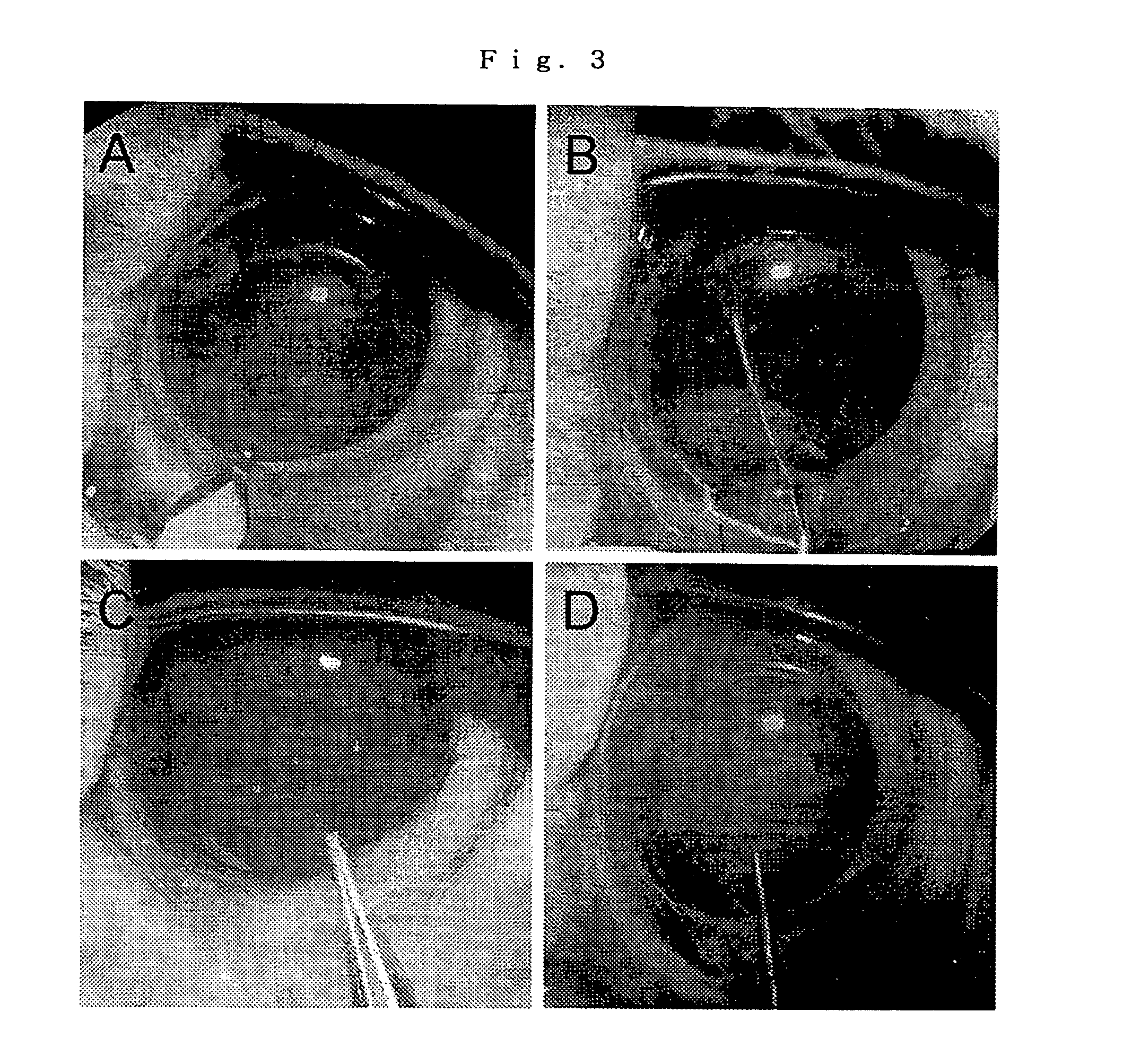
[0078]It is worth commenting on the immunologic rejection of human-to-rabbit xenotransplantation that occurs in this model. In the full-thickness murine corneal xenotransplantation model, no hyperacute rejection occurs, but corneal xenografts are rejected in concordant or discordant combination without exception, on the average over a period of 2 to 16 days after transplantation. In our study, no apparent inflammatory reactions, such as massive cell infiltration, keratic precipitates, or fibrin formation were present in the anterior chamber of rabbit eye under slit lamp microscopy and human DiI-labeled CEC showed no apparent cell loss, evidence that there was no notable episode of acute immunologic rejection. The anterior chamber of the eye is an immune-privileged site and anterior chamber-associated immune deviation (ACAID) permits the long-term acceptance and survival of histoincompatible tissue grafts that otherwise are rejected when transplanted to various anatomic sites. In HCEC sheet transplantation, the transplanted HCECs, which face the anterior chamber, may induce ACAID, thereby avoiding immunologic rejection, the leading cause of graft failure in full-thickness corneal transplantation.

Problems solved by technology

Accordingly, for example, when the injury heals through expanding the surface area of CECs, functioning of the cornea is self-limiting; when torn, disorders such as bullous keratopathy result.
However, in Japan at least, there is an acute shortage of cornea donors.
There is also the problem of tissue compatibility when conducting a transplant.
Treatment by corneal transplant is thus far from being an ideal solution.
However, as stated above, the CECs have a poor ability to proliferate, so that when the cornea is stored for an extended period, there is a problem in that the endothelial cells decrease.
That is, in the case of a cornea is transplanted, the method of storage may lead to a problem in decrease of endothelial cells.
Thus, there is a problem in that the CECs have an extremely poor ability to proliferate within the organism.
However, since the above-described method presents problems in that it requires a cornea from which the endothelial cells have been removed, the method does not provide a solution for a shortage of cornea donors, and for an inability to handle the rejection accompanying a transplant.
However, while the corneal endothelial cell layer is being reconstructed, the patient must do without sight and is greatly burdened in his daily life.

Method used


Image

  • Laminate of cultured human corneal endothelial cells layer and method for manufacturing same
  • Laminate of cultured human corneal endothelial cells layer and method for manufacturing same
  • Laminate of cultured human corneal endothelial cells layer and method for manufacturing same

Examples


examples

[0044]The present invention is further described below in view of examples.

[0045][Media and Culture Conditions for HCECs]

[0046]All the primary cultures and serial passages of HCECs were done in growth medium consisting of low-glucose Dulbecco's modified Eagle's medium (DMEM) supplemented with 15% fetal bovine serum (FBS), 2.5 mg / L fungizone (Gibco BRL, Grand Island, N.Y.), 2.5 mg / L doxycycline, and 2 ng / mL basic fibroblast growth factor (bFGF; Sigma, St. Louis, Mo.). Cells were maintained in a humidified incubator at 37° C. and 10% CO2. Medium used to produce bovine extracellular matrix (ECM) consisted of low-glucose DMEM with 10% FBS, 5% calf serum (Gibco BRL), 2.5 mg / L fungizone, 2.5 mg / L doxycycline, 2 ng / mL bFGF, and 2% dextran (Sigma). Media for the HCEC and bovine CEC cultures were changed every 2-3 days.

[0047][Primary Culture of HCECs]

[0048]Primary HCEC cultures were made as described elsewhere (Miyata K, Drake J, Osakabe Y, et al. Cornea. 2001;20:59-63). Briefly, cultures we...


PUM

PropertyMeasurementUnit
thicknessaaaaaaaaaa
isoelectric pointaaaaaaaaaa

Abstract

A laminate comprising a transparent collagen I type sheet and a human corneal endothelial cell culture layer provided on the sheet. An endothelial cell culture layer laminate usable in transplantation is provided.

Description

TECHNICAL FIELD[0001]The present invention relates to a laminate of cultured human corneal endothelial cells layer and a method for manufacturing the same.BACKGROUND TECHNOLOGY[0002]Corneal endothelial cells (CECs) play a highly important role in maintaining transparency of the cornea. However, CECs are said to proliferate and regenerate little in an organism (the human body). When CECs are injured, the injured area does not fill with proliferating cells; instead, the injury heals by a mechanism where the surface area of the cells is increased to fill the gap through the extensional migration and compensatory expansion of cells. As a result, the density of CECs per unit of surface area decreases. Accordingly, for example, when the injury heals through expanding the surface area of CECs, functioning of the cornea is self-limiting; when torn, disorders such as bullous keratopathy result. In that case, the conventional treatment has been a cornea transplant. However, in Japan at least,...

Claims


Application Information

Patent Timeline
Patent Type & Authority Patents(United States)
IPC IPC(8): A61F2/00A61L27/00C12N5/071
CPCC12N5/0621C12N2533/54
Inventor YAMAGAMI, SATORUMIMURA, TATSUYAAMANO, SHIROTANAKA, KEISUKEHATTORI, SHUNJIIRIE, SHINKICHIOSAKABE, YASUHIRO
Owner YAMAGAMI SATORU
Learn More