

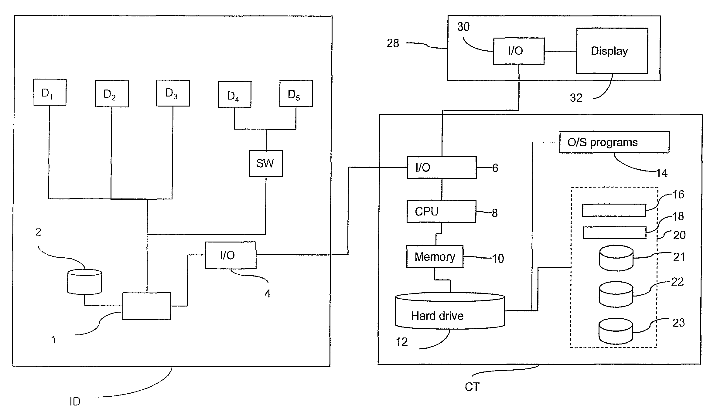
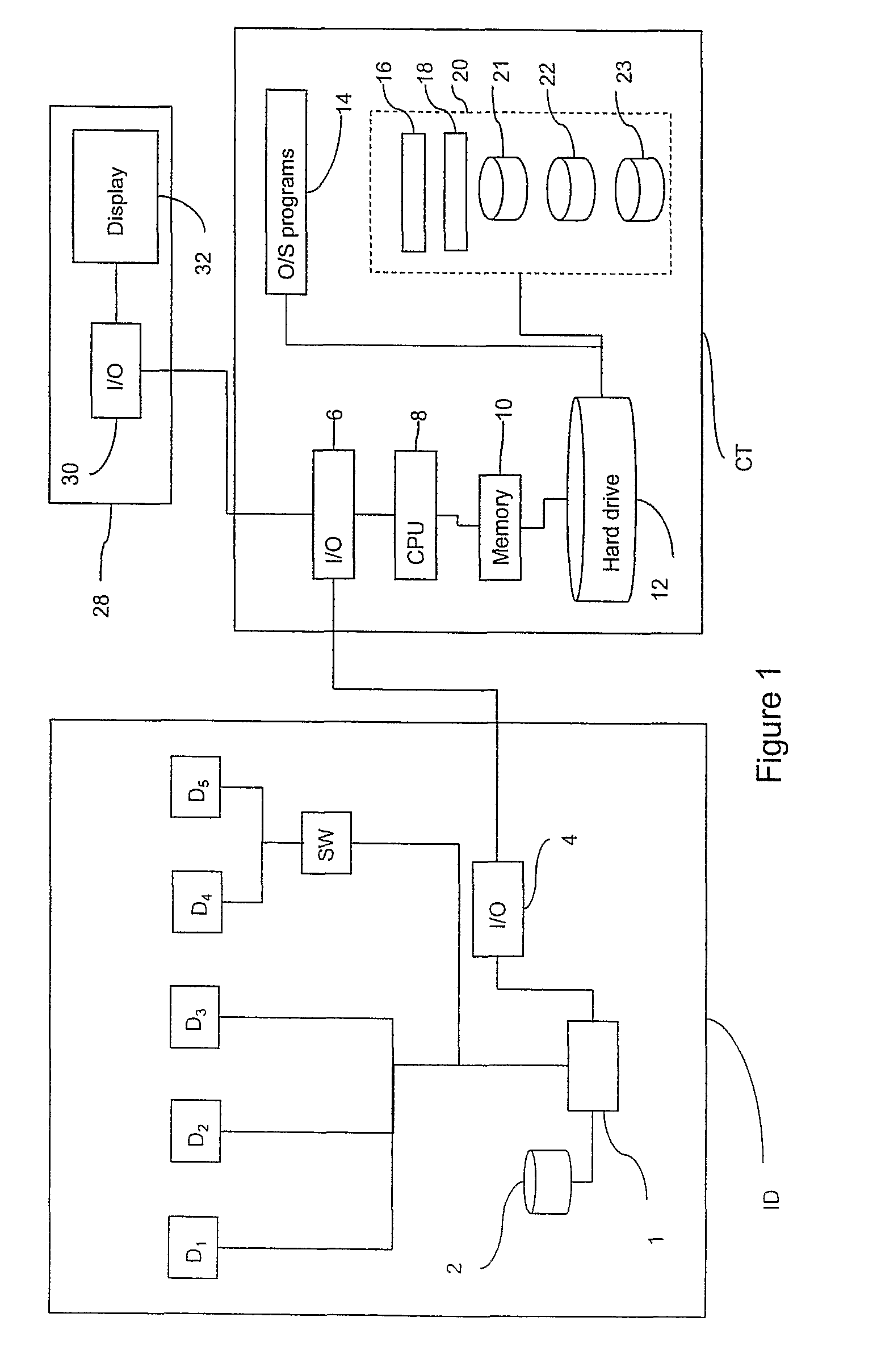
Method of, and apparatus and computer software for, performing image processing
a technology of image processing and computer software, applied in the field of computer-implemented methods of performing image processing and population analysis, can solve the problems of inefficient use of biocellular analysis technology, time-consuming, complex and expensive procedures, poor output data, etc., and achieve the effect of reducing the time taken to set up the image processing apparatus, reducing the cost of assaying cell samples, and improving the efficiency of processing cellular images
- Summary
- Abstract
- Description
- Claims
- Application Information
AI Technical Summary
Benefits of technology
Problems solved by technology
Method used
Image

Examples
example implementation
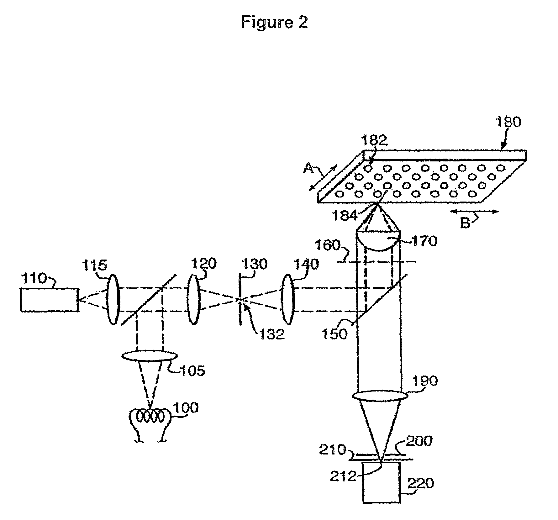
[0151]In this example an image of a G1S Cell Cycle Phase Marker Assay was recorded and the corresponding image data were processed in order to identify cells being in either S-phase or a complement S-phase. The G1S Cell Cycle Phase Marker Assay is available from GE Healthcare Bio-Sciences (product number 25-9003-97) and is the subject of international patent publication number WO 2006 / 008542.
[0152]The G1S Cell Cycle Phase Marker sensor has been developed using functional elements from the human helicase B gene fused to Enhanced Green Fluorescent Protein (EGFP). In G1-phase cells, the sensor protein is predominantly localized within the nucleus. However, increasing Cdk2 / cyclin E activity phosphorylates the sensor late in G1. Consequent exposure of a nuclear export sequence causes the sensor to translocate to the cytoplasm prior to S-phase entry. Cell cycle status can be determined by interrogating the subcellular location and intensity of the green fluorescent protein.
[0153]The G1S C...
PUM
 Abstract
Description
Claims
Application Information