Image data subtraction system suitable for use in angiography

a subtraction system and image data technology, applied in the field of image data subtraction system, can solve the problems of too late enhancement of image quality provided by new and more accurate mask image selection, burden of selecting a new mask image frame, etc., to enhance vessel visualization, remove background image detail, and emphasize vessel structure

Active Publication Date: 2012-01-03
SIEMENS HEALTHCARE GMBH
View PDF17 Cites 10 Cited by
  • Summary
  • Abstract
  • Description
  • Claims
  • Application Information

AI Technical Summary

Benefits of technology

[0005]A system provides substantially real-time automatic, accurate selection of a mask image frame in response to image content while a patient is undergoing a medical procedure. An image data subtraction system suitable for use in Angiography or other medical procedure enhances vessel visualization. The system comprises an imaging system for acquiring, during a medical procedure, data representing multiple temporally sequential individual images of vessels of a portion of patient anatomy. The sequential individual images encompass introduction of a contrast agent. An image processor automatically processes the data representing the multiple temporally sequential individual images to identify a first image indicating presence of the contrast agent and a second image preceding the first image by comparing a difference between measures representative of luminance content of the first and second image, with a threshold. The second image is substantially exclusive of an indication of presence of the contrast agent. The image processor, in response to the difference exceeding the threshold, automatically selects the second image as a mask image and subtracts data representing the mask image from data representing images of the temporally sequential individual images to remove background image detail and emphasize vessel structure in providing processed image data for display. A user interface presents the processed image data for display while the patient is undergoing the medical procedure.

Problems solved by technology

The Mask image frame in known systems is selected in a virtually arbitrary fashion based upon laboratory conditions irrespective of image quality and may often be less than optimal, resulting in the burden of selection of a new Mask image frame.
Therefore, in known systems the enhanced image quality provided by new and more accurate Mask image selection occurs too late to improve guidance to a physician performing a radiological procedure on a patient.

Method used


Image

  • Image data subtraction system suitable for use in angiography
  • Image data subtraction system suitable for use in angiography
  • Image data subtraction system suitable for use in angiography

Examples


Embodiment Construction

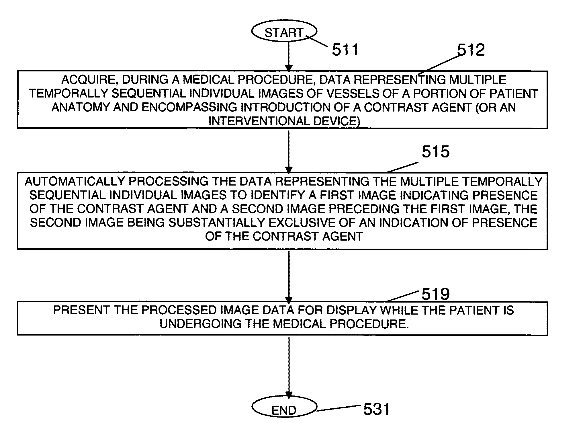
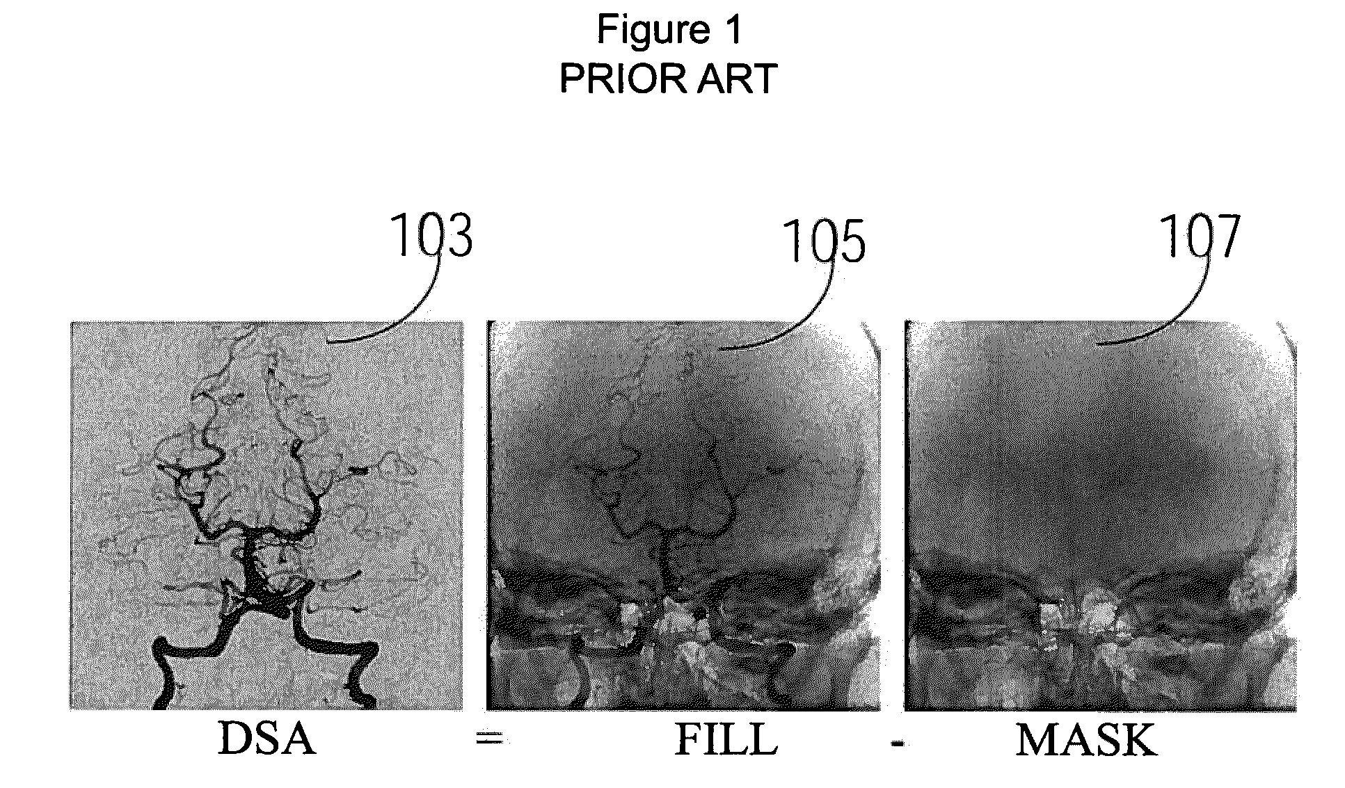
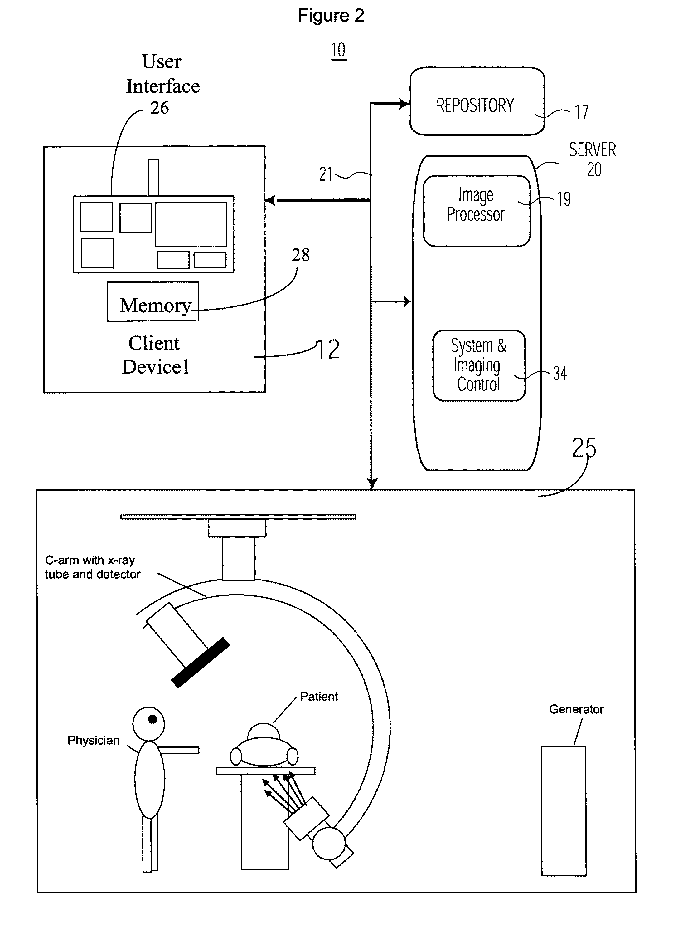
[0011]A system provides substantially real-time automatic, accurate selection of a mask image frame in response to image content while a patient is undergoing a medical procedure. The system automatically detects an image frame by identifying a medical imaging modality device image including an artifact (e.g., indicating presence of a contrast agent, catheter or stent, for example). In response to the image detection, the system selects an image excluding the artifact that precedes the detected image for use as a Mask image while a patient is undergoing a medical procedure. The system advantageously subtracts data representative of the selected Mask image from data representative of medical images used by a physician in performing a medical procedure (such as an Angiography procedure), while a patient is undergoing the procedure. In contrast in known systems, a non-optimal Mask image is selected or a Mask image is selected in an image post-processing operation after performance of a...


PUM


Abstract

An image data subtraction system suitable for use in Angiography or other medical procedure enhances vessel visualization. The system comprises an imaging system for acquiring, during a medical procedure, data representing multiple temporally sequential individual images of vessels of a portion of patient anatomy. The sequential individual images encompass introduction of a contrast agent. An image processor automatically processes the data representing the multiple temporally sequential individual images to identify a first image indicating presence of the contrast agent and a second image preceding the first image by comparing a difference between measures representative of luminance content of the first and second image, with a threshold. The second image is substantially exclusive of an indication of presence of the contrast agent. The image processor, in response to the difference exceeding the threshold, automatically selects the second image as a mask image and subtracts data representing the mask image from data representing images of the temporally sequential individual images to remove background image detail and emphasize vessel structure in providing processed image data for display. A user interface presents the processed image data for display while the patient is undergoing the medical procedure.

Description

[0001]This is a non-provisional application of provisional application Ser. No. 60 / 981,226 filed Oct. 19, 2007, by G. F. Kramp et al.FIELD OF THE INVENTION[0002]This invention concerns an image data subtraction system suitable for use in Angiography to enhance vessel visualization involving automatic identification and subtraction of a mask image from other images to remove background image detail and emphasize vessel structure while a patient is undergoing Angiography, for example.BACKGROUND OF THE INVENTION[0003]Digital Subtraction Angiography (DSA) is a known process employed in angiography imaging applications in radiology to remove background anatomical data which distracts from clinically relevant vessel structure of a patient. For example, in neuro-radiological imaging studies, a physician may desire to see the vessel structure of a patient brain without being obscured by the boney structure of the skull. In performing a DSA process, a single mask frame (containing background...

Claims


Application Information

Patent Timeline
Patent Type & Authority Patents(United States)
IPC IPC(8): G06K9/00
CPCA61B6/481A61B6/504G06T7/0012G06T7/0022G06T2207/10121G06T2207/30104G06T7/97
Inventor KRAMP, GEORGE F.MARTUCCI, GARY S.
Owner SIEMENS HEALTHCARE GMBH
Why Eureka
Try Eureka