Apparatus and method for diagnosing breast cancer including examination table

a technology of examination table and breast cancer, which is applied in the field of medical examination apparatus and methods for detecting breast cancer, can solve the problems of difficult to treat breast cancer, low screening cost, and large health problems of women, and achieve the effects of safe, simple, comfortable, convenient and effective treatmen

Active Publication Date: 2012-01-10
INTERSTITIAL SYST
View PDF20 Cites 51 Cited by
  • Summary
  • Abstract
  • Description
  • Claims
  • Application Information

AI Technical Summary

Benefits of technology

[0014]The present invention provides for an apparatus including an examination table to be used in a clinical setting, to provide screening and imaging of breast anomalies in a way that is safe, simple, comfortable, convenient and effective. In an embodiment, the apparatus is configured as an examination table. In an embodiment, the present invention provides for an apparatus for screening or diagnosing cancer or other pathological disorders, such as in a breast of a patient, comprising a table having a horizontal upper surface upon w

Problems solved by technology

Breast cancer is a major health problem for women.
While early detection leads to improved treatment and increased longevity, the existing low-cost screening methods leave much to be desired.
Some 20 million women annually fail to comply with the American Cancer Society recommendations for annual mammograms and about 120,000 of these women develop late stage, difficult-to-treat breast cancer.
The reasons for the non-compliance are, in part, that mammography is physically uncomfortable, provides uncertain results and poses an ionizing radiation risk.
The physical discomfort from mammograms arises because 40% of the tumors occur near the armpit or axilla.
The excess treatment costs for these 120,000 unscreened women are on the order of several billion dollars and the social costs are high in terms of reduced longevity and impaired life style.
However, these have not considered how the patient can be comfortably, safely and reliably examined by physicians on a routine basis to screen women or diagnose women for breast cancer.
While some of these systems have been demonstrated theoretically or in small-scale laboratory tests, no provision for large-scale, reliable and comfortable examinations has been noted.
This technique required physically manipulating the breast and this introduced uncertainties as to the location of the electrodes and lead to uncertain results.
These uncertain results caused this low-frequency met

Method used


Image

  • Apparatus and method for diagnosing breast cancer including examination table
  • Apparatus and method for diagnosing breast cancer including examination table
  • Apparatus and method for diagnosing breast cancer including examination table

Examples


Embodiment Construction

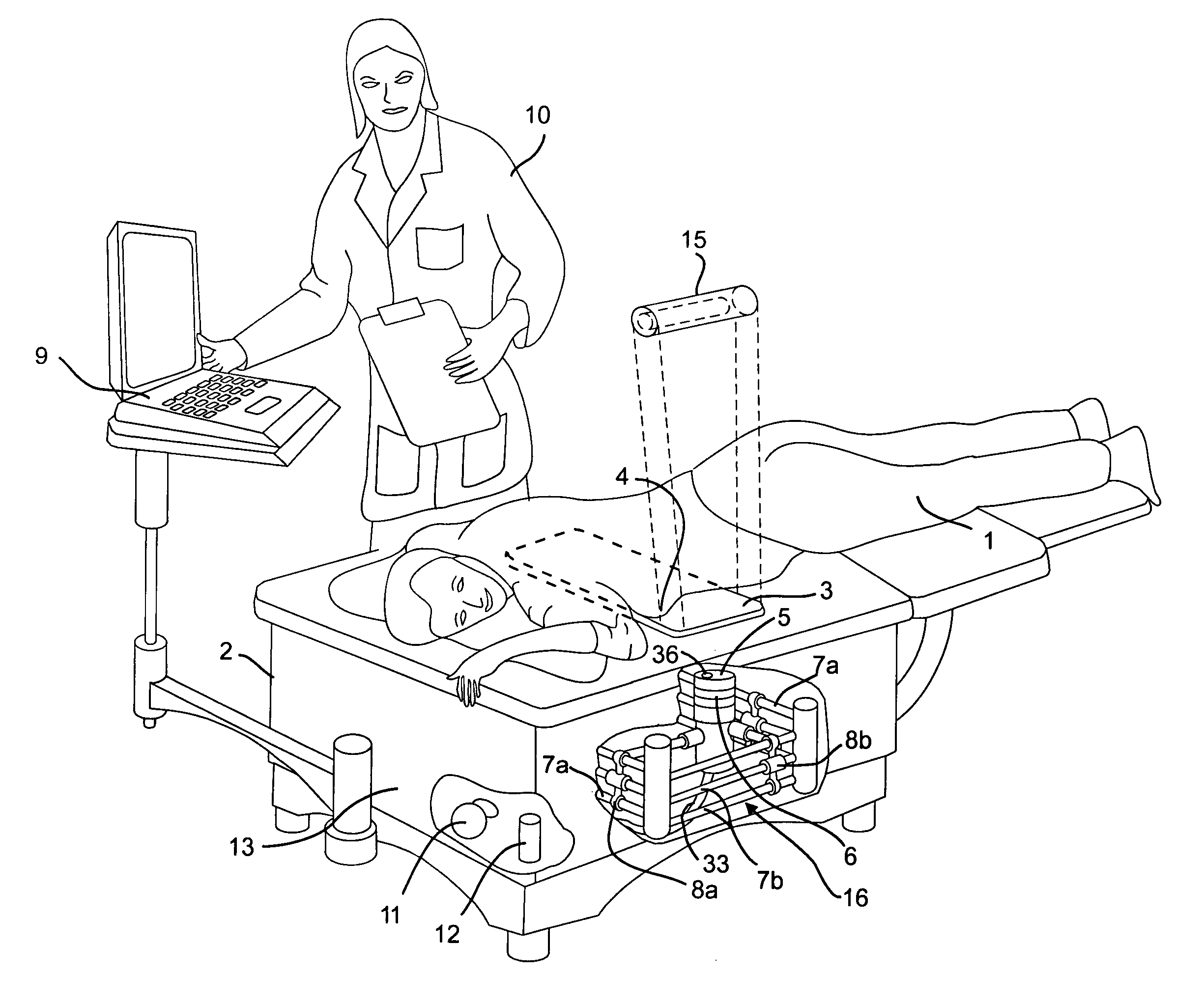
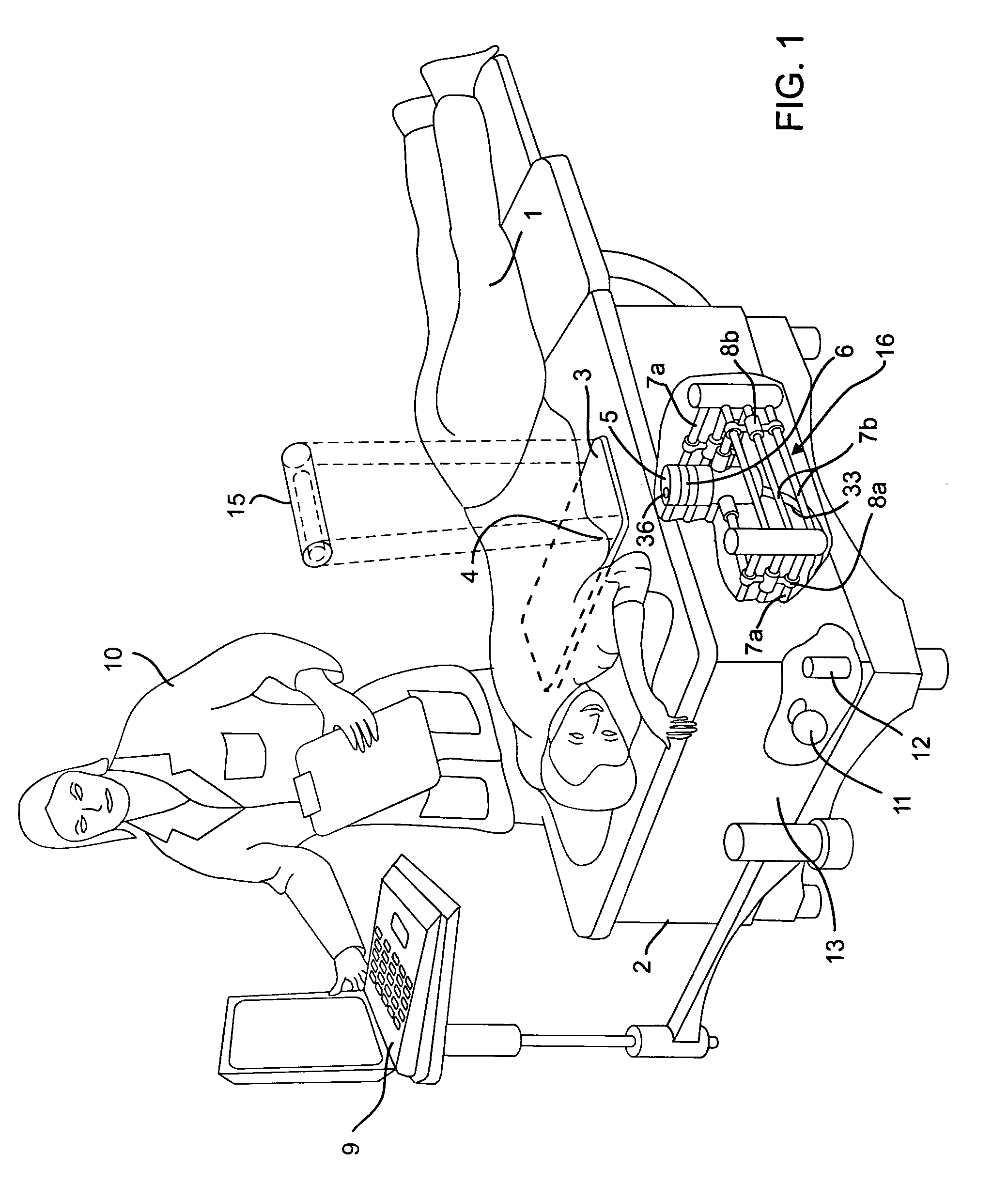
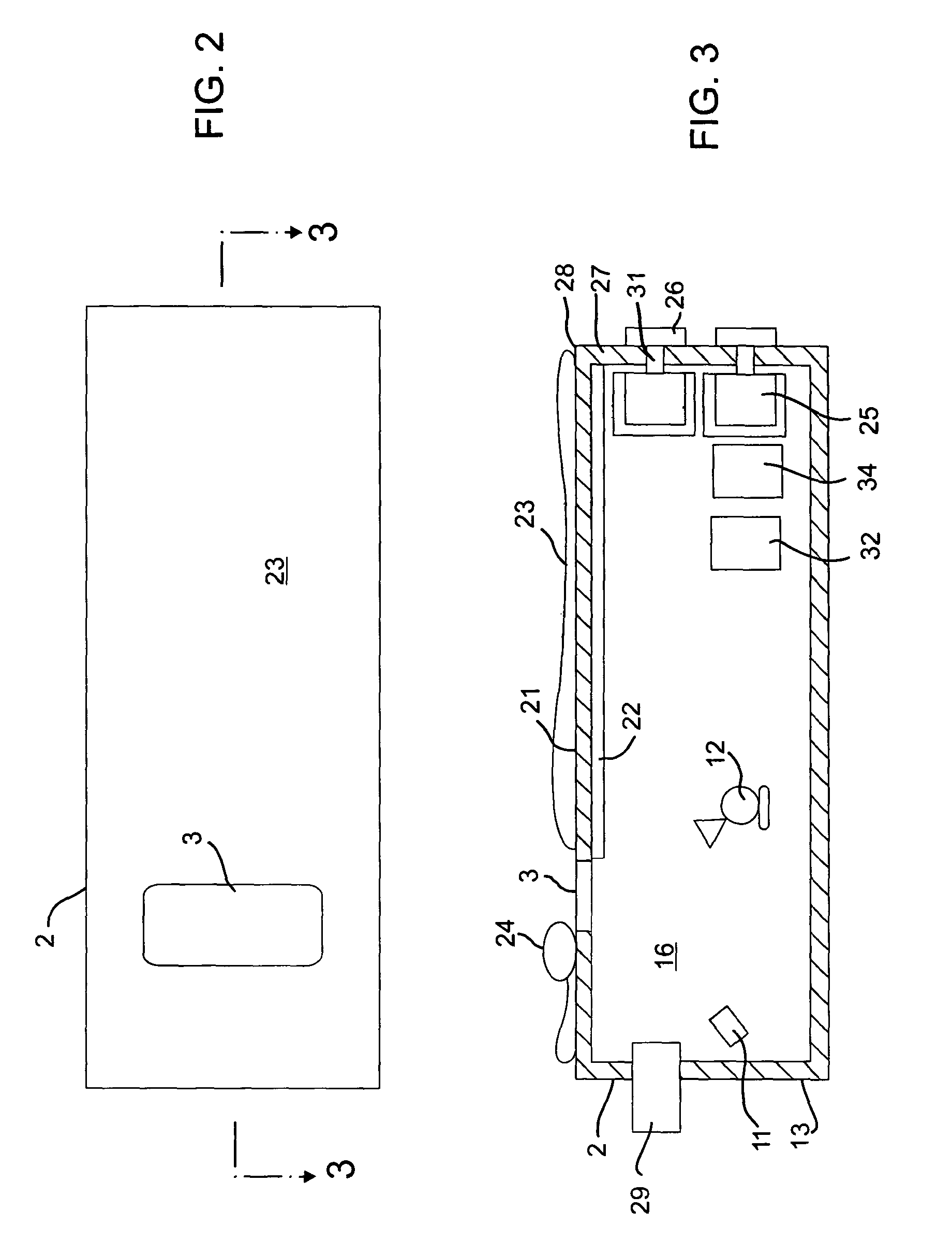
[0037]An embodiment of the present invention is illustrated with reference to FIGS. 1-8. FIGS. 9-10 disclose an alternate embodiment discussed below. FIG. 1 illustrates a breast cancer radar screening system with a patient 1 lying prone on an examination table 2 of the present invention, with her breasts 4 pressed against a microwave and optically transparent scan plate 3. An upper horizontal face 5 of an antenna assembly 6 is in close proximity to the scan plate 3 with an air gap that avoids abrasion with the upper face 6 of the antenna assembly and also use of the scan plate 3 avoids contact with the patient 1. In an embodiment, the upper face 5 of the antenna 6 may be within approximately 1-3 mm of the scan plate 3.

[0038]A scanning subsystem 7a, 7b, 8a, 8b is located in an enclosure 16 formed within the table 2. The scanning subsystem 7a, 7b, 8a, 8b includes a motorized system that moves the antenna assembly 6 over the bottom of the scan plate in a predetermined pattern (see FIG....


PUM


Abstract

A microwave breast cancer imaging method that includes an examination table that is both comfortable and reliable is provided that includes a support system and an orientation system such that breasts can remain in a fixed position to allow for scanning. A horizontal microwave and optically transparent scan plate forms part of the top of the examination table. The imprint of the breasts on the scan plate may be visually displayed to aid in the orienting of each breast such that all volumes within the breast are scanned. Microwave power is then scanned upward through the scan plate to develop a microwave response that is indicative of the presence of a lesion. After scanning, the visual imprint of the breast is recorded. As needed, microwave equipment can be included within a microwave shielded enclosure that also forms part of the scan table. Spurious leakage of microwave power may be further suppressed by use of microwave-absorbing materials, within the enclosure and, in the padding that covers the surface of the examination table and removable pads.

Description

[0001]This application claims the benefit of the filing date of co-pending provisional application Serial No. 60 / 402,511, filed Aug. 9, 2002.BACKGROUND[0002]The present invention pertains to a medical examination apparatus and method for detecting breast cancer. In particular, the present invention pertains to an examination table having scanning equipment, including microwave equipment and features to aid in screening or diagnosis of breast cancer.[0003]Breast cancer is a major health problem for women. While early detection leads to improved treatment and increased longevity, the existing low-cost screening methods leave much to be desired. For example, as many as 10 to 30% of the malignant tumors in mammogram-screened women go undetected. The result is that about 16,000 women who annually have a mammogram will have a malignant tumor and not know it. Some 20 million women annually fail to comply with the American Cancer Society recommendations for annual mammograms and about 120,0...

Claims


Application Information

Patent Timeline
IPC IPC(8): A61B5/00A61BA61B8/02
CPCA61B5/0091A61B5/0507A61B5/4312A61B5/704A61B5/0068
Inventor SMITH, RANDALL M.BRIDGES, JACK E.JONES, II, DENIS E.HENRY, TODD R.
Owner INTERSTITIAL SYST
Why Eureka
Try Eureka