
Method to inhibit airway hyperresponsiveness using aerosolized T cell receptor antibodies

a technology of t cell receptor and anti-airway, which is applied in the direction of antibody medical ingredients, aerosol delivery, spray delivery, etc., can solve the problems of drug-induced lung disease, and increased susceptibility to infection, so as to reduce the airway hyperresponsiveness of the mammalian body

Inactive Publication Date: 2012-05-15
NAT JEWISH HEALTH
View PDF11 Cites 68 Cited by
  • Summary
  • Abstract
  • Description
  • Claims
  • Application Information

AI Technical Summary

Benefits of technology

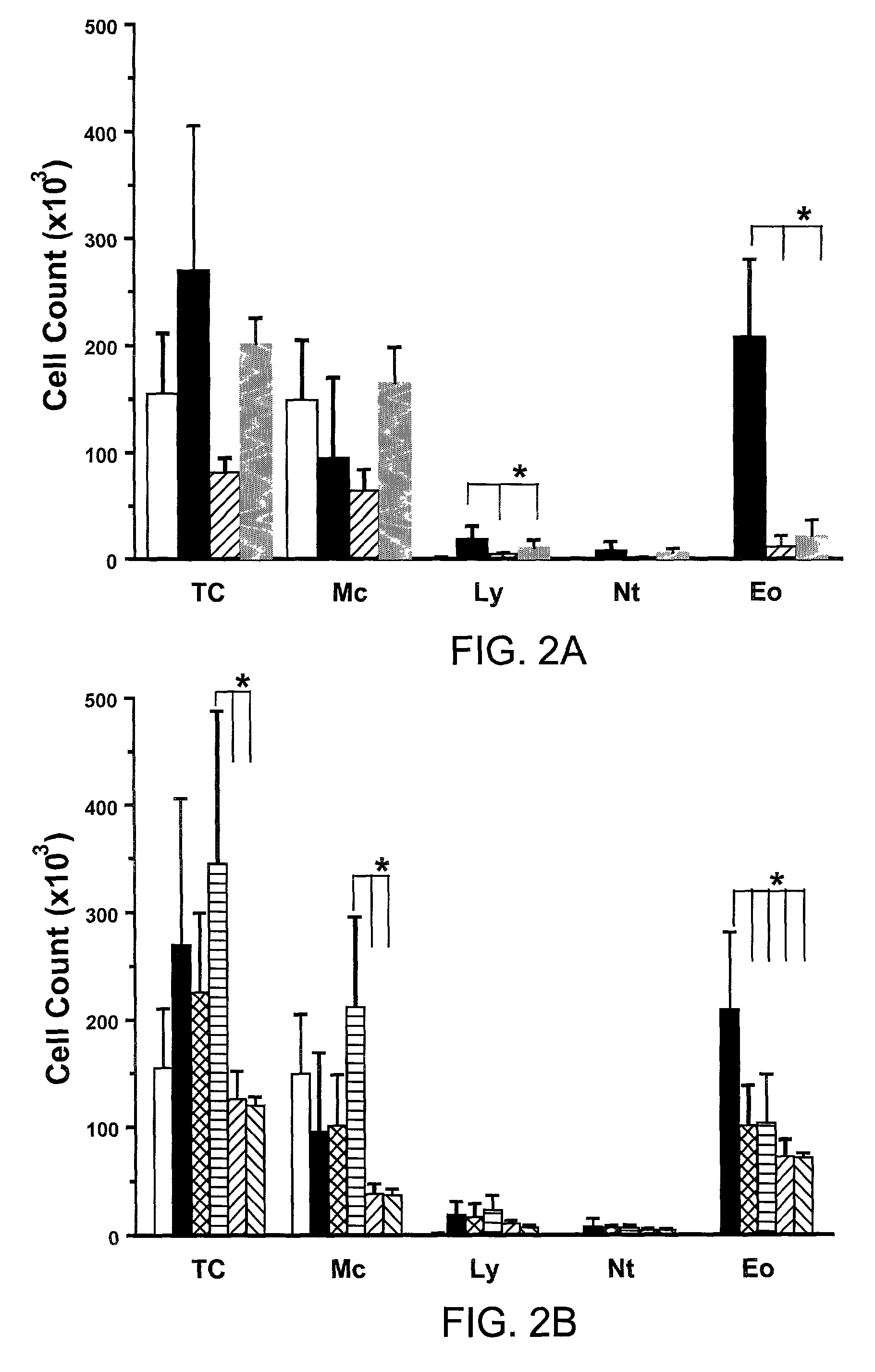
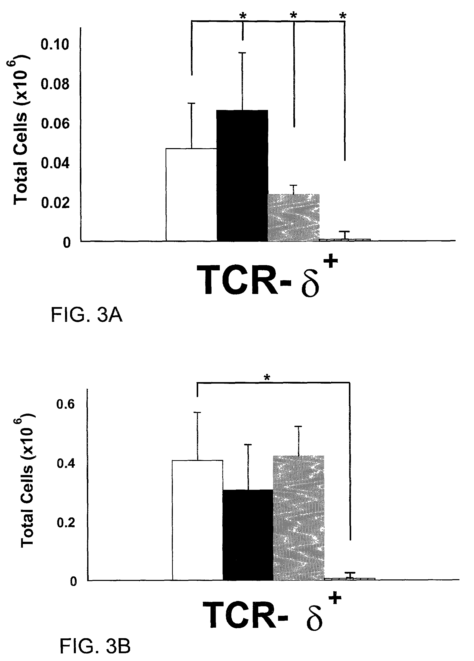
[0011]In difference to previously described clinical approaches for the treatment of diseases of the airways, the present inventors have used aerosolized antibodies against T cell receptors to modify diseases in the lung. The present inventors have shown that this method is highly effective at reducing airway hyperresponsiveness, targets pulmonary T cell populations in the absence of any substantial effect on peripheral T cells, and most surprisingly, the antibodies can be administered at extremely low doses which are believed to be at least about 1000-fold lower than systemic doses of antibody required to achieve the same effect. Therefore, the present inventors have discovered a novel method for treating immune related diseases in the lung without significantly affecting systemic immune responses of the host.
[0017]In one embodiment, the mammal has been sensitized to an allergen and has been exposed to, or is at risk of being exposed to, an amount of the allergen that is sufficient to induce airway hyperresponsiveness (AHR) in the mammal in the absence of the aerosolized antibody formulation. Preferably, the aerosolized antibody formulation is administered within a time period of between 48 hours or less prior to exposure to an AHR provoking stimulus that is sufficient to induce AHR, and within 48 hours or less after the detection of the first symptoms of AHR. In one aspect, the aerosolized antibody formulation is administered upon the detection of the first symptoms of acute onset AHR. In another aspect, the aerosolized antibody formulation is administered within 1 hour after the detection of the first symptoms of acute onset AHR. In another aspect, the aerosolized antibody formulation is administered within 12 hours or less prior to exposure to a AHR provoking stimulus that is sufficient to induce acute onset AHR. In another aspect, the aerosolized antibody formulation is administered within 2 hours or less prior to exposure to a AHR provoking stimulus that is sufficient to induce acute onset AHR. In another aspect, the aerosolized antibody formulation reduces the airway hyperresponsiveness of the mammal such that the FEV1 value of the mammal is improved by at least about 5%. Preferably, the administration of the aerosolized antibody formulation does not substantially affect peripheral immune function in the mammal.

Problems solved by technology

However, the mechanisms leading to AHR are still poorly understood and can be attributed to both immune-dependent and immune-independent mechanisms.
These agents, however, have the potential of serious side effect, including, but not limited to, increased susceptibility to infection, liver toxicity, drug-induced lung disease, and bone marrow suppression.
Thus, such drugs are limited in their clinical use for the treatment of lung diseases associated with airway hyperresponsiveness.
The use of anti-inflammatory and symptomatic relief reagents is a serious problem because of their side effects or their failure to attack the underlying cause of an inflammatory response.
However, in practice, at least with regard to airway responses in the lung, attempts to use aerosolized therapeutic antibodies have not generally met with success.
Fahy et al. concluded that the observed lack of efficacy was probably due to the inability of the aerosol route of delivery to result in high enough concentrations of antibody in the lung tissue compartments to neutralize IgE.

Method used


Image

  • Method to inhibit airway hyperresponsiveness using aerosolized T cell receptor antibodies
  • Method to inhibit airway hyperresponsiveness using aerosolized T cell receptor antibodies
  • Method to inhibit airway hyperresponsiveness using aerosolized T cell receptor antibodies

Examples


example 1

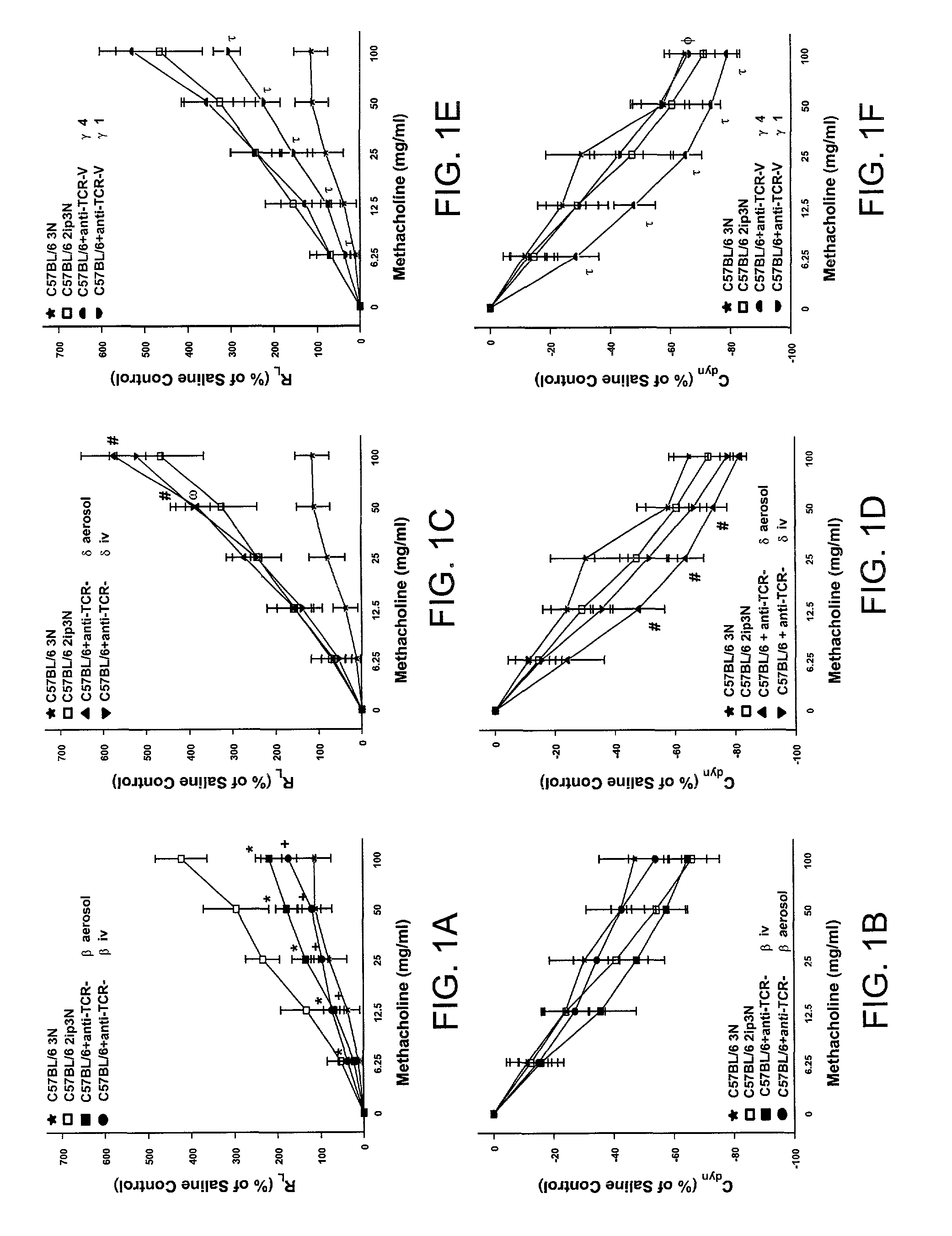
[0096]The following example describes the comparison of aerosolized versus intravenously given mAbs against TCR-β in ovalbumin (OVA) sensitized and challenged mice.

[0097]The classical animal model of allergic inflammation (see, e.g., Takeda et al., (1997). J. Exp. Med. 186, 449-454; Renz et al., 1992, J. Allergy Clin. Immunol. 89:1127-1138; Larsen et al., 1992, J. Clin. Invest. 89:747-752; and Saloga et al., 1993, J. Clin. Invest. 91:133-141) was used to assess the ability of aerosolized mAbs to influence AHR. The present inventors and others have previously shown that αβ T cells are necessary for the development of allergic inflammation and airway hyperresponsiveness (AHR) (Holt, J. Exp. Med. 183:1297-1301 (1996); Lahn et al., Nature Med. 5:1150-6 (1999)). In this experiment, C57BL / 6 mice were injected twice with Alum / OVA intraperitoneally (sensitization) before a three day exposure to aerosolized OVA (challenge). The exact procedure has been previously published (Lahn et al., Natu...

example 2

[0102]The following example describes the comparison of aerosolized versus intravenously given mAbs against TCR-δ in ovalbumin (OVA) sensitized and challenged mice.

[0103]In this example, experiments were conducted as described in Example 1, but with the use of a mAb against TCR-δ (a 1:1 mixture of mAbs GL3 (Goodman et al., J. Exp. Med. 170:1569 (1989)) and 403A10 Itohara et al., Proc. Natl. Acad. Sci. USA 86:5094-5098 (1989)). As previously published (Lahn et al., Nature Med. 5:1150-6 (1999)), the intravenous injection of mAbs against TCR-δ (▾ in FIGS. 1C and 1D) caused only a slight increase in AHR when compared to 2ip3N treated C57BL / 6 mice in the absence of antibody (□ in FIGS. 1C and 1D). The significant differences between 2ip3N treated mice and 2ip3N mice treated with i.v. anti-TCR-δ are shown by “ω” in FIG. 1C. Again, the addition of 10 μg / ml of anti-TCR-δ to the last OVA aerosol application (▴ in FIGS. 1C and 1D) had a similar effect as the intravenous injection of the same ...

example 3

[0104]The following example describes the comparison of aerosolized mAbs against TCR-Vγ1 and TCR-Vγ4 in ovalbumin (OVA) sensitized and challenged mice.

[0105]Finally, the γδ T cell subsets Vγ1 and Vγ4 were examined for their contribution to the regulation of AHR. Experiments were performed as described in Examples 1 and 2 above, but with the use of mAbs against γδ TCR subsets having Vγ1 or Vγ4.

[0106]The aerosol application of mAbs against Vγ1 showed a significant reduction in AHR of 2ip3N treated C57BL / 6 mice (top in FIGS. 1E and 1F). The significant differences between 2ip3N treated mice and 2ip3N treated mice with aerosol anti-Vγ1 are indicated by “τ” in FIGS. 1E and 1F). This result indicates that the removal of this particular subset of γδ T cells, which may be recruited during allergic inflammation to the lung, has a beneficial effect on regulating AHR associated with allergic inflammation. Thus, an aerosol of a particular subset of γδ T cells in humans, presumably the Vγ9 subs...


PUM

PropertyMeasurementUnit
body weightaaaaaaaaaa

Abstract

Disclosed is a method to reduce airway hyperresponsivesness in an animal by the direct delivery to the lungs of aerosolized antibodies against T cell receptors. The method is particularly useful for treating airway hyperresponsiveness associated with allergic inflammation, is effective at extremely low doses of antibody, and does not have a substantial effect on the peripheral immune system.

Description

GOVERNMENT RIGHTS[0001]This invention was supported, in part, by Grant Nos. R01 HL-65410; AI-40611; HL-36557; HL-61005; and R01 AI-44920, all awarded by the National Institutes of Health, and Grant No. R825702, awarded by the Environmental Protection Agency. The government has certain rights to this invention.FIELD OF THE INVENTION[0002]This invention generally relates to the use of aerosolized antibodies against T cell receptors for the inhibition of airway hyperresponsiveness (AHR). Specifically, aerosolized antibodies against receptors on both αβ and γδ T cells are disclosed for the treatment of AHR.BACKGROUND OF THE INVENTION[0003]A variety of inflammatory agents can provoke airflow limitation, including allergens, cold air, exercise, infections and air pollution. In particular, allergens and other agents in allergic or sensitized mammals (i.e., antigens and haptens) cause the release of inflammatory mediators that recruit cells involved in inflammation. Such cells include lymph...

Claims


Application Information

Patent Timeline
Patent Type & Authority Patents(United States)
IPC IPC(8): A61K39/395C07K16/28A61K9/00
CPCA61K9/0073C07K16/2809C07K16/2812C07K16/2815A61K2039/505A61K2039/544Y10S424/81Y10S530/868A61P11/00
Inventor LAHN, MICHAEL F.BORN, WILLI K.KANEHIRO, ARIHIKOGELFAND, ERWIN
Owner NAT JEWISH HEALTH
Learn More