
Image processing method and image processing apparatus, and program

a technology of image processing applied in the field of image processing method and image processing apparatus, and program, can solve the problems of increasing the burden on the operator and lacking in correct boundary separation

Inactive Publication Date: 2012-05-15
GE MEDICAL SYST GLOBAL TECH CO LLC
View PDF24 Cites 7 Cited by
  • Summary
  • Abstract
  • Description
  • Claims
  • Application Information

AI Technical Summary

Benefits of technology

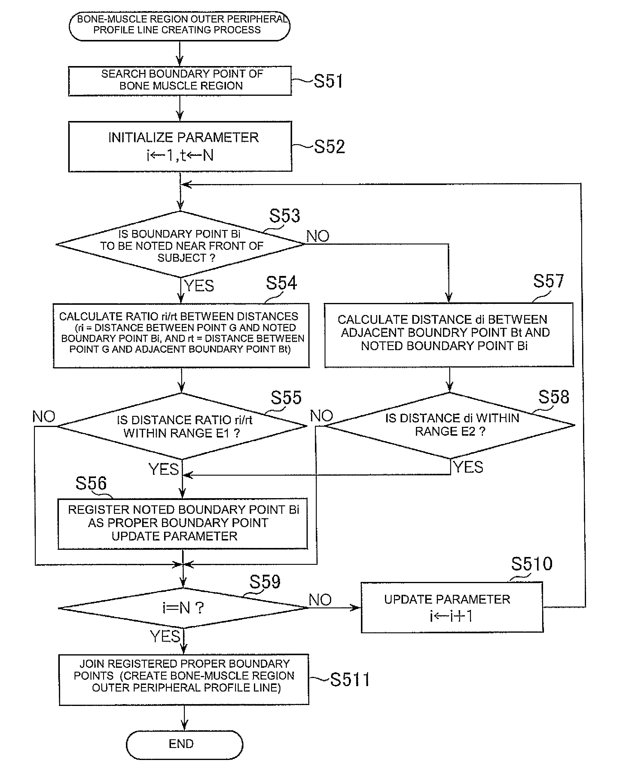
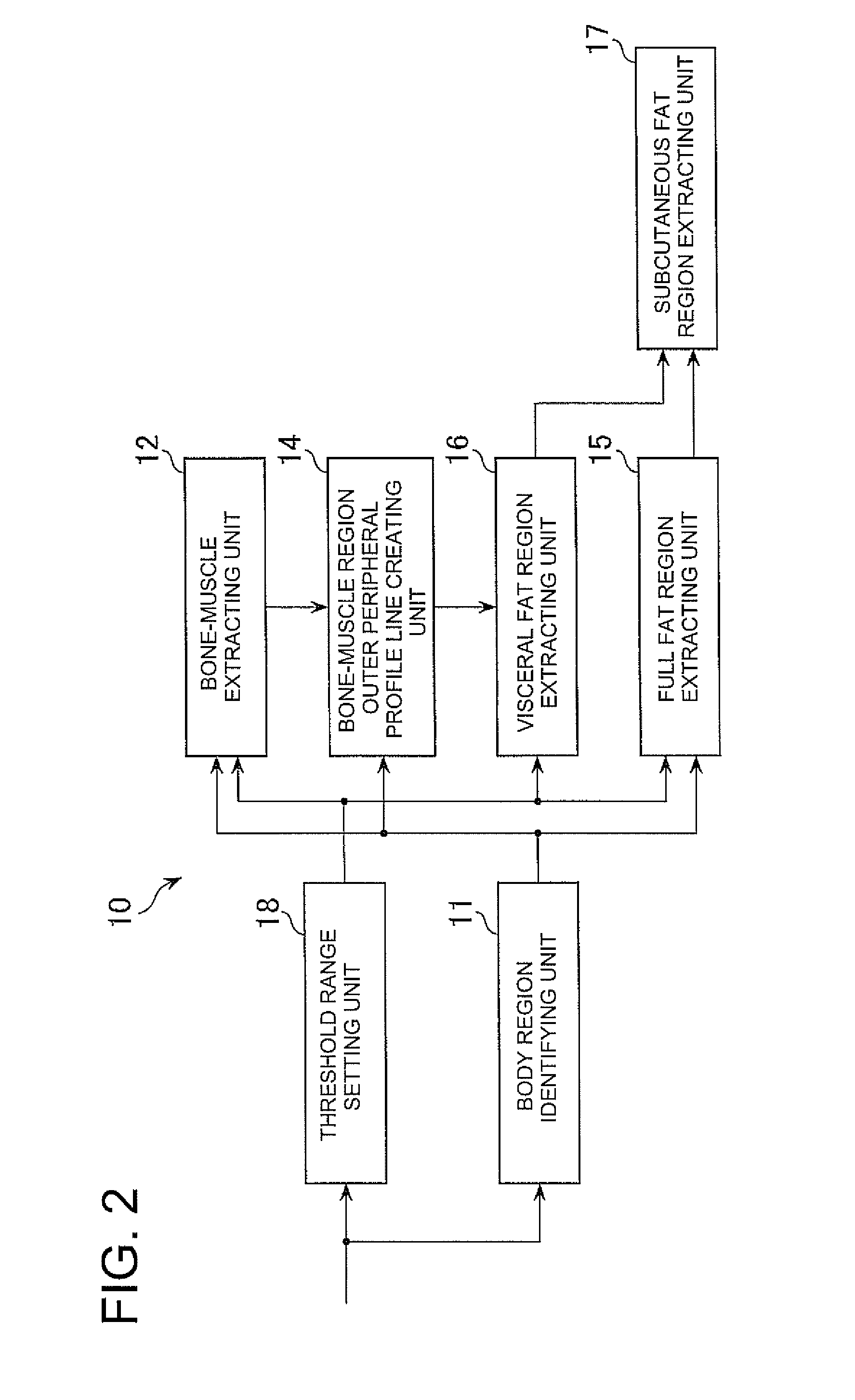
[0031]According to the invention, a bone muscle region constituted of bone muscle pixels having pixel values corresponding to bones or muscles, of a body region of a subject at a tomographic image is extracted. Boundary points of the bone muscle region are searched from outside to inside. Of the searched boundary points, only boundary points at which each index related to at least one of the length and slope of each line segment connecting between the adjoining boundary points meets or satisfies a predetermined condition, are joined together to create a bone-muscle region outer peripheral profile line. A visceral fat region constituted of fat pixels having pixel values corresponding to fat at a region lying inside the bone-muscle region outer peripheral profile line is extracted. Therefore, a profile or contour of a region considered to contain only the visceral fat region as a fat region can be determined by detecting only boundary points having a high possibility that they will be disposed on the profile, by a relatively simple process and joining these together. A subject's visceral fat region at the tomographic image can be separated and extracted from a subcutaneous fat region in a short period of time and more accurately.

Problems solved by technology

Since, however, this method increases the burden on the operator, a method capable of separating the visceral fat region and the subcutaneous fat region from each other simpler has been expected.
Therefore, the subcutaneous fat region is expanded inside by its expansion, whereby there is a possibility of lacking in correctness in the boundary separation between the visceral fat and the subcutaneous fat.

Method used


Image

  • Image processing method and image processing apparatus, and program
  • Image processing method and image processing apparatus, and program
  • Image processing method and image processing apparatus, and program

Examples


Embodiment Construction

[0052]An embodiment of the invention will be explained below with reference to the accompanying drawings.

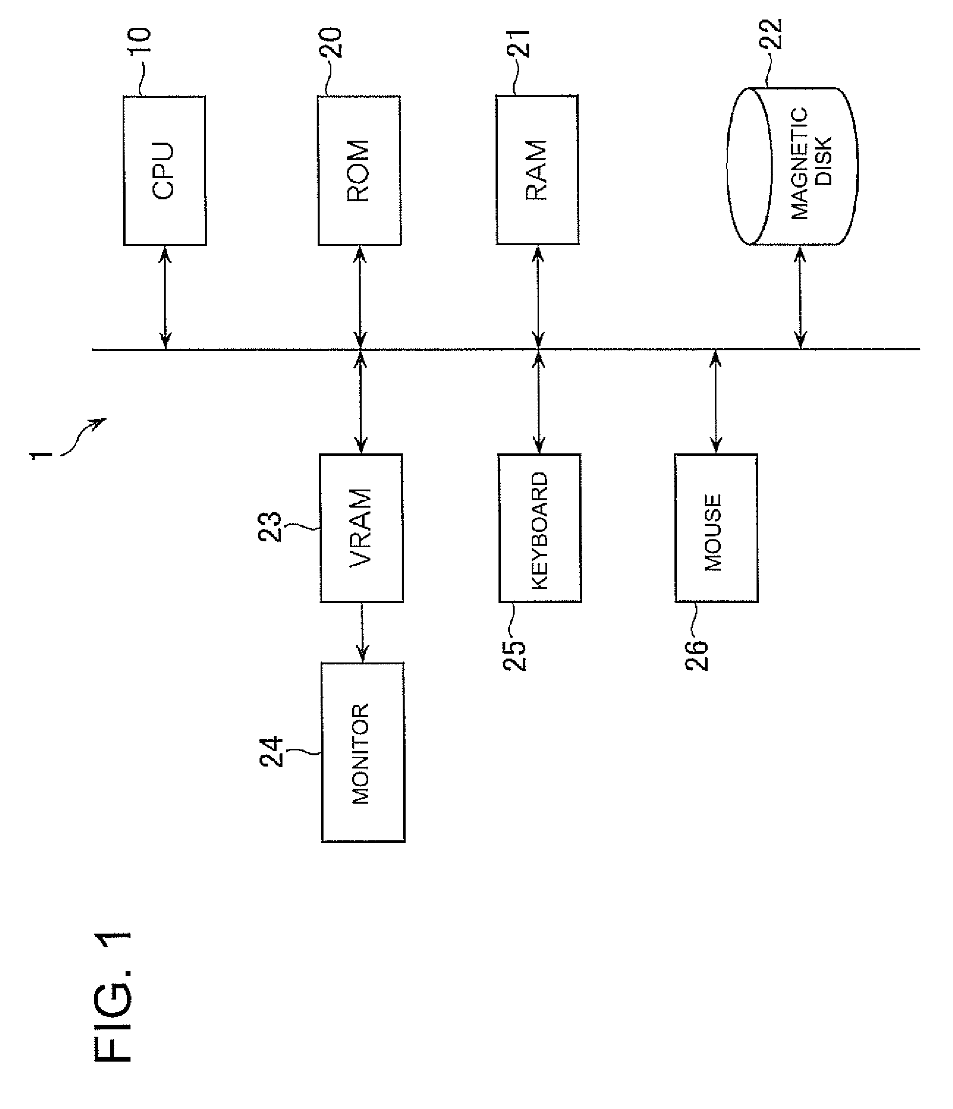
[0053]FIG. 1 is a block diagram showing an overall configuration of a tomographic image processing apparatus (an image processing apparatus) 1 illustrative of one embodiment of the invention. The tomographic image processing apparatus 1 separates and extracts a visceral fat region of a subject and a subcutaneous fat region thereof from a tomographic image of the subject, which is obtained by modality of an X-ray CT apparatus, an MRI apparatus, an ultrasonic apparatus or the like.

[0054]As shown in the drawing, the tomographic image processing apparatus 1 includes a central processing unit (CPU) 10 which takes charge of control on the entire apparatus, a ROM (Read Only Memory) 20 which stores a boot program and the like therein, a RAM (Random Access Memory) 21 which functions as a main storage device, an OS (Operating Software), a magnetic disk 22 which stores an image processing p...


PUM


Abstract

An image processing method includes identifying a body region in a tomographic image based on pixel values within the tomographic image, extracting a bone muscle region corresponding to essential parts of bones and muscles based on bone muscle pixels having pixel values corresponding to the bones or muscles in the body region, searching boundary points of the bone muscle region from outside a region surrounding the extracted bone muscle region to inside the region, creating a bone-muscle region outer peripheral profile line by joining only boundary points at which each index related to at least one of a length and a slope of a line segment connecting between the adjoining boundary points of the searched boundary points satisfies a predetermined condition, and extracting a visceral fat region comprised of fat pixels having pixel values corresponding to fat in a region lying inside the bone-muscle region outer peripheral profile line.

Description

CROSS REFERENCE TO RELATED APPLICATIONS[0001]This application claims the benefit of Japanese Patent Application No. 2007-253898 filed Sep. 28, 2007, which is hereby incorporated by reference in its entirety.BACKGROUND OF THE INVENTION[0002]The subject matter disclosed herein relates to an image processing method and an image processing apparatus both of which extract a fat region from a tomographic image of a subject, and a program used therefore.[0003]A threshold process of pixel values is performed on tomographic images such as a CT (Computed Tomography) image, an MRI (Magnetic Resonance Imaging) image, etc., thereby making it possible to extract each fat region. A decision as to whether a metabolic syndrome is suspected, is made with a visceral fat area as one index clinically. Since, however, full fat regions are extracted simply by performing the threshold process of the pixel values, there is a need to separate a full fat region into a visceral fat region and a subcutaneous fa...

Claims


Application Information

Patent Timeline
Patent Type & Authority Patents(United States)
IPC IPC(8): G06K9/00A61B6/00
CPCG06T7/0083G06T2207/10072G06T2207/20136G06T2207/30008G06T7/12G06T7/181
Inventor OMI, YASUOHAQUE, HASNINE AKTER
Owner GE MEDICAL SYST GLOBAL TECH CO LLC
Learn More