Integrated breast X-ray and molecular imaging system

a breast x-ray and molecular imaging technology, applied in the field of breast imaging, can solve the problems of increasing the difficulty of distinguishing cancerous lesions from breast tissue, affecting the diagnosis speed and accuracy, and affecting the sensitivity of the mammogram, so as to facilitate the registration of images, and increase the speed and accuracy of diagnosis.

Active Publication Date: 2012-07-10
HOLOGIC INC
View PDF6 Cites 15 Cited by
  • Summary
  • Abstract
  • Description
  • Claims
  • Application Information

AI Technical Summary

Benefits of technology

[0009]The present invention fuses tomosynthesis imaging capability and molecular imaging capabilities into a single breast imaging device. Integrating the different imaging modalities into a single imaging device may increase the speed and accuracy of diagnosis by enabling the radiologist to tune the diagnostic workflow according to the particular needs of each patient. For example the radiologist may choose to use one modality over another depending upon a known anatomical structure (i.e., density) of the patient's breast. Or the radiologist may use the two diagnostic imaging methods in sequence to obtain additional information as needed. For example, if a routine mammogram such as that shown in FIG. 1A, obtained using the x-ray components, suggests the presence of a lesion 10, the radiologist may opt to obtain a molecular image of the patient's breast during the patient's visit. The molecular image verifies the presence of lesion 10, while also highlighting additional calcifications 12, 14, which may have been missed in the x-ray image. The present invention enables tomosynthesis images and molecular images to be viewed together on a single display, either side by side (for example providing both FIGS. 1A and 1B on a display), overlaid, in cine mode, etc. The ability to obtain the molecular image with the same equipment during a single office visit facilitates registration of the images, allows comparison of the anatomical and physiological information of the breast at a slice granularity, allows comparison of CAD results of the images, increases the speed and accuracy of the diagnosis and thereby reduces unnecessary biopsies and concomitant patient anxiety.

Problems solved by technology

However, mammograms suffer in both the area of sensitivity and specificity.
However, because mammograms are two dimensional representations of a three dimensional structure, the sensitivity of a mammogram is compromised due to overlapping structures in the compressed breast.
In addition, the similarity of x-ray attenuation characteristics between breast tissue and cancerous tissue increases the difficulty of differentiating cancerous lesions from breast tissue, particularly when imaging dense breast tissue.
While breast tomosynthesis methods greatly improve the sensitivity of x-ray cancer screening, specificity issues associated with dense breasts remain an issue.
However the sensitivity of the MRI modality negatively affects its specificity.
In addition, the cost of the MRI devices limits their general deployment.

Method used


Image

  • Integrated breast X-ray and molecular imaging system
  • Integrated breast X-ray and molecular imaging system
  • Integrated breast X-ray and molecular imaging system

Examples


Embodiment Construction

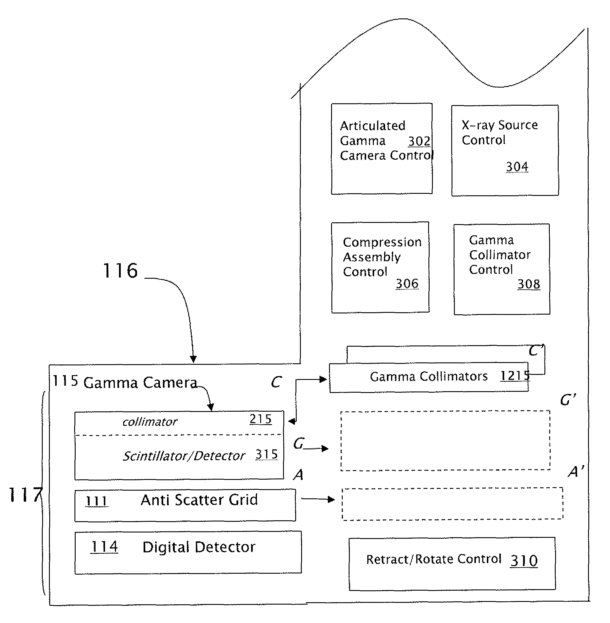
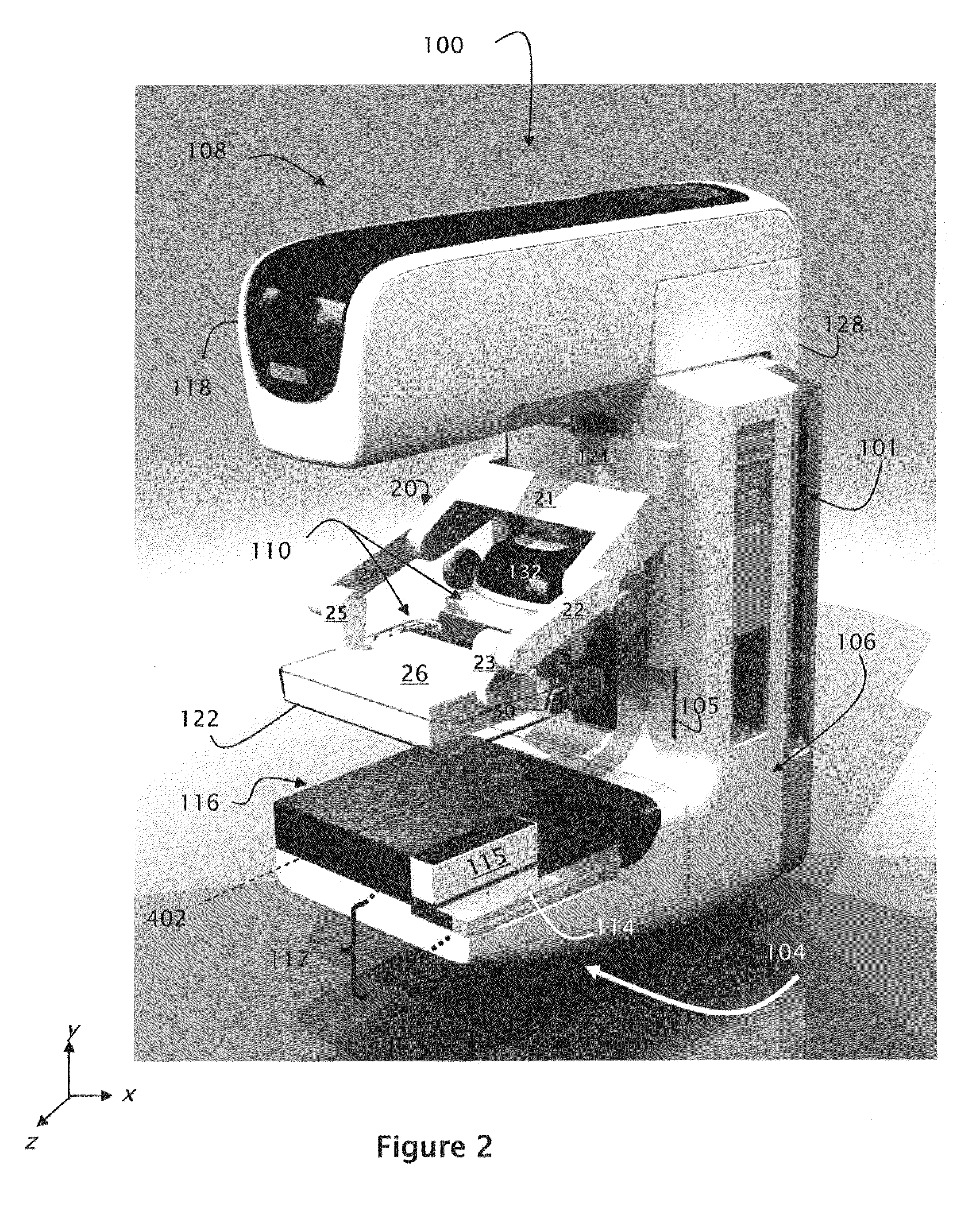
[0030]FIG. 2 illustrates an exemplary embodiment of an integrated Tomosynthesis / Molecular Breast Imaging (T / MBI) device 100 of the present invention. The T / MBI device 100 integrates x-ray components with molecular imaging components to provide a breast imaging system having increased sensitivity and specificity.

[0031]The T / MBI device 100 of FIG. 2 is shown to include a generally C shaped gantry comprised of an x-ray tube assembly 108, a gantry base 106 and a receptor housing 104, each of which is described in detail below. The C-shaped gantry is slideably mounted on a stand 140 (FIG. 12) via tracks 101 for movement along a Y axis to selectively position the gantry for breast imaging.

[0032]X-Ray Tube Assembly 108

[0033]The x-ray tube assembly 108 includes an x-ray tube head 118 and a x-ray support arm 128. The x-ray support arm is pivotably mounted on the gantry base 106 to enable movement of the x-ray tube head 118 about a horizontal axis 402 for tomosynthesis imaging. For example, d...


PUM

PropertyMeasurementUnit
sizeaaaaaaaaaa
sizeaaaaaaaaaa
energyaaaaaaaaaa

Abstract

An integrated tomosynthesis / molecular breast imaging device having improved sensitivity includes tomosynthesis imaging components and molecular breast imaging components. The imaging components may be used individually or in combination to provide a system with improved sensitivity and specificity. Molecular imaging components may be smoothly advanced or withdrawn depending upon the desired imaging mode. The system supports both PET and SPECT imaging and enables SPECT collimation to be modified in accordance with image capture requirements.

Description

FIELD OF THE INVENTION[0001]This invention relates generally to the field of breast imaging and more particularly to a system and process for breast imaging which integrates multiple imaging modalities such as tomosynthesis imaging and molecular breast imaging into a single imaging device.BACKGROUND OF THE INVENTION[0002]The quality of a breast cancer imaging technique is frequently evaluated in terms of its sensitivity and specificity. Sensitivity is the ability of the imaging technology to detect a cancerous lesion. Specificity is the ability of the imaging technology to ignore objects in images which merely appear similar to lesions. It is thus desirable to use a breast cancer imaging technology that is both sensitive (to ensure that cancerous lesions are not missed) and specific (to reduce the number of medical procedures when no cancer is present).[0003]Mammography is currently the most frequently utilized FDA approved method for breast cancer screening. However, mammograms suf...

Claims


Application Information

Patent Timeline
Patent Type & Authority Patents(United States)
IPC IPC(8): G01N23/02
CPCA61B6/025A61B6/037A61B6/0414A61B6/4411A61B6/502A61B6/5235A61B6/4291A61B6/06A61B6/4441A61B6/4021
Inventor STEIN, JAYREN, BAORUISMITH, ANDREW P.BROOKS, KENNETH F.ZHENXUE, JINGSHAW, IANDEFREITAS, KEN
Owner HOLOGIC INC
Why Eureka
Try Eureka