Needle device

a needle and needle tip technology, applied in the field of multi-lumen needle devices, can solve the problems of prolonging and complicating the procedure, blind percutaneous insertion of the needle device entails prolonging the procedure, and prolonging the risk of damage to blood vessels and body organs. , to achieve the effect of reducing the risk of damage to blood vessels and body organs

Active Publication Date: 2012-10-02
BOSTON SCI SCIMED INC
View PDF36 Cites 42 Cited by
  • Summary
  • Abstract
  • Description
  • Claims
  • Application Information

AI Technical Summary

Benefits of technology

[0007]It is an object of the present invention to provide a versatile diagnostic and treatment needle device useful for inspection and treatment of internal body tissues, including removing material within a body such as calculi, hydrodissection of tissue planes, tissue cutting, flushing and debridement, and percutaneous injection of bulking materials, while substantially reducing the risk of damage to blood vessels and body organs.
[0008]Further, it is an object of the present invention to provide a versatile diagnostic and treatment needle device capable of percutaneous approaches to body cavities while providing continuous visual supervision of the surgical field, control of the insertion of the device, and performance of a therapeutic procedure through an operative channel of the device.
[0009]Finally, it is an object of the present invention to provide a versatile diagnostic and treatment needle device which is miniature enough to be acceptable for introduction into and visualization of tissue areas, which are difficult to otherwise access without resorting to open surgery techniques or to essentially blind guidance techniques.
[0010]Accordingly, a low profile needle device for penetrating body tissues while substantially reducing the risk of damage to blood vessels, nerves, and body organs by enabling the visualization and control of the operative tip during insertion and the procedure, is disclosed herein. The needle device of the present invention has a large and diverse applicability to a number of medical procedures because it enables internal visual inspection of body tissues and cavities at the treatment site without resorting to open surgery or to the simultaneous introduction or supplemental penetrating of visualization devices at the treatment site.
[0012]The present invention enables the physician to observe the advance of the operative pointed tip of the device, thereby assisting the physician in controlling the path of the needle device and rate of its advance, as well as permits the physician to observe the tissues in front of the tip in order to avoid damage to vessels, nerves, and organs. In addition, the invention permits the physician to visualize the opening of the operative channel during the procedure. Because of its convenient size and low profile, the device of the present invention performs the above-described functions without compromising maneuverability and ease of use.

Problems solved by technology

Despite the use of preliminary exploratory measuring procedures such as ultrasound and X-ray, it is extremely difficult for a medical professional to determine the positional relationship of the tip of the needle to an internal organ or body cavity to be treated.
Moreover, blind percutaneous insertion of the needle device entails the risk of damaging blood vessels, puncturing organs, or tearing tissue as the needle is directed toward the target.
This may prolong and complicate the procedure, as well as cause inconvenience to a patient.
Furthermore, as described above, initial unguided insertion of a puncture needle increases a risk of damaging blood vessels and internal organs.
While it is desirable that surgical instruments have a minimum diameter, the small diameter of instruments heretofore resulted in functional limitations.
Such additional instruments, however, are typically too large to be successfully used in many medical procedures involving percutaneous devices.

Method used


Image

  • Needle device
  • Needle device
  • Needle device

Examples


Embodiment Construction

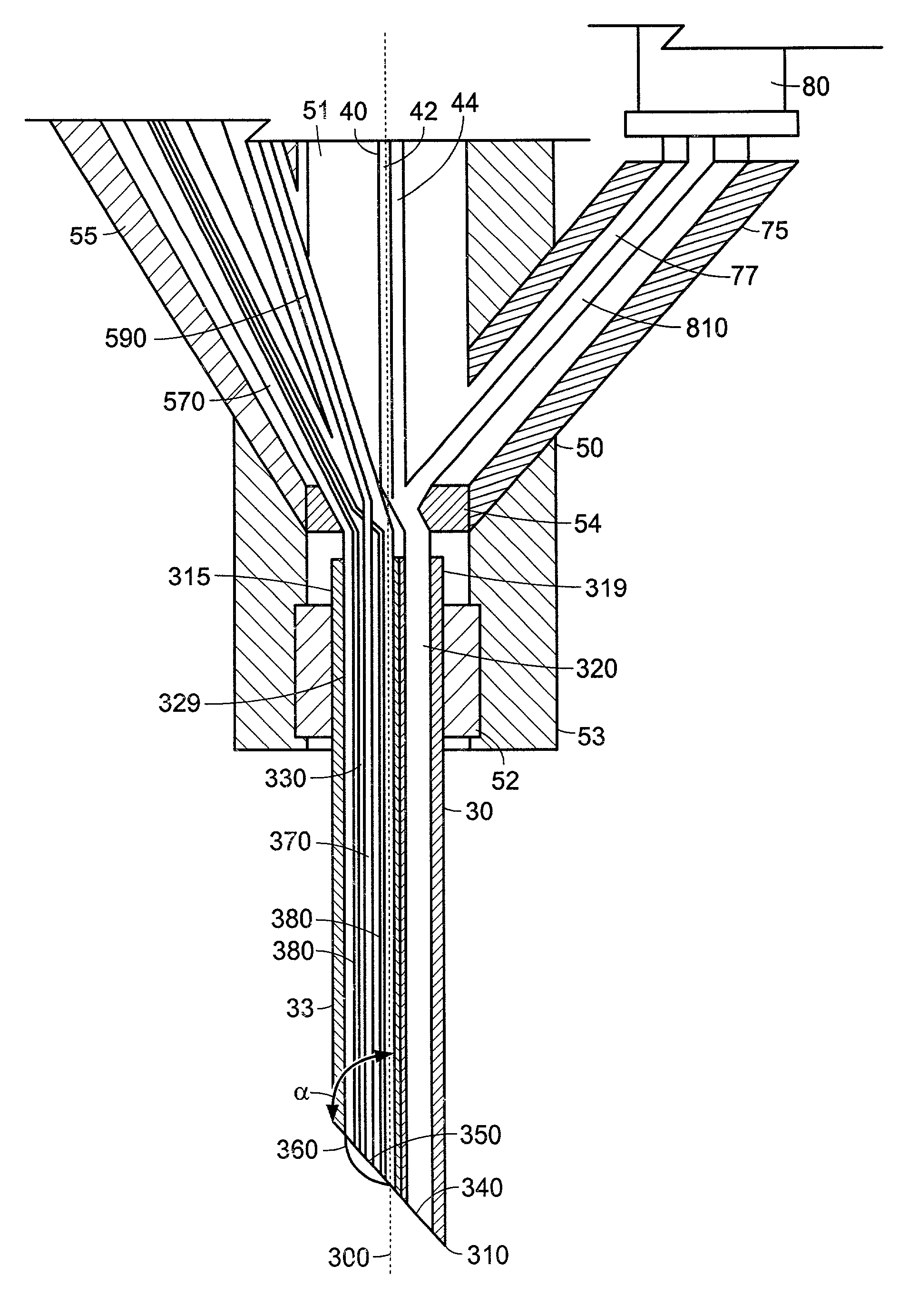
[0036]A feature common to each of the embodiments of the needle device according to the invention described below is a multi-lumen nozzle with a tissue-piercing point. At least one lumen in the nozzle is a working channel, for example, for aspiration, flushing or introduction of a surgical instrument. At least one other lumen has an optical system. The optical system is positioned in the nozzle to enable an operator to view the tissue piercing point during a surgical procedure.

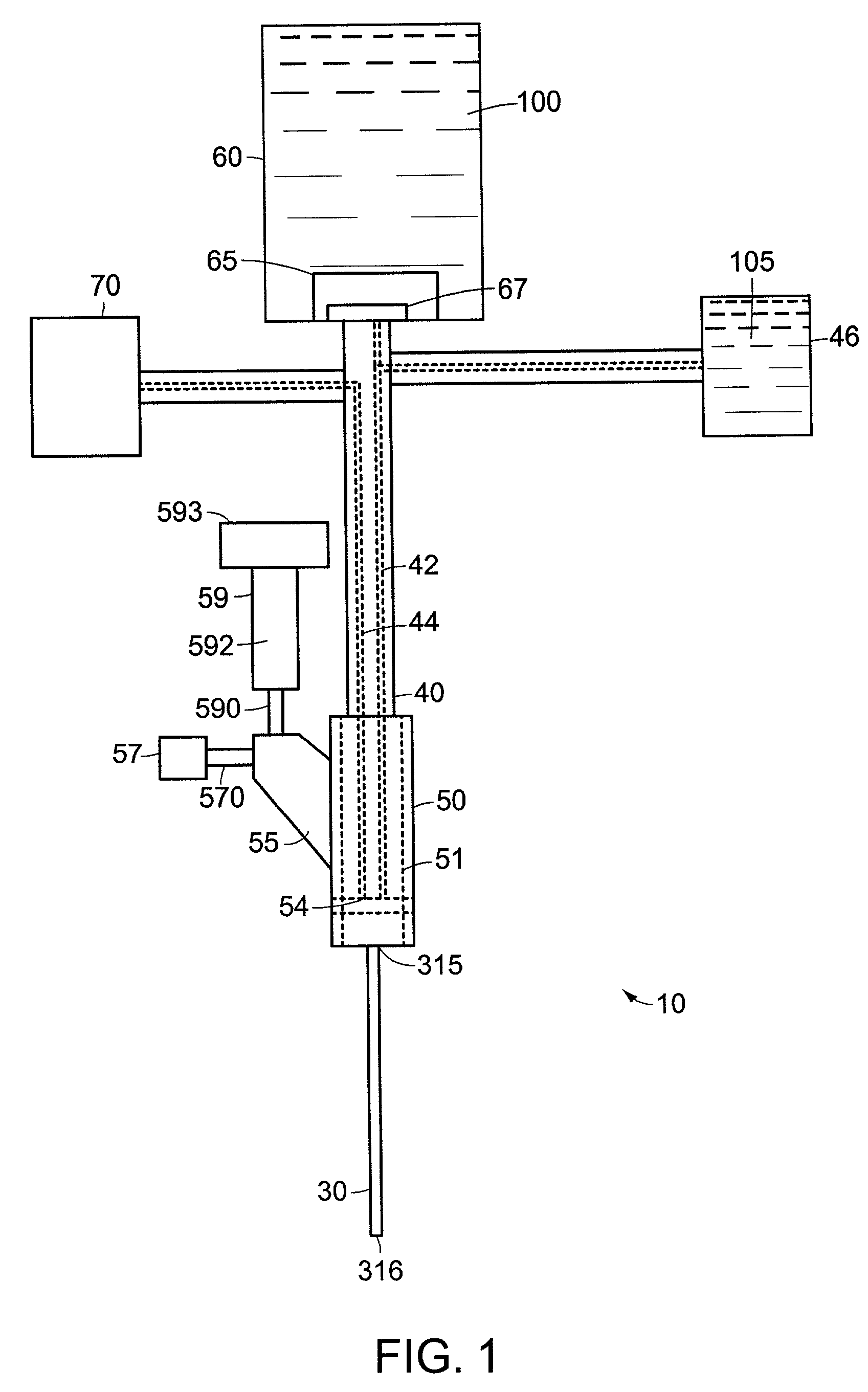
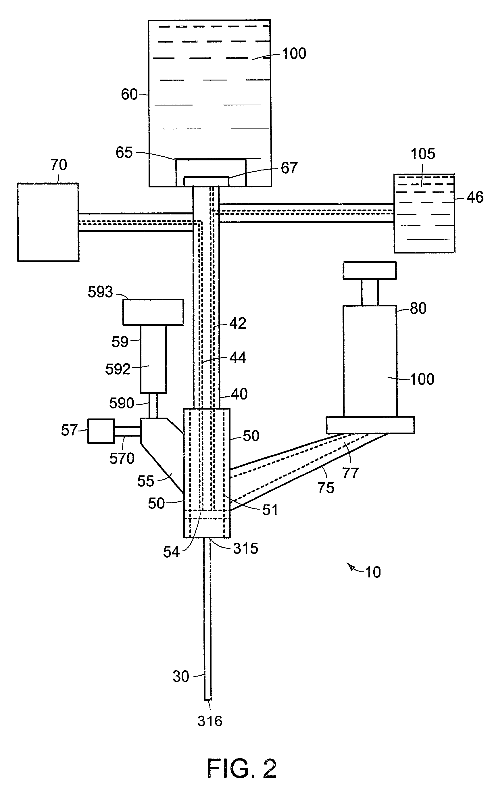
[0037]Referring to FIG. 1, a needle device 10 includes a multi-lumen nozzle 30 connected to a reservoir 60 by a supply tube 40. The reservoir 60 supplies fluid 100 under pressure through the supply tube 40 to the nozzle 30, which directs the pressurized fluid 100 onto a target body site in the patient. In one embodiment of the present invention, the needle device 10 also includes a suction apparatus 70 connected to the nozzle 30, for example, through the supply tube 40 to enable aspiration through the nozzle 3...


PUM


Abstract

A needle device for penetrating body tissues while substantially reducing the risk of damage to blood vessels and body organs by using visual control of the operative tip during insertion and the procedure, is disclosed herein. The needle device of the present invention has a large and diverse applicability to a number of medical procedures by enabling internal visual inspection of body tissues and cavities during treatment without open surgery or supplemental penetrating or visualization devices.

Description

TECHNICAL FIELD[0001]The present invention generally relates to devices for percutaneous diagnostic and therapeutic procedures, and, more particularly, to a multi-lumen needle device incorporating an optical element for continuously visualizing placement of the operative tip of the device during the procedure.BACKGROUND INFORMATION[0002]Percutaneous needle devices are used in a number of medical procedures, where access to body cavities or organs is desired. Such needle devices typically have a hollow shaft ending in a point, which serves to pierce the body tissue. After inserting the device to the target body cavity or organ, the hollow shaft may be used as a channel to take a fluid sample from the target site, aspirate, irrigate or deliver medicaments and other materials to the target site.[0003]Despite the use of preliminary exploratory measuring procedures such as ultrasound and X-ray, it is extremely difficult for a medical professional to determine the positional relationship ...

Claims


Application Information

Patent Timeline
Patent Type & Authority Patents(United States)
IPC IPC(8): A61M37/00A61B1/015A61B5/15A61B19/00A61M5/158A61M23/00
CPCA61B1/015A61B1/313A61B1/00167A61B5/6848A61B2019/5206A61B2019/5217A61M5/1582A61B2090/3614A61B2090/306
Inventor GELLMAN, BARRY N.
Owner BOSTON SCI SCIMED INC
Why Eureka
Try Eureka