Methods and devices for providing access into a body cavity

a technology for providing surgical access and body cavity, which is applied in the direction of surgery, cannulas, trocars, etc., can solve the problems of damage to the tissue, difficult positioning of the surgical access device in the incision, and the absence of existing surgical access devices

Inactive Publication Date: 2013-04-09
CILAG GMBH INT
View PDF144 Cites 10 Cited by
  • Summary
  • Abstract
  • Description
  • Claims
  • Application Information

AI Technical Summary

Benefits of technology

[0008]The present invention generally provides methods and devices for providing surgical access into a body cavity. In one embodiment, an adjustable access device is provided and can include a housing having a cannula extending distally therefrom defining a working channel extending therethrough for receiving surgical instruments. The housing can have one or more ports formed therein for receiving surgical instruments, and at least one seal can be disposed in the working channel and can be configured to form at least one of a seal around an instrument disposed through the working channel and a seal across the working channel when no instrument is disposed through the working channel. The surgical access device can also include an actuator rotatably disposed on the housing such that rotation of the actuator relative to the housing is effective to move the cannula between an insertion configuration in which the cannula has a first outer diameter and a first length, and a deployed configuration in which the cannula has a second outer diameter and a second length. In an exemplary embodiment, the first outer diameter can be less than the first length of the cannula and the second outer diameter can be greater than the first outer diameter and the second length being less than the first length. The second outer diameter can be less than the second length of the cannula.
[0010]In other exemplary embodiments, a proximal portion of the flexible band can be mated to the actuator. Rotation of the actuator to move the cannula to the insertion configuration can be effective to wind the proximal portion of the flexible band around the actuator to cause the cannula to increase in length and decrease in diameter to facilitate insertion of the cannula into and removal of the cannula from a tissue opening. Rotation of the actuator to the deployed configuration can be effective to unwind the proximal portion of the flexible band from around the actuator to cause the cannula to decrease in length and increase in diameter. In one embodiment, the cannula can be configured to engage a tissue opening in the deployed configuration to thereby stabilize the access device.
[0013]Exemplary methods are also provided and in one embodiment, a method of providing access through tissue to a body cavity is provided and can include positioning a cannula in an opening in tissue such that a working channel of the cannula provides access through the tissue and into a body cavity. The method can also include rotating an actuator to move the cannula from an insertion configuration to a deployed configuration in which the cannula decreases in length and increases in diameter, and rotating the actuator to move the cannula to the insertion configuration in which the cannula increases in length and decreases in diameter to increase a size of the opening in the tissue. The method can further include removing the cannula from the opening in tissue. In some embodiments, rotating the actuator to move the cannula to the deployed configuration can anchor the cannula against the tissue through which it extends.
[0014]Rotating the actuator to move the cannula to the insertion configuration can wind a proximal end of a flexible band around the actuator to tighten a helix formed by the flexible band. Furthermore, rotating the actuator to move the cannula to the deployed configuration can unwind a proximal end of a flexible band from around the actuator to loosen a helix formed by the flexible band.
[0018]In some embodiments, a first group of fibers from the weave of fibers can be coupled to the actuator, and a second group of fibers from the weave of fibers can not be coupled to the actuator. Rotation of the actuator in the first direction can be effective to shorten a length of the first group of fibers, and the second group of fibers can be configured to maintain an original length when the actuator is rotated in the first direction. The actuator can be rotatable in a second opposite direction to move the anchor portion of the cannula from the deployed configuration to an insertion configuration in which the cannula has a substantially constant outer diameter.
[0021]The cannula can be formed of any material, but in one embodiment, the cannula is formed of a plurality of fibers woven into a mesh material and rotating the actuator to move the cannula to the deployed configuration rotates a first group of the plurality of fibers to shorten the fibers relative to a second group of the plurality of fibers to invert the cannula. In addition, rotating the actuator to move a cannula longitudinally through the housing can telescope the cannula longitudinally through the housing.

Problems solved by technology

One problem with existing methods and devices is that existing surgical access devices do not retract tissue beyond the initial incision to any appreciable degree.
It can thus be difficult to position a surgical access device in the incision, particularly in minimally invasive surgical procedures where the incision is relatively small.
It can also be difficult as an initial matter to choose an appropriately sized access device to position within the incision during the stress and time constraints of surgery.
Removal of an access device from an incision in tissue can present further challenges when the access device is snugly positioned therein, requiring an amount of pullout force that can cause damage to the tissue and / or prolong length of the surgical procedure.
Such forceful removal of the access device can also increase the size of the incision, thereby reducing the healing and cosmetic benefits of a minimally invasive surgical procedure.

Method used


Image

  • Methods and devices for providing access into a body cavity
  • Methods and devices for providing access into a body cavity
  • Methods and devices for providing access into a body cavity

Examples


Embodiment Construction

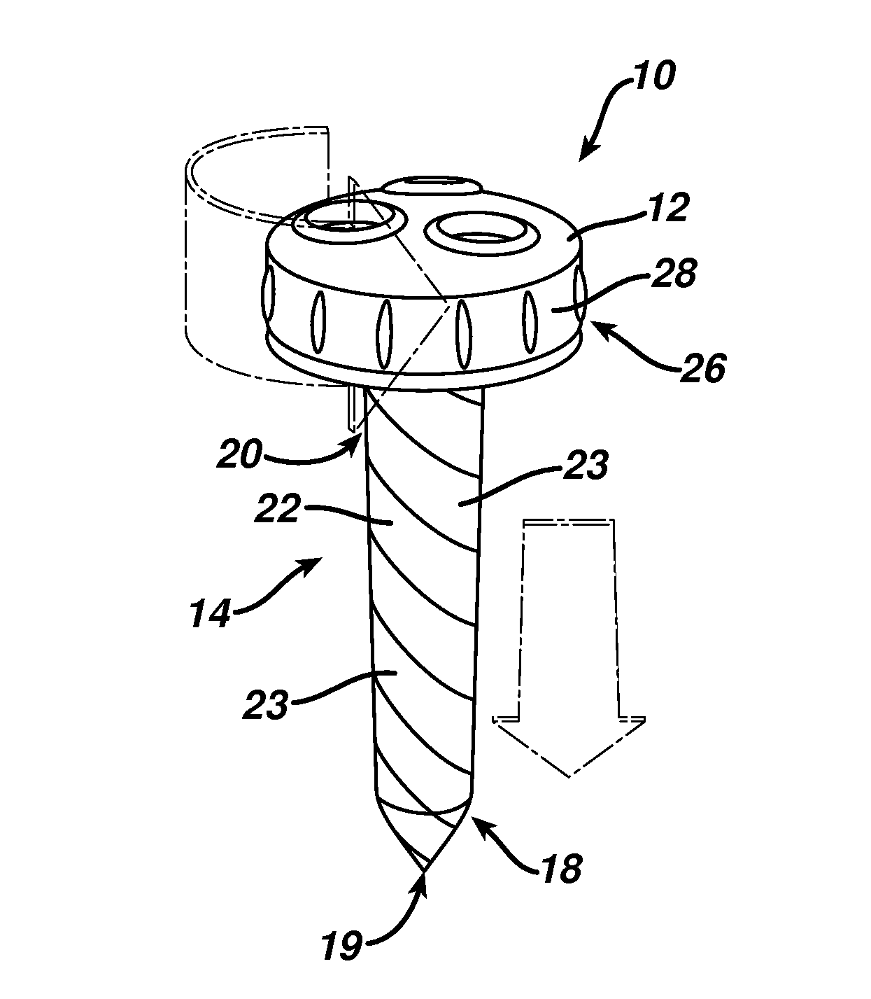
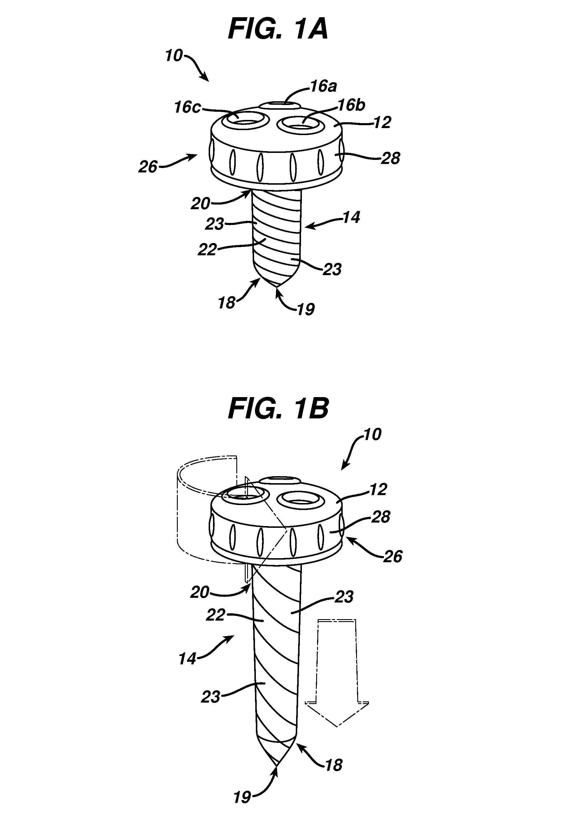
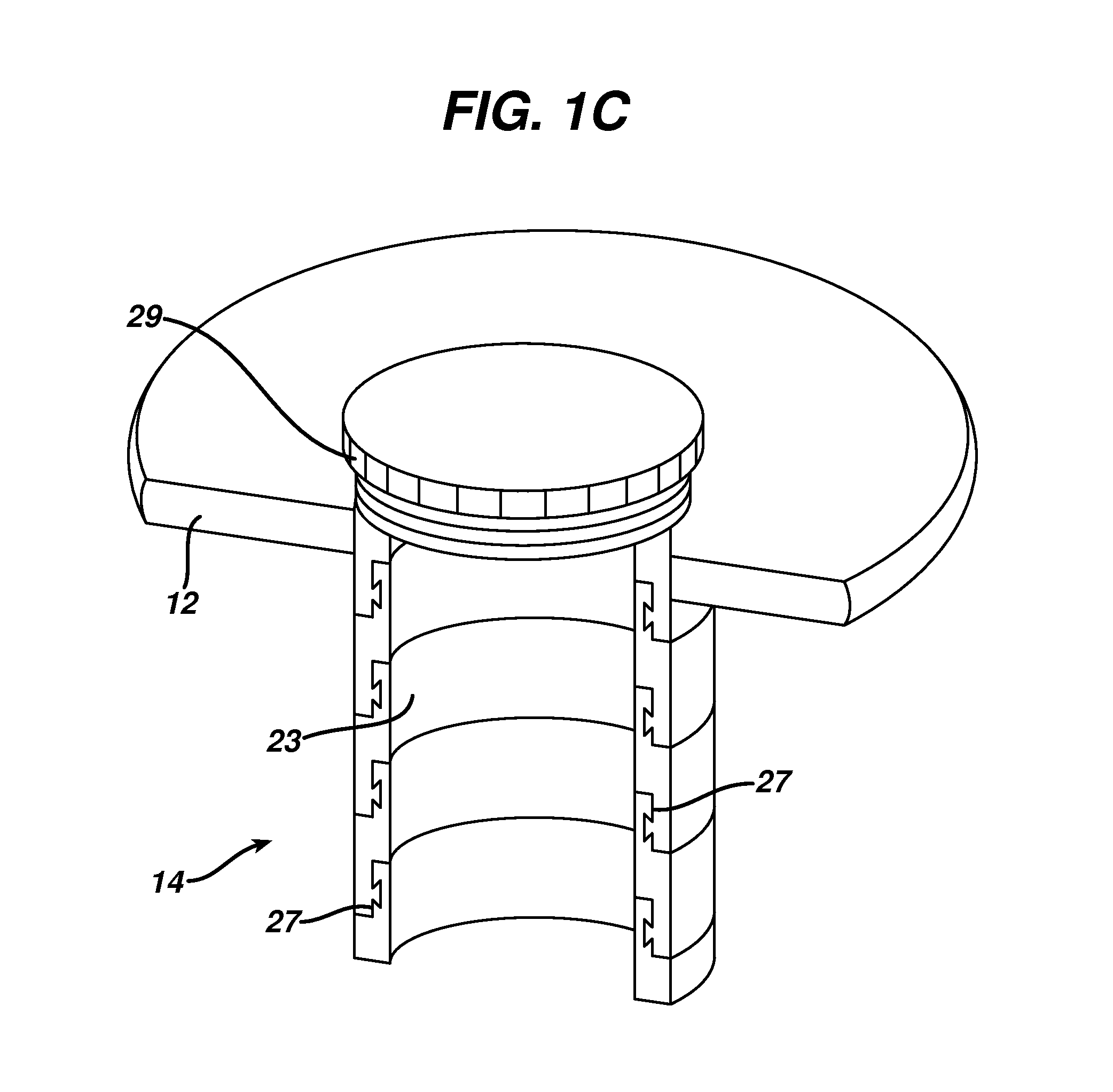
[0044]Certain exemplary embodiments will now be described to provide an overall understanding of the principles of the structure, function, manufacture, and use of the devices and methods disclosed herein. One or more examples of these embodiments are illustrated in the accompanying drawings. Those skilled in the art will understand that the devices and methods specifically described herein and illustrated in the accompanying drawings are non-limiting exemplary embodiments and that the scope of the present invention is defined solely by the claims. The features illustrated or described in connection with one exemplary embodiment may be combined with the features of other embodiments. Such modifications and variations are intended to be included within the scope of the present invention.

[0045]Various exemplary methods and devices are provided for surgically accessing a body cavity. In general, the methods and devices allow a surgical access device to be securely positioned within an ...


PUM


Abstract

Methods and devices are provided for providing surgical access into a body cavity. In general, the methods and devices allow a surgical access device to be securely positioned within an opening in tissue to provide access to a body cavity underlying the tissue. An actuator can be rotatably disposed on or in a housing of a surgical access device such that rotation of the actuator relative to the housing is effective to move a cannula of the surgical access device between an insertion configuration in which the cannula has a reduced profile enabling it to easily be inserted into a tissue opening, and an expanded profile enabling it to form an anchor against and / or within the tissue opening.

Description

CROSS-REFERENCE TO RELATED APPLICATIONS[0001]The present application is being filed with U.S. application Ser. No. 12 / 635,762 entitled “Methods and Devices For Providing Access Into a Body Cavity,” which is incorporated by reference herein in its entirety.FIELD OF THE INVENTION[0002]The present invention relates to methods and devices for providing surgical access into a body cavity.BACKGROUND OF THE INVENTION[0003]Access ports are widely used in medical procedures to gain access to anatomical cavities ranging in size from the abdomen to small blood vessels, such as veins and arteries, epidural, pleural and subarachnoid spaces, heart ventricles, and spinal and synovial cavities. The use of access ports has become more common as they provide minimally invasive techniques for establishing a portal for a number of procedures, such as those involving the abdominal cavity. Reduced postoperative recovery time, markedly decreased post-operative pain and wound infection, and improved cosmet...

Claims


Application Information

Patent Timeline
Patent Type & Authority Patents(United States)
IPC IPC(8): A61B1/32
CPCA61B1/32A61B17/3421A61B17/3423A61B17/3431A61B17/3439A61B2017/3443A61B2017/3466A61B2017/3484
Inventor FARRELL, THEODORE R.MORGAN, JEROME R.SHELTON, IV, FREDERICK E.
Owner CILAG GMBH INT
Why Eureka
Try Eureka