Plectin-1 targeted agents for detection and treatment of pancreatic ductal adenocarcinoma

a technology of pancreatic ductal adenocarcinoma and targeted agents, which is applied in the direction of instruments, peptide/protein ingredients, drug compositions, etc., can solve the problem that cross-sectional abdominal imaging has proven to be unreliable in detecting early-stage pdac in high-risk patients

Active Publication Date: 2014-09-09
THE GENERAL HOSPITAL CORP
View PDF18 Cites 0 Cited by
  • Summary
  • Abstract
  • Description
  • Claims
  • Application Information

AI Technical Summary

Benefits of technology

[0043]Also described herein are rapid and efficient methods for the identification of binding molecules that exhibit selective affinity for one or more peptide ligands of interest. The methods are advantageous in that they allow the simultaneous screening of multiple binding molecules against multiple peptide ligands of interest. Moreover, very little information is required regarding the identity or function of either the binding molecule or the ligand for use in the present inventions. For example, diverse populations of binding molecules can be simultaneously screened against diverse populations of peptide ligands to rapidly identify numerous molecules exhibiting a desired binding specificity. The methods described herein can therefore be advantageously applied for the discovery of specific reagents, such as peptide ligands and biomarkers, for diagnosis and treatment of human diseases.Definitions
[0075]The terms “specific binding” or “specifically binding” when used in reference to the interaction of a peptide (ligand) and a receptor (molecule) also refers to an interaction that is dependent upon the presence of a particular structure (i.e., an amino sequence of a ligand or a ligand binding domain within a protein); in other words the peptide comprises a structure allowing recognition and binding to a specific protein structure within a binding partner rather than to molecules in general. For example, if a ligand is specific for binding pocket “A,” in a reaction containing labeled peptide ligand “A” (such as an isolated phage displayed peptide or isolated synthetic peptide) and unlabeled “A” in the presence of a protein comprising a binding pocket A the unlabeled peptide ligand will reduce the amount of labeled peptide ligand bound to the binding partner, in other words a competitive binding assay.
[0090]As used herein, the term “endothelial cells”, as used herein, refers to any cell that may provide a lining for a bodily organ comprising a lumen (e.g., blood vessels, intestines, lymphatic vessels or ducts etc.). Usually, endothelial cells provide physical and chemical protection as well a selective absorption of nutrients or other metabolically active compounds.
[0113]As used herein, the term “reducing cancer in a patient” in reference to a treatment refers to any treatment for decreasing the number of cancer cells in a patient, slowing the growth of cancer cells in a patient, reducing the metastasis of cancer cells in a patient and includes any type of response for either relieving cancer symptoms or increasing the life-span of a patient.

Problems solved by technology

In addition, cross-sectional abdominal imaging has proven to be unreliable to detect early-stage PDAC in high-risk patients (Pelaez-Luna et al., Am J Gastroenterol 102:2157-2163 (2007)).

Method used


Image

  • Plectin-1 targeted agents for detection and treatment of pancreatic ductal adenocarcinoma
  • Plectin-1 targeted agents for detection and treatment of pancreatic ductal adenocarcinoma
  • Plectin-1 targeted agents for detection and treatment of pancreatic ductal adenocarcinoma

Examples


examples

[0238]The following examples serve to illustrate certain embodiments and aspects of the present invention and are not to be construed as liming the scope thereof.

[0239]In the experimental disclosures which follow, the following abbreviations apply: N (normal); M (molar); mM (millimolar); μM (micromolar); mol (moles); mmol (millimoles); μmol (micromoles); nmol (nanomoles); pmol (picomoles); g (grams); mg (milligrams); pg (micrograms); ng (nanograms); pg (picograms); L and 1 (liters); ml (milliliters); μl (microliters); cm (centimeters); mm (millimeters); μm (micrometers); nm (nanometers); U (units); min (minute); s and sec (second); k (kilometer); deg (degree); ° C. (degrees Centigrade / Celsius), colony-forming units (cfu), plaque forming units (PFU), optical density (OD; o.d.), internal diameter (i.d.), and polymerase chain reaction (PCR).

example i

[0240]This example describes exemplary materials and methods for assays used during the development of the present inventions.

Cell Culture

[0241]Primary mouse pancreatic ductal cells from wildtype mice were isolated and cultivated using published methods (Schreiber, et al., (2004) Gastroenterology 127:250-260). Early passage PDAC cell lines were isolated from tumors arising in Pdx1-Cre LSL-KrasG12D p53L / L mice (designated Kras / p53L / L) (Bardeesy, et al. (2006) Proc. Natl. Acad. Sci. U.S.A. 103:5947-5952). For the phage display experiments, PDAC cells were first grown in the primary duct cell media (F12 medium supplemented with 5 mg / mL D-glucose (Sigma), 0.1 mg / mL soybean trypsin inhibitor type I (Sigma), 5 mL / L insulin-transferrin-selenium (ITS+; BD Biosciences, Palo Alto, Calif.), 25 μg / mL bovine pituitary extract (BD Biosciences), 20 ng / mL epidermal growth factor (BD Biosciences), 5 nmol / L 3,3′,5-triiodo-L-thyronine (Sigma), 1 μmmol / L dexamethasone (Sigma), 100 ng / mL cholera toxin (...

example ii

In vitro Selection and Validation of PDAC-specific Peptides

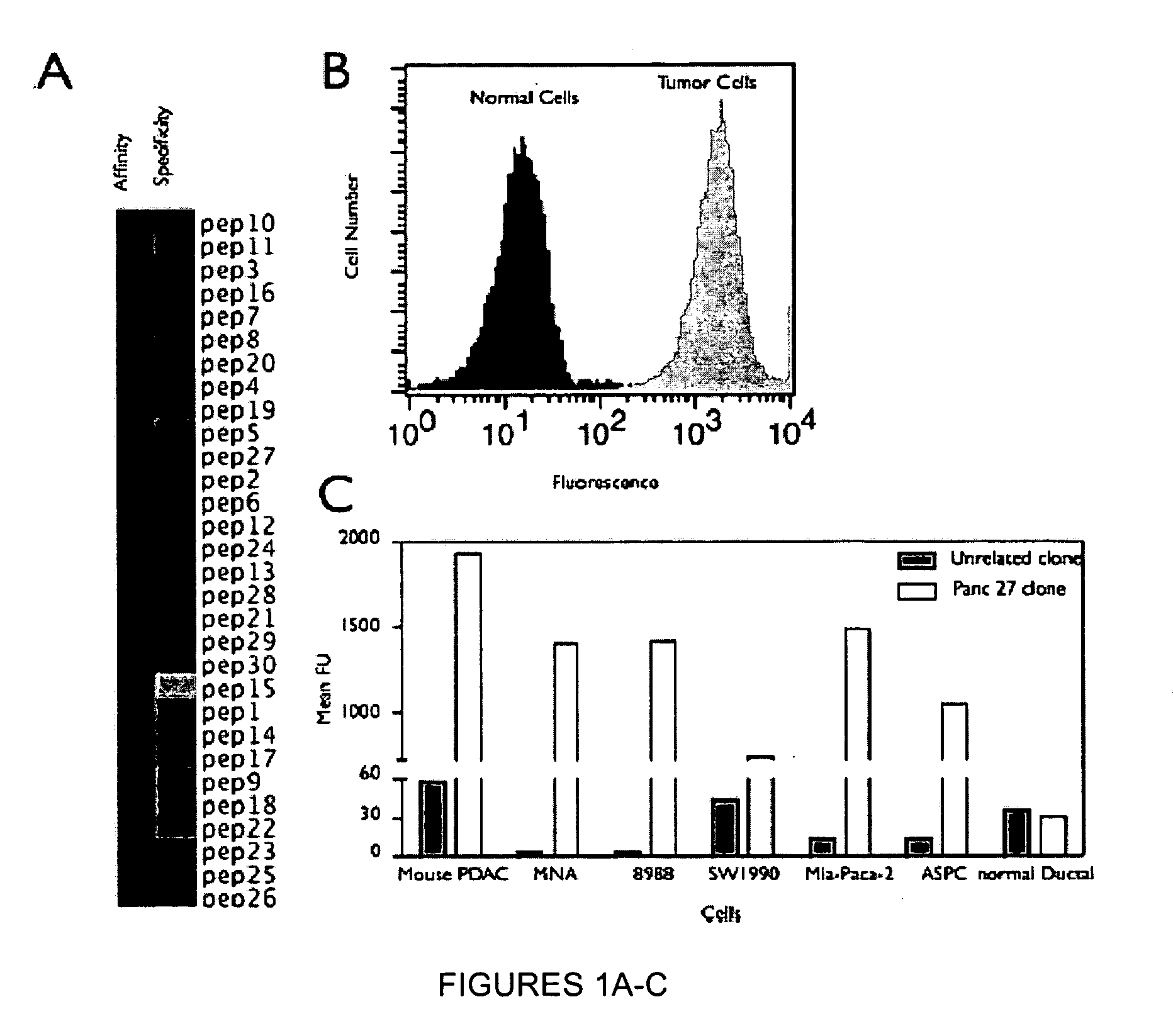
[0261]A genetically engineered mouse model of PDAC that recapitulates many of the histopathological, genomic and molecular features of the human disease was used (Carrière et al., Proc Natl Acad Sci USA. 104(11):4437-42 (2007); and Bardeesy et al. Proc Natl Acad Sci USA 103:5947-5952 (2006)). The Kras / p53 L / L model (Bardeesy et al., Proc Natl Acad Sci USA 103:5947-5952 (2006)) and wildtype controls served as a source of well-defined early passage PDAC cell lines and normal pancreatic ductal cells, respectively, for use in phage display selection and subtraction procedures to identify a pool of phage peptides specific for PDAC cells. Subsequent to selection procedures, the inventors isolated thirty individual phage plaques and performed an ELISA to identify the most selective phage for PDAC cells. The results of two experiments performed in triplicate are presented in the heat map shown in FIG. 1A and in the bar graph shown i...


PUM

PropertyMeasurementUnit
pHaaaaaaaaaa
pHaaaaaaaaaa
pHaaaaaaaaaa

Abstract

Described herein are compositions and methods for cancer cell biomarkers, such as pancreatic ductal adenocarcinoma (PDAC) cell biomarkers, and binding molecules for diagnosis and treatment of cancer, e.g., PDAC. Methods of identifying “accessible” proteomes are disclosed for identifying cancer biomarkers, such as plectin-1, a PDAC biomarker. Additionally, imaging compositions are provided comprising magnetofluorescent nanoparticles conjugated to peptide ligands for identifying PDACs.

Description

CLAIM OF PRIORITY[0001]This application is a 371 application of PCT / US2009 / 040480, filed on Apr. 14, 2009, and claims the benefit of U.S. Provisional Patent Application Ser. No. 61 / 044,818, filed on Apr. 14, 2008, the entire contents of which are hereby incorporated by reference.GOVERNMENT SUPPORT[0002]This invention was made with government support from the United States National Institutes of Health (NIH) under grant numbers P50-CA86355, PO1-CA117969-01, K01 CA104647-03, EB004626, ROI-HL078641, ROI-HL36436, HL080731, and P01-AI 054904. The United States Government may have certain rights in the invention.FIELD OF THE INVENTION[0003]The present invention relates to compositions and methods for providing cancer cell biomarkers, such as pancreatic ductal adenocarcinoma (PDAC) cell biomarkers, and binding molecules for diagnosis and treatment. Specifically, methods of identifying “accessible” proteomes are disclosed for providing biomarkers, such as plectin-1, for identifying PDACs. A...

Claims


Application Information

Patent Timeline
Patent Type & Authority Patents(United States)
IPC IPC(8): C07K7/06C07K7/08A61K38/08A61K38/10A61K38/00A61K47/48A61K49/00A61K49/18B82Y5/00G01N33/531G01N33/533G01N33/574
CPCG01N33/57438A61K49/0032B82Y5/00A61K47/48246G01N33/533C07K7/06G01N33/531A61K49/0002A61K49/1818A61K49/0056A61K38/00A61K49/14C07K16/18A61K47/64A61K38/08A61P1/18A61P35/00G01N33/54326G01N33/54346
Inventor KELLY, KIMBERLYWEISSLEDER, RALPHBARDEESY, NABEEL
Owner THE GENERAL HOSPITAL CORP
Why Eureka
Try Eureka