Biocompatible glaucoma drainage device

a glaucoma and drainage device technology, applied in the field of implants, can solve the problems of high intraocular pressure, relatively high complication and failure rate, and achieve the effect of minimal tissue disruption

Inactive Publication Date: 2014-09-16
SNYDER BIOMEDICAL
View PDF21 Cites 5 Cited by
  • Summary
  • Abstract
  • Description
  • Claims
  • Application Information

AI Technical Summary

Benefits of technology

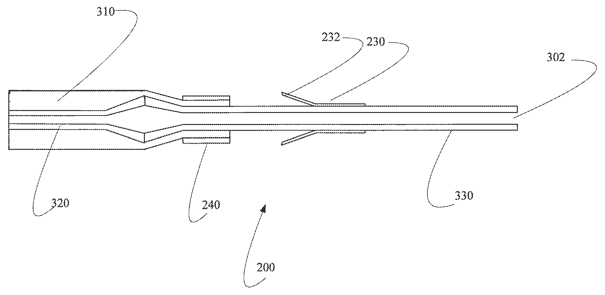
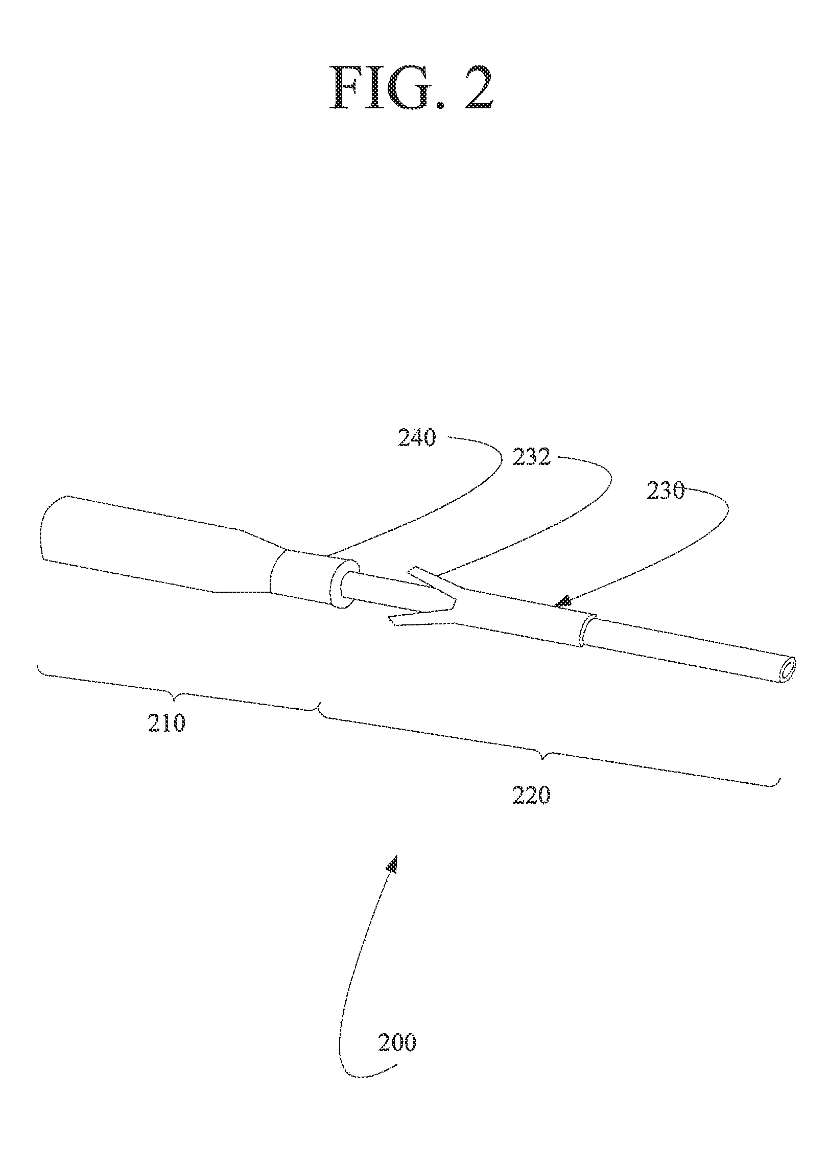
[0005]A glaucoma drainage device is presented, wherein that device is biocompatible and physically shaped such that it can be placed with minimal tissue disruption and is self-secured in an appropriate position. The surgical procedure benefits of this design include: (1) a small tangential conjunctival and scleral incisions are made rather than requiring a circumferential incision, and (2) there is minimal disruption of the blood or lymph channels which carry fluid away from a reservoir or distal end.

Problems solved by technology

When these methods fail to control intraocular pressure, ophthalmic surgeons may use aqueous drainage devices or “glaucoma shunts” to facilitate movement of aqueous humor from the anterior chamber to outside the eye and thus reduce high levels of intraocular pressure.
Currently, these devices are used as a secondary intervention because some believe they have a relatively high complication and failure rate.

Method used


Image

  • Biocompatible glaucoma drainage device
  • Biocompatible glaucoma drainage device
  • Biocompatible glaucoma drainage device

Examples


example

[0069]This example describes the implantation of a denucleated ePTFE shunt of the present invention and its performance, including the healing and functional characteristics of the novel drainage device of this invention.

[0070]A 10 year-old neutered male Boston terrier had developed angle closure glaucoma secondary to uveitis with collapse of the ciliary cleft following earlier phacoemulsification with placement of an intraocular lens (IOL). Despite maximal glaucoma medications including topical carbonic anhydrase inhibitors and prostaglandin F2 alpha analogs (latanoprost) the intraocular pressure could not be controlled and implantation of the denucleated ePTFE device was elected in an attempt to preserve the eye. Routine preoperative preparation of the eye was conducted as for intraocular surgery. The device was placed in a subconjunctival tunnel with the tube passed into the anterior chamber via a tunneled incision in the superotemporal limbal sclera. The conjunctiva was closed w...


PUM


Abstract

A biocompatible glaucoma drainage device, comprising a non-porous element, a porous element, an anchoring assembly disposed around the non-porous element, wherein the anchoring assembly comprises a plurality of barbs extending outwardly therefrom.

Description

CROSS-REFERENCE TO RELATED APPLICATIONS[0001]This Non-Provisional application claims priority to a U.S. Provisional Application filed Oct. 21, 2011 and having Ser. No. 61 / 627,954.FIELD OF THE INVENTION[0002]The present invention is directed to implants, and in particular to a glaucoma drainage device. In addition, the invention is further directed to a method to form Applicants' glaucoma drainage device. Furthermore, the invention is directed to a method to implant into a subject in need thereof Applicants' glaucoma drainage deviceBACKGROUND OF THE INVENTION[0003]Glaucoma is a disease where the nerve fibers that form the optic nerve degenerate over time and cause loss of sight corresponding to the areas of the retina from which the nerve fibers transmit the photopic stimulation. Elevated intraocular pressure (IOP) is the primary risk factor for degeneration of these nerve fibers and reduction of the interocular pressure is the primary goal of treatment to prevent further loss of opt...

Claims


Application Information

Patent Timeline
Patent Type & Authority Patents(United States)
IPC IPC(8): A61F9/00A61F2/14A61F9/007A61F9/008A61M27/00
CPCA61F9/00781A61F9/007A61F9/0017A61F2/14A61M27/002A61F2009/00891A61F2210/0057A61F2210/0076A61F2220/0008A61F2220/0016Y10S623/905A61F2250/0024
Inventor SNYDER, ROBERT W.MARTENS, JOHN A.MUNGER, ROBERT
Owner SNYDER BIOMEDICAL
Why Eureka
Try Eureka