

Methods for repairing and regenerating human dura mater
a technology of human dura mater and regenerative therapy, which is applied in the field of repairing and regenerating human dura mater, can solve the problems of increasing surgical trauma, affecting the safety of patients, and affecting the safety of patients, and achieves excellent risk-reduction and volume stability
- Summary
- Abstract
- Description
- Claims
- Application Information
AI Technical Summary
Benefits of technology
Problems solved by technology
Method used
Image

Examples
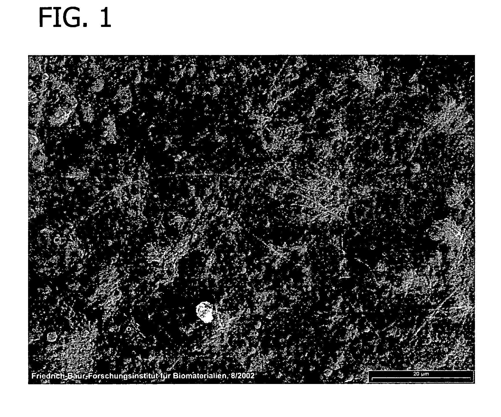
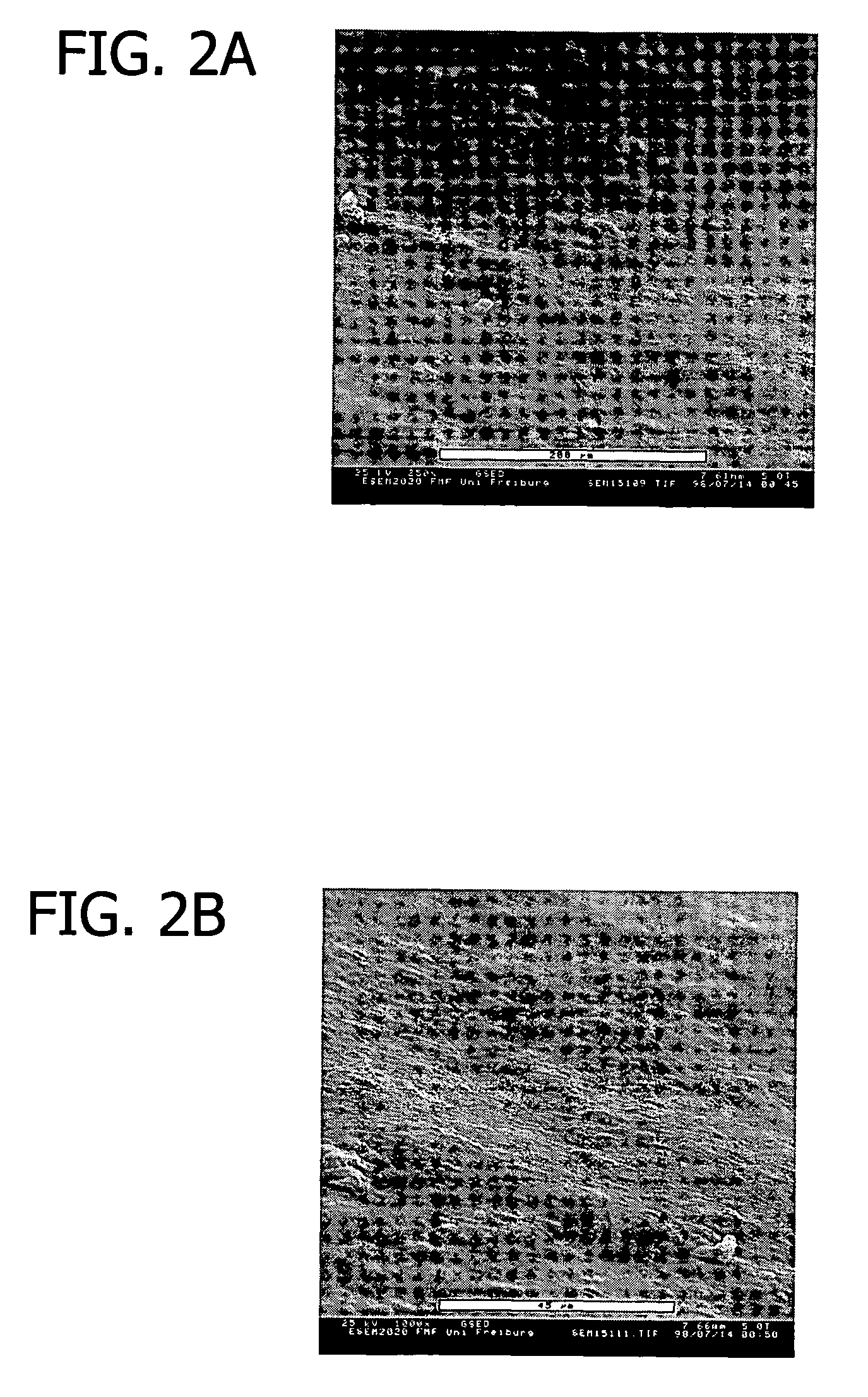
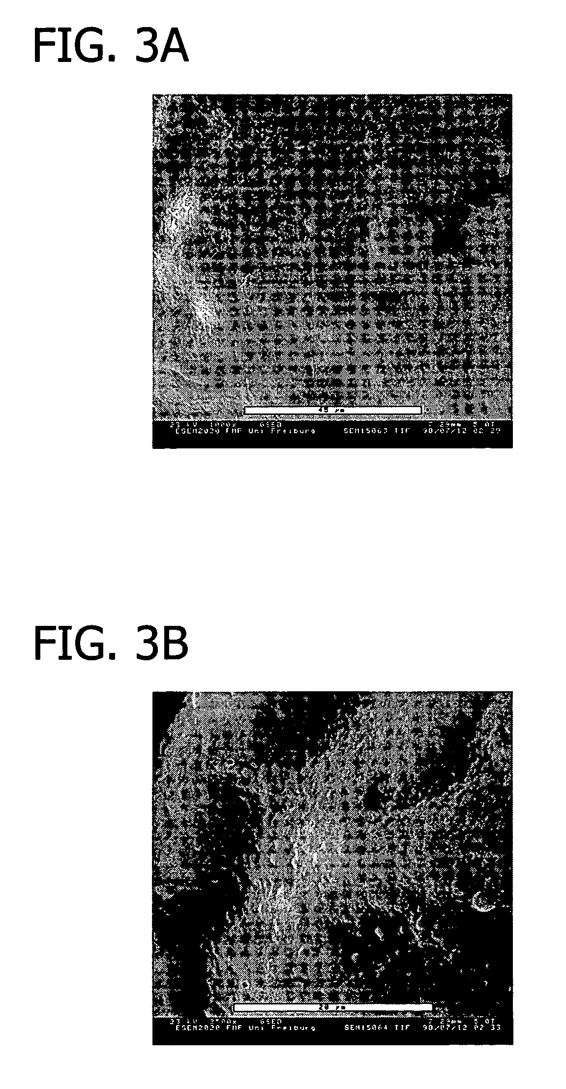
example 1
[0116]This example presents the results of experiments in sheep to evaluate an equine collagen foil for its suitability as a dura mater substitute used to repair dura mater tissue and as a biomatrix for dura regeneration.
[0117]An experiment was conducted to assess the properties of an equine collagen foil for use as cranial dura mater substitute as investigated in a sheep model. The equine collagen foil comprises native equine collagen fibrils (5.6 mg / cm2) purified from minced equine Achilles tendon and contains no cellular components.
[0118]The reference product used was preserved human cadaveric dura (Tutoplast® Dura). Both products were attached in position using fibrin glue only (Tissucol Duo S Immuno, Baxter AG, Vienna, Austria).
[0119]The following items were studied:[0120]Macroscopic aspects of incorporation of the two grafts;[0121]Reactions of adjacent tissue structures (inflammation, adhesion, fibrosis, necrosis); and[0122]Histological assessment of the incorporation process ...
example 2
Swelling Capacity of Equine Collagen Foil
[0193]The investigations concerning the swelling capacity of the equine collagen foil were carried out as follows:[0194]1) The equine collagen foil was first cut into 1 cm quadratic pieces.[0195]2) Samples of these cut pieces were examined under a conventional scanning microscope to determine the gross morphologic consistency and the thickness of the material as a reference for the swelling procedures.[0196]3) These pieces were then brought into plastic culture dishes.[0197]4) The capacity of fluid uptake and swelling capacity was then examined by successive titration with physiologic saline solution, administered with Eppendorf micropipettes using ascending amounts of fluid, starting with 10 μl / cm2 up to 150 μl / cm2 (see Table 3).
[0198]
TABLE 3Amount of Fluid Administered to the Equine Collagen FoilTime10 μl / cm220 μl / cm230 μl / cm240 μl / cm250 μl / cm275 μl / cm2100 μl / cm2125 μl / cm2150 μl / cm21 HrNo swellingFluidFluidFluid10 μl25 μl50 μl75 μl100 μleff...
example 3
Increase of Length of Hydrated Equine Collagen Foil
[0204]Seven dry pieces of equine collagen foil measuring 1.0 cm2 were hydrated for 1 hour in isotonic sodium chloride. The average length extension resulting from hydration of dry pieces of equine collagen foil was approximately 3.4 percent.
[0205]
TABLE 4Piece No.Length Dry (mm)Length Hydrated (mm)117.518217.718.331717.9417.818.5518.118.7618.619.5717.718.2Average17.818.4
PUM
| Property | Measurement | Unit |
|---|---|---|
| ultimate tensile force | aaaaa | aaaaa |
| ultimate tensile force | aaaaa | aaaaa |
| diameter | aaaaa | aaaaa |
Abstract
Description
Claims
Application Information