
Colorimetric substrates, colorimetric sensors, and methods of use

a colorimetric sensor and substrate technology, applied in the field of colorimetric substrates, colorimetric sensors, and methods of use, can solve the problems of unintended effect of breeding resistant strains of bacteria, many symptoms only evident, and severe, even life-threatening infections, and achieve rapid and low-cost detection, reduce the negative side effects of their us

Inactive Publication Date: 2015-01-06
WOUNDCHEK LAB US
View PDF19 Cites 0 Cited by
  • Summary
  • Abstract
  • Description
  • Claims
  • Application Information

AI Technical Summary

Benefits of technology

The present invention is a method for detecting proteins, enzymes, and microorganisms, including those that cause infections. This technology can be used in a healthcare setting to identify the presence of microorganisms in a wound and provide prophylactic measures before the infection is established. It also allows patients to monitor the condition of a wound after discharge from a hospital and take appropriate measures if a pathogenic microorganism is detected. The invention is a rapid, low-cost detection method that does not require expensive equipment and can be performed in a portable manner.

Problems solved by technology

Infection of wounds is a major source of healthcare expenditure in the United States.
Staphylococci are usually carried as harmless commensals, however given a breach in the epidermis, they can cause severe, even life threatening infection.
While generally effective, this strategy has the unintended effect of breeding resistant strains of bacteria.
Unfortunately, many symptoms are only evident after the infection is already established.
Furthermore, after a patient is discharged from the hospital he / she becomes responsible for monitoring their own healthcare, and the symptoms of infection may not be evident to the unskilled patient.

Method used


Image

  • Colorimetric substrates, colorimetric sensors, and methods of use
  • Colorimetric substrates, colorimetric sensors, and methods of use
  • Colorimetric substrates, colorimetric sensors, and methods of use

Examples


example 1

Some Metal Chelation Embodiments

[0181]In this example, histidine-tagged peptides were purified based on the ability of consecutive histidine residues to bind to a resin (e.g., SEPHAROSE®) containing nickel ions immobilized by covalently attached NTA.

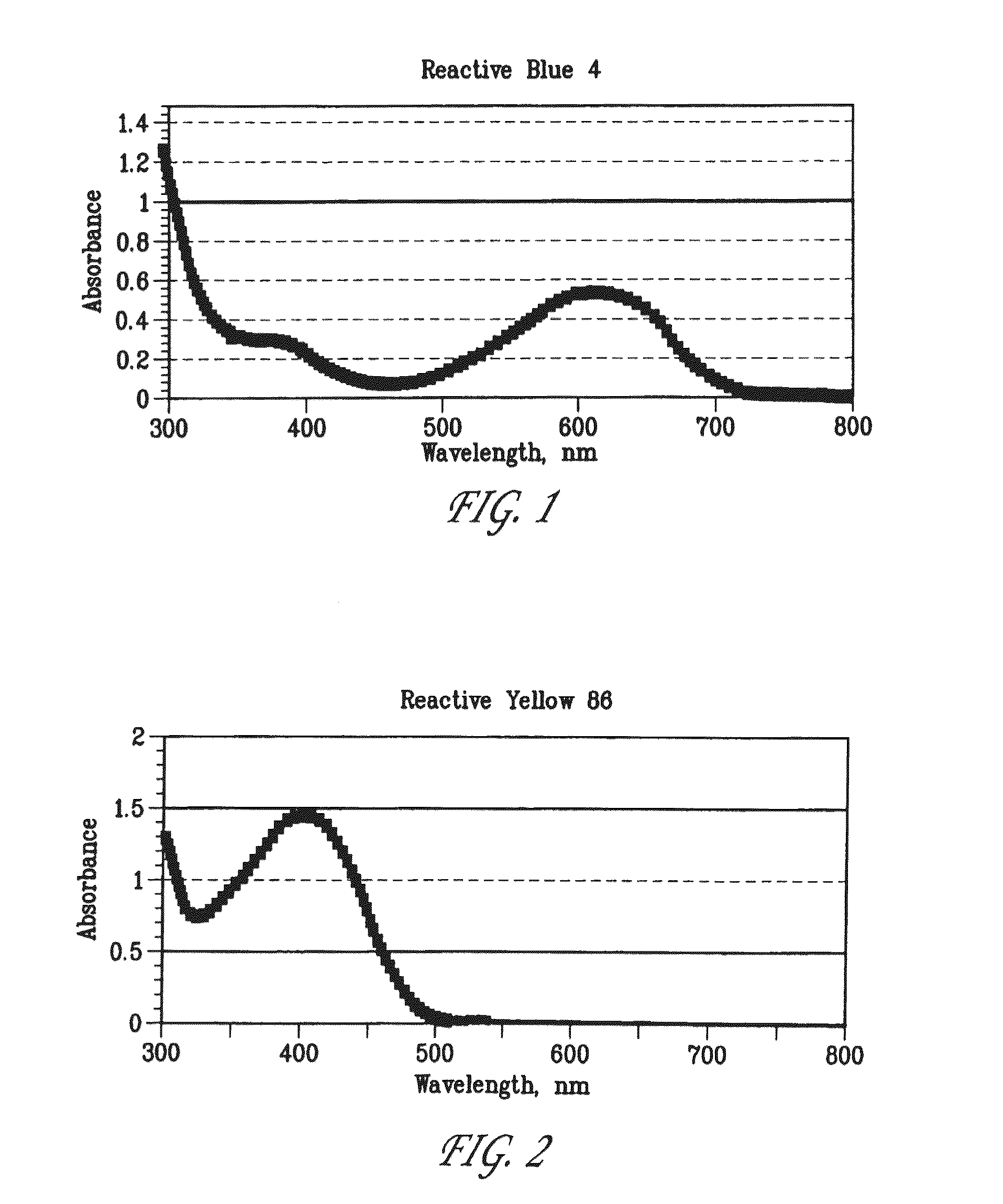
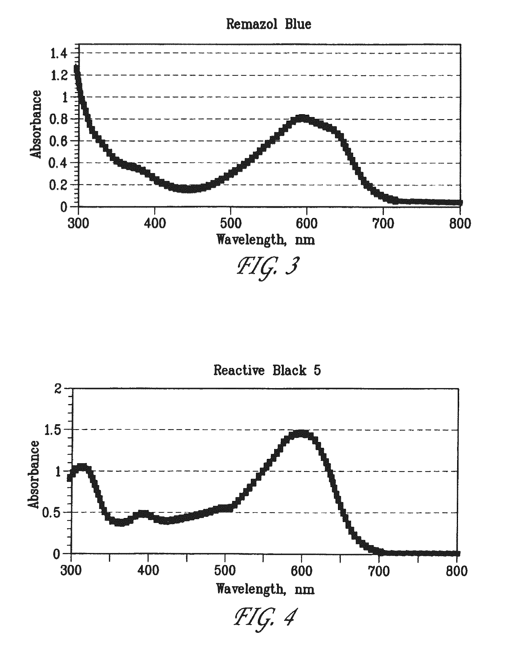
[0182]CPI3, Papa4, and LL3 peptides were labeled on the cysteine site with Reactive Black 5. The SSP3 peptide was labeled on the serine site with Reactive Blue 4. After purification and characterization, the four peptides were bound to NTA resin in phosphate buffered saline (PBS) pH ˜7.4 for ˜2 hours at about room temperature. The resin was then rinsed extensively with PBS. The NTA resin gave a high level of peptide binding (very dark blue) without non-specific binding, as evidenced by very little binding with non-histidine tagged peptides or free reactive dyes.

[0183]The CPI3 and Papa4 peptides on NTA resin were cleaved with Pseudomonas aeruginosa (PA14), the SSP3 peptide was cleaved with Serratia marcescens, and the LL3 peptide was clea...

example 2

Some Charged Membrane Embodiments

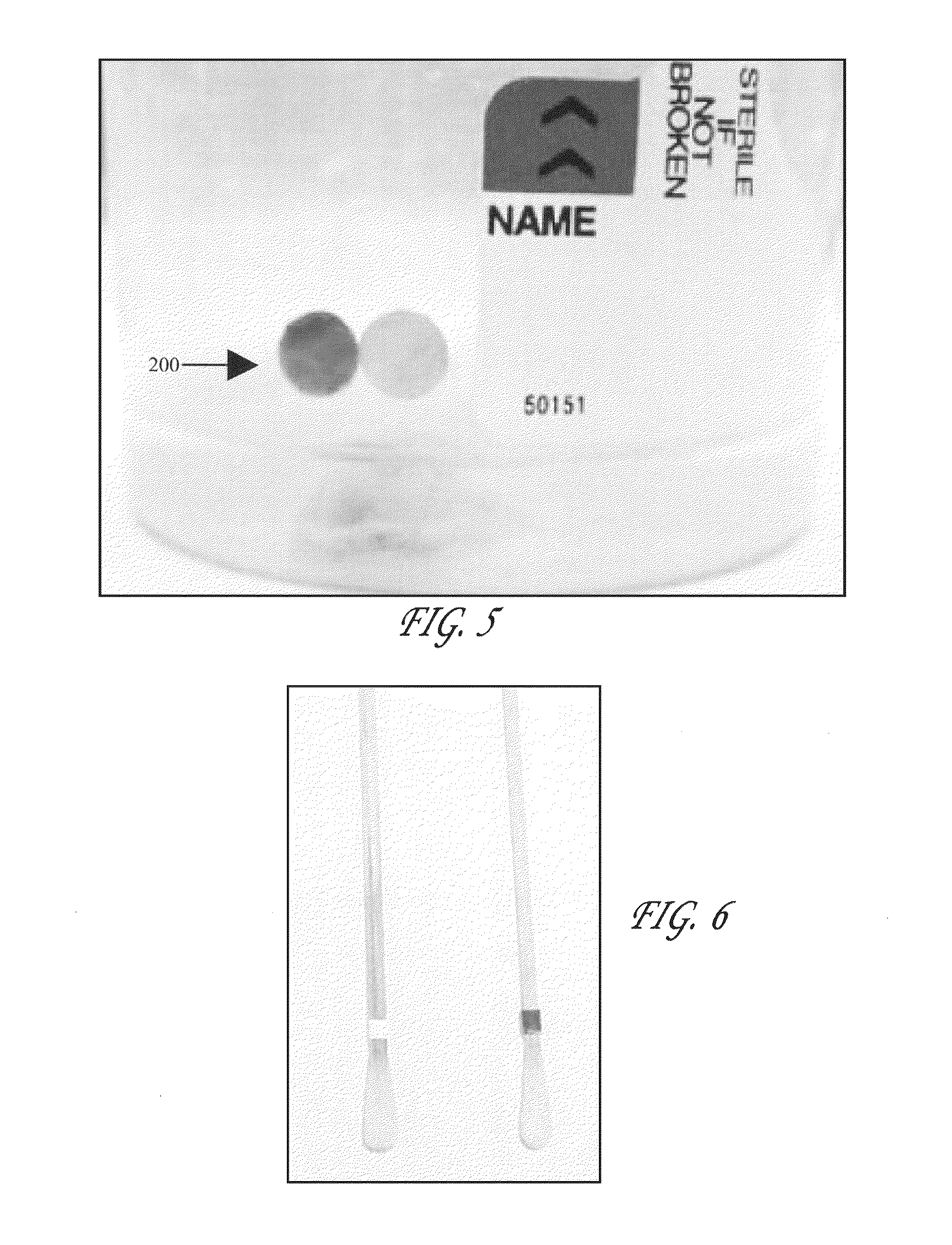
[0185]In this example, a negatively charged membrane (membrane ICE-450, available from Pall Corporation, East Hills, N.Y.) was used to bind peptides that contained a positively charged region placed at the end of the peptide (i.e., the 15 arginines of the peptides). The peptides were labeled with negatively charged dyes which when contained on the cleaved portion of the peptide had low affinity for the negatively charged membrane, but a high affinity for a positively charged collector membrane (membrane SB-6407, available from Pall Corporation, East Hills, N.Y.). This concept is shown in FIG. 12.

[0186]The CPI4, Papa5, and T5 peptides were labeled on the cysteine site with Reactive Black 5. After purification and characterization, the three peptides were bound to the negatively charged ICE-450 membrane in phosphate buffered saline (PBS) pH ˜7.4 overnight. The membranes were then rinsed with PBS. Next, positively charge membranes (membrane SB-6407, ava...

example 3

Labeling of Peptides with Reactive Dyes

[0189]Many of the peptide substrates were labeled with reactive dyes. Vinyl sulfone dyes were used to target cysteine groups on the peptides. Mono- and di-chlorotriazine dyes were used to target serine groups on the peptides. Sulfonyl chloride and N-hydroxysulfosuccinimidly (sulfo-NHS) ester dyes were used to target amine groups on the peptides.

[0190]An example of a labeling procedure for the labeling of the peptide CPI4 with Reactive Black 5 is shown in Table 1. CPI4 contains two cysteine groups that are targeted by Reactive Black 5 (a REMAZOL® dye) under basic conditions. The table below demonstrates that different levels of labeling (as evidenced by the dye-to-peptide ratio) can be obtained by varying the labeling conditions. The peptide was at ˜5 mg / ml in water, Reactive Black 5 was at ˜5 mg / ml in water, and the Na2CO3 was at ˜10 mg / ml in water. Labeling was ˜18 hours at ˜37° C.

[0191]

TABLE 1Dye-to-PeptidePeptide (uL)Dye (uL)Na2CO3 (uL)Ratio...


PUM

PropertyMeasurementUnit
pHaaaaaaaaaa
volumesaaaaaaaaaa
volumesaaaaaaaaaa

Abstract

Described herein is a substrate comprising at least one colorimetric component attached to a peptide, as well as methods for detecting a modification of said substrate. Also described are methods of detecting the presence or absence of a protein, for example, a protein produced by a microorganism, by contacting a substrate with a sample. If the sample contains the protein of interest, the substrate is modified and a visible color change results, indicating the presence or absence of the protein in the sample. The present invention also features biosensors and kits for detecting the presence or absence of microorganisms and / or proteins in a sample.

Description

RELATED APPLICATIONS[0001]This application is the U.S. National Stage of International Application No. PCT / US2004 / 036469, filed Nov. 3, 2004, published in English, and claims priority under 35 U.S.C. $119 or 365 to U.S. Provisional Application No. 60 / 516,688, filed Nov. 3, 2003 as well as U.S. Provisional Application 60 / 578,502, filed Jun. 9, 2004. The entire teachings of the above applications are incorporated herein by reference,BACKGROUND OF THE INVENTION[0002]Infection of wounds is a major source of healthcare expenditure in the United States. Approximately 5% of all surgical wounds become infected with microorganisms, and that figure is considerably higher (˜10-20%) for patients undergoing abdominal surgery. Bacterial species, such as Staphylococci, are the most frequently isolated organisms from infected wounds. This is probably because humans are the natural reservoir for Staphylococci in the environment, with up to 50% of the population colonized at any given time. Colonizat...

Claims


Application Information

Patent Timeline
Patent Type & Authority Patents(United States)
IPC IPC(8): C12Q1/00C12Q1/04C12Q1/37
CPCC12Q1/04C12Q1/37
Inventor SANDERS, MITCHELL C.COLPAS, GERARD J.ELLIS-BUSBY, DIANE L.HAVARD, JENNIFER M.
Owner WOUNDCHEK LAB US
Learn More