System and method for lesion-specific coronary artery calcium quantification

a technology of lesion-specific coronary artery calcium and quantification method, which is applied in the direction of angiography, image enhancement, instruments, etc., to achieve the effect of improving prediction and assessment of cardiac risk and obtaining more data

Active Publication Date: 2015-03-03
VOROS SZILARD +1
View PDF16 Cites 27 Cited by
  • Summary
  • Abstract
  • Description
  • Claims
  • Application Information

AI Technical Summary

Benefits of technology

[0006]Embodiments of the system and method described herein utilize the significantly greater data provided by a non-contrast-enhanced CAC scan; data that is left unused by the “whole-heart” Agatston or volume scores. While the Agatston score and volume score summarize overall coronary calcium burden, they are blind to the number of vessels involved, to the geographic distribution of the lesions, to the size and shape of the individual lesions and to the distance of the lesions from the coronary ostium. However, such information is inherently present in these clinically acquired scans. Embodiments of the systems and methods described herein extract and use the enhanced information provided by 3-D CAC scan data to improve prediction and assessment of cardiac risk.
[0007]Systems and methods described herein demonstrate that utilizing lesion-specific CAC (LS-CAC) and distance-weighted lesion-specific CAC (DWLS-CAC) improves prediction and assessment of obstructive coronary artery disease and prediction of cardiovascular events. Accordingly, these systems and methods significantly increase the clinical predictive value of traditional coronary artery calcium scanning.
[0008]An advantage of the systems and methods described herein is the ability to use standard imaging protocol. Embodiments of the lesion-specific calcium scoring systems and methods work on datasets acquired with typical clinical imaging protocols on conventional CT scanners. There is no need to modify the CT hardware or update the imaging protocol. Accordingly, there is no increased radiation exposure for the patients.
[0009]The described systems and methods also achieve fast semi-automatic calcific lesion-labeling and fully automated geometric and morphologic lesion measurements. Embodiments use a graphical user interface with automatic 3D propagation algorithm, to enable users to analyze a CAC volume rapidly (e.g., within 5 minutes), which is similar to, or even faster than using conventional CAC software while providing enhanced diagnostic capability.

Problems solved by technology

However, such information is inherently present in these clinically acquired scans.

Method used


Image

  • System and method for lesion-specific coronary artery calcium quantification
  • System and method for lesion-specific coronary artery calcium quantification
  • System and method for lesion-specific coronary artery calcium quantification

Examples


Embodiment Construction

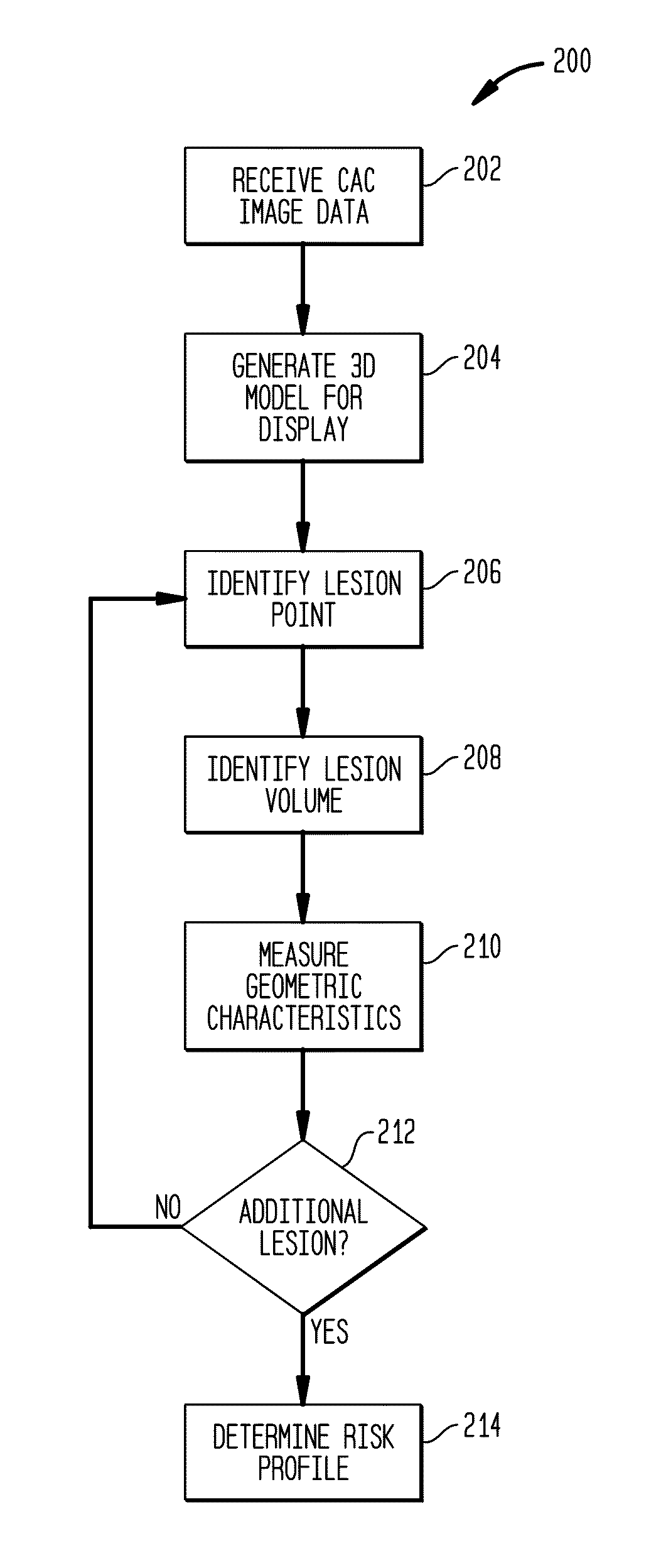
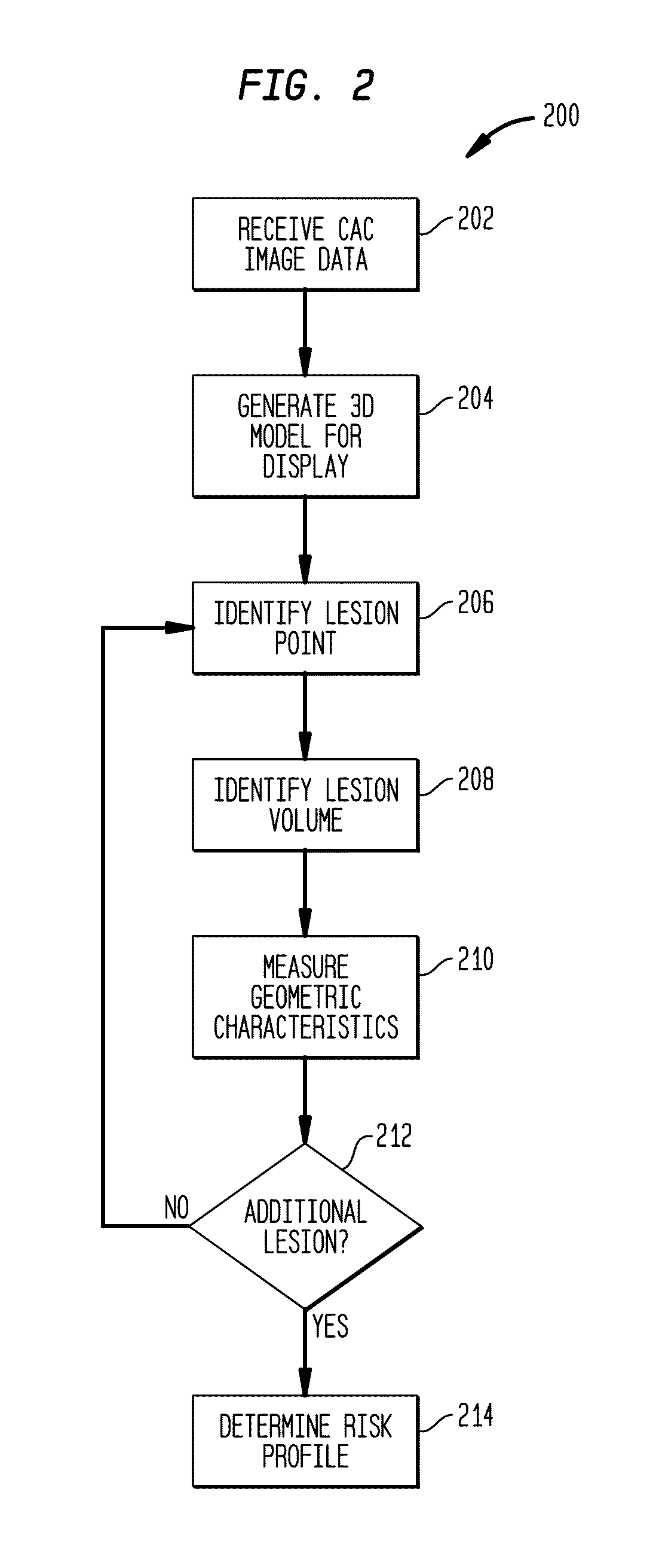
[0025]Conventional CAC image volume analysis reduces the three-dimensional (3D) volume, to a stack of two-dimensional (2D) slices that form the 3D volume to facilitate processing. The stack 2D slices of 3D data are evaluated and used to identify and quantify the presence of artery calcium, indicative of atherosclerosis. Typically, the presence of calcium within the CAC image volume is evaluated as a whole to determine the calcium burden on the heart depicted in the CAC image volume. These conventional techniques ignore the position of the calcium lesions as well as the geometry and size of individual calcium lesions. To minimize processing, risks of atherosclerosis are evaluated based upon these 2D images and averaged data.

[0026]In contrast, systems and methods described herein use the 3D volume of image data without reducing it to 2D data, avoiding potential errors introduced by representation of 3D data using sets of 2D images. For example, when 3D data is treated as a stack of 2D...


PUM


Abstract

Methods and systems utilizing the data provided by a non-contrast-enhanced CAC scan that is left unused by the “whole-heart” Agatston or volume scores. Agatston and volume scores summarize overall coronary calcium burden, but do not show the number of vessels involved, the geographic distribution of the lesions, the size and shape of the individual lesions and the distance of the lesions from the coronary ostium. The methods and systems described herein extract and use the enhanced information provided by 3-D CAC scan data and significantly increases its clinical predictive value by providing vessel and lesion specific CAC scores which are superior to the whole-heart Agatston and volume scores in predicting obstructive Coronary artery disease (CAD).

Description

CROSS-REFERENCE TO RELATED APPLICATIONS[0001]This application claims the benefit of U.S. provisional Application Ser. No. 61 / 139,165, entitled, “System and Method for Lesion-Specific Coronary Artery Calcium Quantification”, filed on Dec. 19, 2008.TECHNICAL FIELD[0002]The present system and method is a lesion-specific quantification tool for coronary artery calcium scans that can enhance or supplant other predictive indicators.BACKGROUND OF THE INVENTION[0003]Atherosclerosis is the leading cause of morbidity and mortality worldwide. It is a complex disease initiated and propagated by lipoprotein deposition and inflammation. Later stages of atherosclerosis are characterized by progressive deposition of calcium in the coronary arterial vessel-wall. Histopathology, computed tomography and intravascular ultrasound studies have confirmed that the extent of coronary calcification is closely correlated to the atherosclerosis plaque burden.[0004]A cardiac tomography (CT) based coronary arter...

Claims


Application Information

Patent Timeline
Patent Type & Authority Patents(United States)
IPC IPC(8): G06T15/00A61B5/02A61B6/00A61B6/03G06F19/00G06T7/00G06T15/08
CPCA61B6/504A61B5/02007G06T7/0012G06T15/08A61B6/032A61B6/5288G06F19/321G06F19/3431G06T2200/24G06T2207/10081G06T2207/30048G16H50/30G16H30/40G16H30/20
Inventor VOROS, SZILARDQIAN, ZHEN
Owner VOROS SZILARD
Why Eureka
Try Eureka