Fluorescence axial localization with nanometer accuracy and precision

a fluorescence axial and precision technology, applied in the field of microscopy, can solve the problems of limiting the resolution of conventional fluorescence microscopes, limiting the applicability of single molecule biological imaging, and challenging the resolution and single molecule localization accuracy along the optical axis

Inactive Publication Date: 2015-08-11
IOWA STATE UNIV RES FOUND
View PDF5 Cites 11 Cited by
  • Summary
  • Abstract
  • Description
  • Claims
  • Application Information

AI Technical Summary

Benefits of technology

[0015]Another aspect of the invention is a method to align the AFM tip with the excitation laser along the optical axis of the microscope, and then align the sample along the optical axis. The method first determines, with nanometer precision, the location of the center of the AFM tip. The AFM tip is imaged by precisely moving it through the fixed excitation laser beam and measuring the light reflected off the AFM tip at each location. The center of the tip is determined by finding the centroid of the optical intensity of the reflected light in the image. This allows moving the tip into precise alignment with the excitation beam. The method can also determine the position(s) of target fluorescent molecules(s) or other structures in a sample by similarly scanning the sample ...

Problems solved by technology

However, the resolution of conventional fluorescence microscopes is limited by the diffraction of light: with a high numerical aperture objective and visible excitation, resolution is about 200 nm in the lateral direction and 500 nm along the optical axis.
Unlike imaging in the x- and y-direction, improving resolution and single molecule localization accuracy along...

Method used


Image

  • Fluorescence axial localization with nanometer accuracy and precision
  • Fluorescence axial localization with nanometer accuracy and precision
  • Fluorescence axial localization with nanometer accuracy and precision

Examples


Embodiment Construction

[0053]Overview

[0054]For a better understanding of the invention, several exemplary embodiments which illustrate aspects of the invention will now be described in detail. It is to be understood that these are but several examples of forms the invention can take and are neither inclusive nor exclusive. Variations obvious to those skilled in the art will be included within these examples. But the invention and its aspects can take many other and different forms of embodiments.

[0055]Example of Apparatus

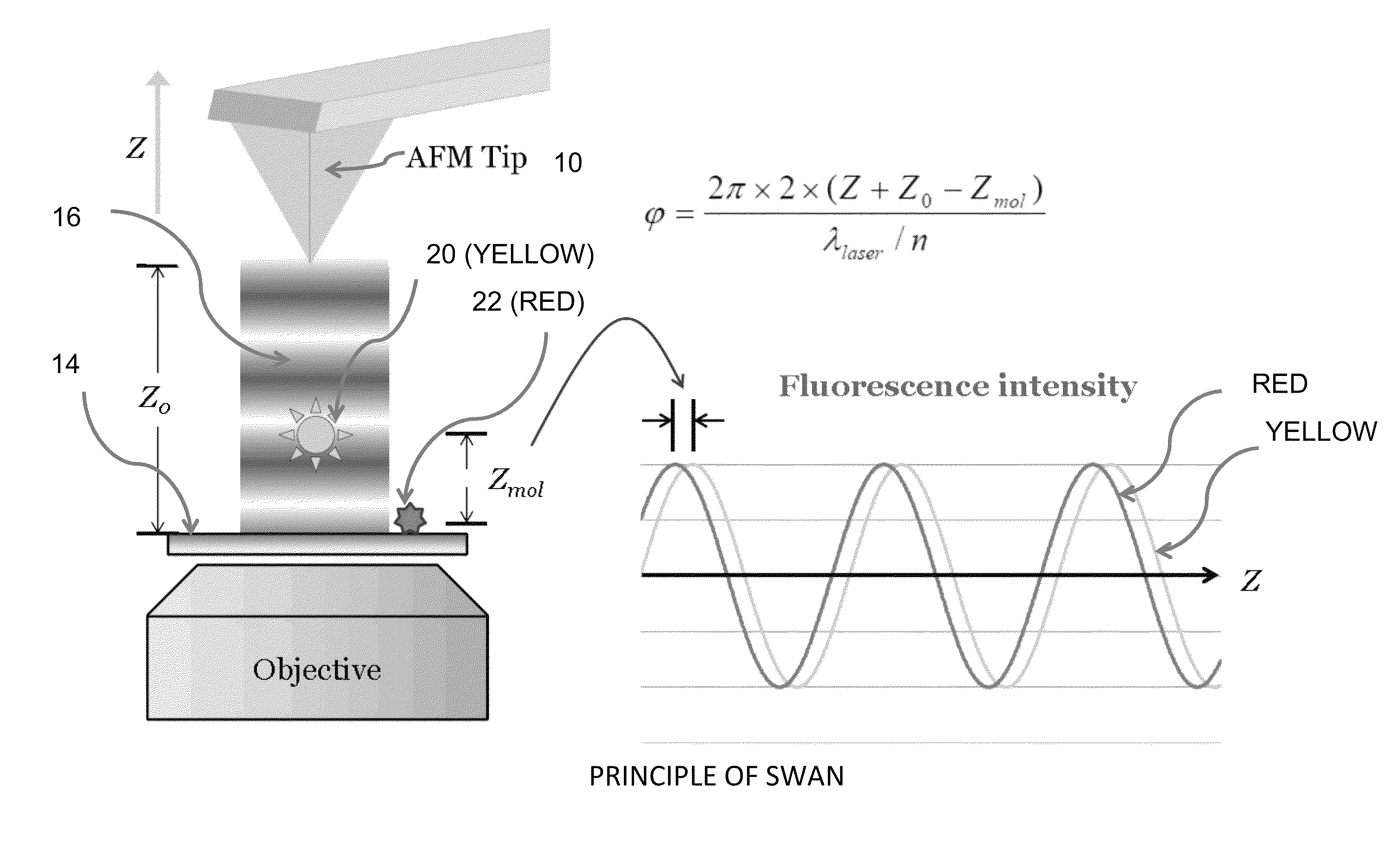
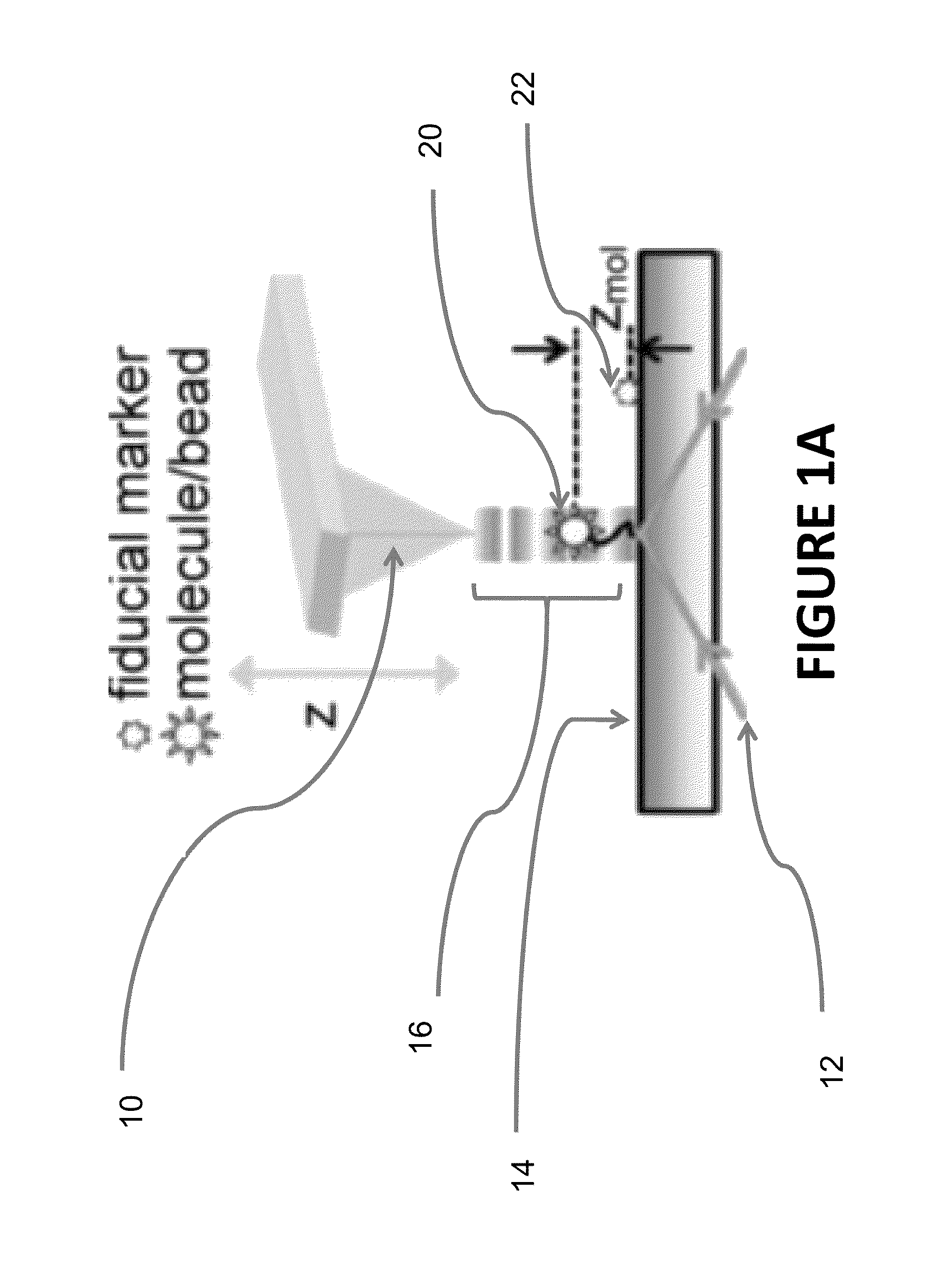
[0056]An example of an apparatus for SWAN is shown at FIG. 4.

[0057]At a general level, the apparatus is a combination of a confocal fluorescence microscope and an AFM microscope. This includes components for AFM functions as well as for fluorescent measurements. Such systems are commercially available or can be assembled for operation by those skilled in the art.

[0058]Such systems would include a user interface, processor(s), and actuators to perform AFM / fluorescence microscopy functions,...


PUM


Abstract

We describe a new technique, standing wave axial nanometry (SWAN), to image the axial location of a single nanoscale fluorescent object with sub-nanometer accuracy and 3.7 nm precision. A standing wave, generated by positing an atomic force microscope tip over a focused laser beam, is used to excite fluorescence; axial position is determined from the phase of the mission intensity.

Description

CROSS-REFERENCE TO RELATED APPLICATIONS[0001]This application claims priority under 35 U.S.C. §119 to provisional application Ser. No. 61 / 796,641 filed Nov. 16, 2012, herein incorporated by reference in its entirety.BACKGROUND OF THE INVENTION[0002]1. Field of the Invention[0003]The present invention relates to microscopy and, in particular, to apparatus, methods, and systems for resolving axial position of nanoscale objects, including but not limited to single molecules or other nanoscale structures.[0004]We disclose a new technique, sometimes called standing wave axial nanometry (SWAN), to determine the axial location of single nanoscale fluorescent objects with sub-nm accuracy and 3.7 nanometer (nm) precision. A standing-wave excitation pattern is generated by positioning an Atomic Force Microscope (AFM) tip or other reflecting surface (e.g. mirror) over a focused laser beam. A fluorescent nanoparticle or a single fluorescent molecule is positioned within the standing-wave and it...

Claims


Application Information

Patent Timeline
IPC IPC(8): G01J1/58G01N21/64G01B11/14
CPCG01N21/6404G01B11/14G01Q60/24G01Q30/025G01N21/6458G02B21/0076
Inventor SIVASANKAR, SANJEEVILI, HUIYEN, CHI-FU
Owner IOWA STATE UNIV RES FOUND
Why Eureka
Try Eureka