
Circular coils for deep transcranial magnetic stimulation
a transcranial magnetic stimulation and coil technology, applied in magnetotherapy, magnetotherapy using coils/electromagnets, magnetotherapy, etc., can solve the problems of low effect of affecting deeper neuronal structures, increase the risk of seizures, physiological damage to tissue, etc., and achieve higher absolute electric field intensity, better depth penetration profile, and higher relative electric field intensity
Patent Information
- Authority / Receiving Office
- US · United States
- Patent Type
- Patents(United States)
- Current Assignee / Owner
- Publication Date
- 2016-02-02
Smart Images
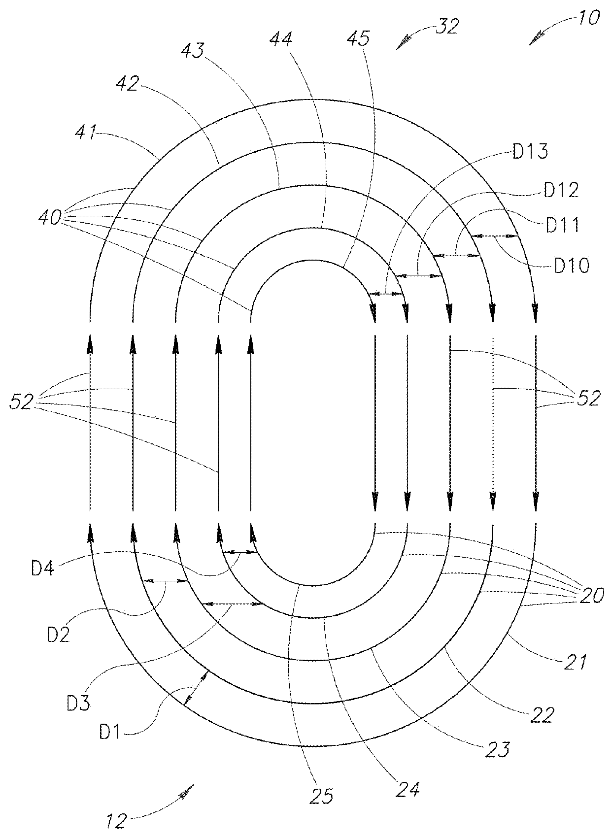
Figure 1 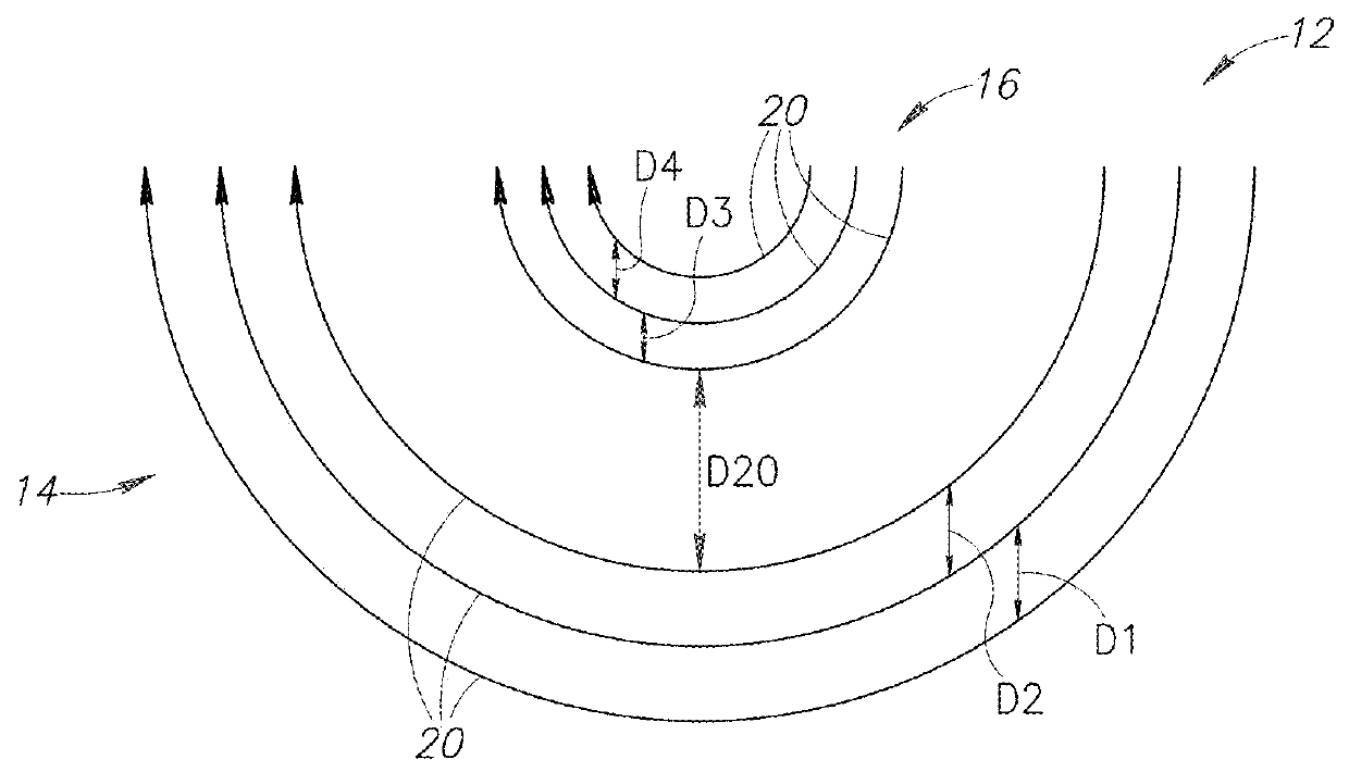
Figure 2 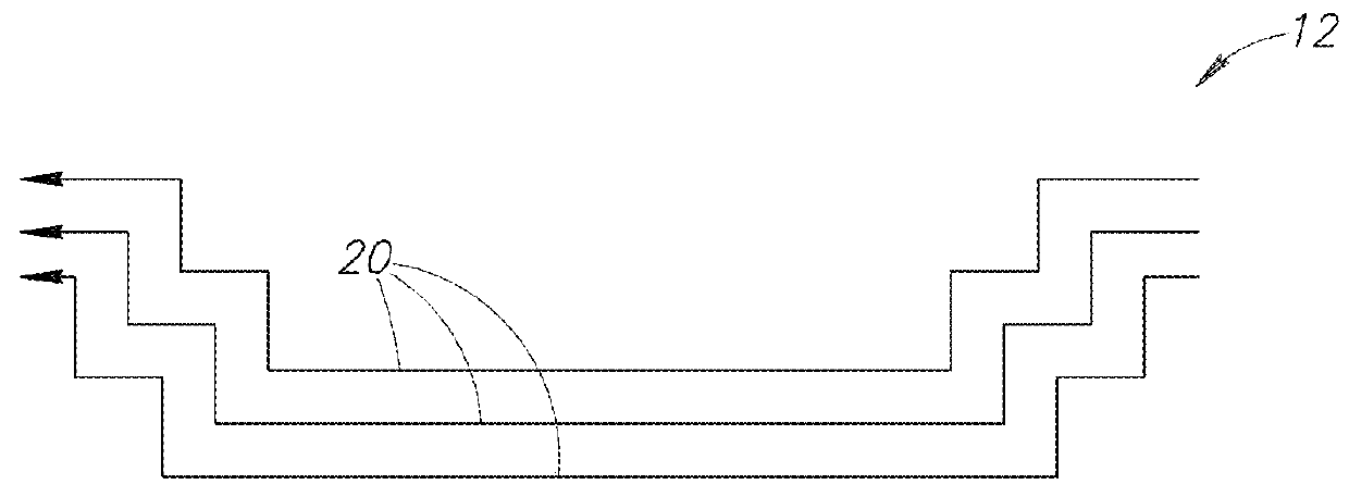
Figure 3
Abstract
Description
FIELD OF THE INVENTION
[0001] The present invention relates to a family of deep transcranial magnetic stimulation (TMS) coils, wherein a stimulating portion of the coils is at least partially circular and is configured to encircle at least a section of a body organ.BACKGROUND OF THE INVENTION
[0002] Transcranial magnetic stimulation (TMS) is a noninvasive technique used to apply brief magnetic pulses to the brain, or to other human organs, and to thereby activate neuronal structures. The pulses are administered by passing high currents by a stimulator through an electromagnetic coil externally placed upon the patient (for example, placed on the scalp for brain treatment), inducing electrical currents in the underlying tissue, thereby producing a localized axonal depolarization. This technique has become a major tool in central nervous system research, as well as a potentially promising treatment option for various neurobehavioral and neurological disorders.
[0003] Most known TMS coils sti...
Examples
examples
[0056]The field distribution produced by coil 110 of FIG. 6 was measured in a human head phantom model. A probe was moved in three directions inside the phantom model using a displacement system with 1 mm resolution, and the field distribution of coil 110 was measured in the whole head model volume with 1 cm resolution. Axial and coronal field maps were produced. The field maps were superimposed on anatomical T1-weighted MRI coronal slices, to show the induced field in each anatomical brain region.
[0057]Reference is now made to FIG. 9, which is an illustration of electric field distribution maps of coil 110 as measured in the human head phantom model. The field maps are shown for stimulator output set at 120% of threshold. The dark pixels indicate field magnitude above the threshold for neuronal activation. The threshold was set to 100 V / m, which is within the accepted range of thresholds required for hand motor activation. The intensity of stimulator power output used for drawing t...