Method for producing corneal endothelial cell

a stem cell and corneal technology, applied in the field of stem cell production of corneal endothelial cells, can solve the problems of corneal stroma edema, reduced corneal transparency, and serious influence on visual function, and achieve the effect of producing more efficiently

Active Publication Date: 2016-05-24
KEIO UNIV
View PDF12 Cites 18 Cited by
  • Summary
  • Abstract
  • Description
  • Claims
  • Application Information

AI Technical Summary

Benefits of technology

[0021]According to the production method of the present invention, a corneal endothelial cell can be produced more efficiently from a stem cell. The corneal endothelial cell obtained by the production method can be used as a medicament for the treatment of a disease caused by functional disorder of corneal endothelial cell, such as a corneal sheet for corneal transplantation and the like, or for cellular therapy for treating such disease.

Problems solved by technology

Therefore, the turbidity in the cornea caused by damage and the like has a serious influence on the visual function.
The above-mentioned function of corneal endothelial cell is impaired by a damage to the corneal endothelium such as a decrease in the corneal endothelial cell and the like, thus resulting in the edema of the corneal stroma.
This decreases transparency of the cornea, and reduces the eyesight.
While the penetrating keratoplasty is an established technique, the supply of cornea is short in Japan as the situation stands, and the rejection reaction poses a problem.
Since the number of donor of cornea is limited, the problem of donor shortage cannot be overcome, like penetrating keratoplasty.
Furthermore, since corneal endothelial cell is difficult to culture, preparation of cultured cells in a number sufficient for transplantation places a large burden in terms of time and cost.

Method used


Image

  • Method for producing corneal endothelial cell
  • Method for producing corneal endothelial cell
  • Method for producing corneal endothelial cell

Examples


example 1

Differentiation Induction of Mouse COPs into Corneal Endothelial Cells

(Material and Method)

1. Separation of Mouse COPs

[0075]Mouse COPs were separated from mouse cornea according to a publication (non-patent document 4). The corneal stromal disc was cut out from mouse (C57BL / 6 mouse), processed into small pieces and digested with 0.05% trypsin (Sigma-Aldrich, St. Louis, Mo.) at 37° C. for 30 min. Then, they were treated with 78 U / ml collagenase (Sigma-Aldrich) and 38 U / ml hyaluronidase (Sigma-Aldrich) at 37° C. for 30 min. Parenchymal cells were mechanically dissociated to give single cells, and cultured in DMEM / F-12 (1:1) medium containing 20 ng / ml EGF (Sigma-Aldrich), ng / ml FGF2 (Sigma-Aldrich), B27 supplement (Invitrogen, Carlsbad, Calif.), and 103 U / ml LIF (Chemicon International, Temecula). The cells were cultured at plating density of 1×10⁵ cells / ml under humidified atmosphere containing 5% CO2 at 37° C. The first culture was performed in a 35 mm dish, and thereafter in a 25 cm...

example 2

Differentiation Induction of Human iPS Cell-Derived Neural Crest Stem Cell into Corneal Endothelial Cell—(1)

(Material and Method)

1. Preparation of Human iPS Cell-Derived Neural Crest Stem Cell

[0078]Based on a publication (Nature Protocols, 2010 vol. 5, No. 4, 688-701), neural crest stem cells were obtained from human iPS cell. This Example is different from the above-mentioned publication in that floating culture was employed without using Matrigel for culturing iPS cells. Suspension culture enabled more efficient induction of differentiation into neural crest stem cells. The human iPS cells used were 201B7 (provided by Prof. Shinya Yamanaka (Kyoto University) and Prof. Hideyuki Okano (Keio University)).

2. Preparation of Differentiation Induction Medium

[0079]The medium was prepared in the same manner as the differentiation induction medium used in Example 1.

(Results)

[0080]Human iPS cell-derived neural crest stem cells were plated in a gelatin-coated 35 mm dish (Asahi Techno Glass) a...

example 3

Evaluation of Corneal Endothelial Cell (Corneal Endothelial Cell Marker Measurement)

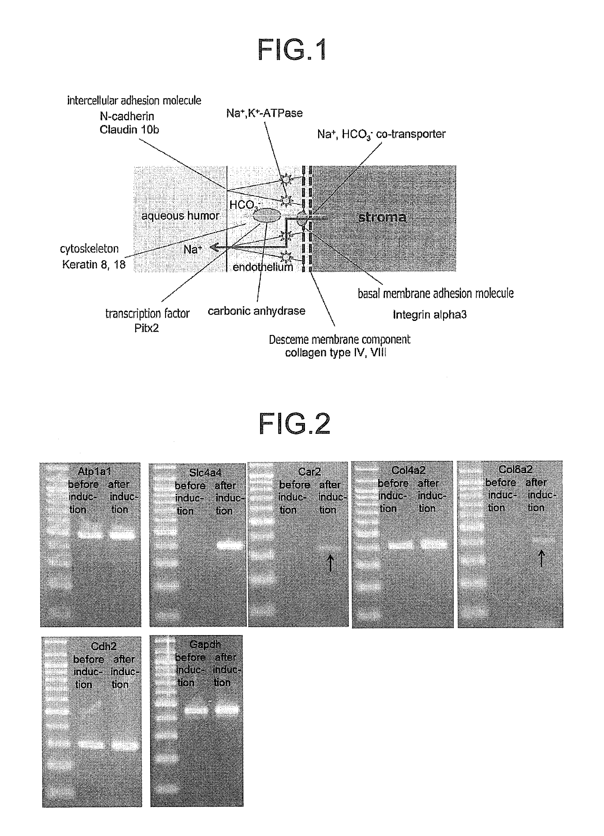
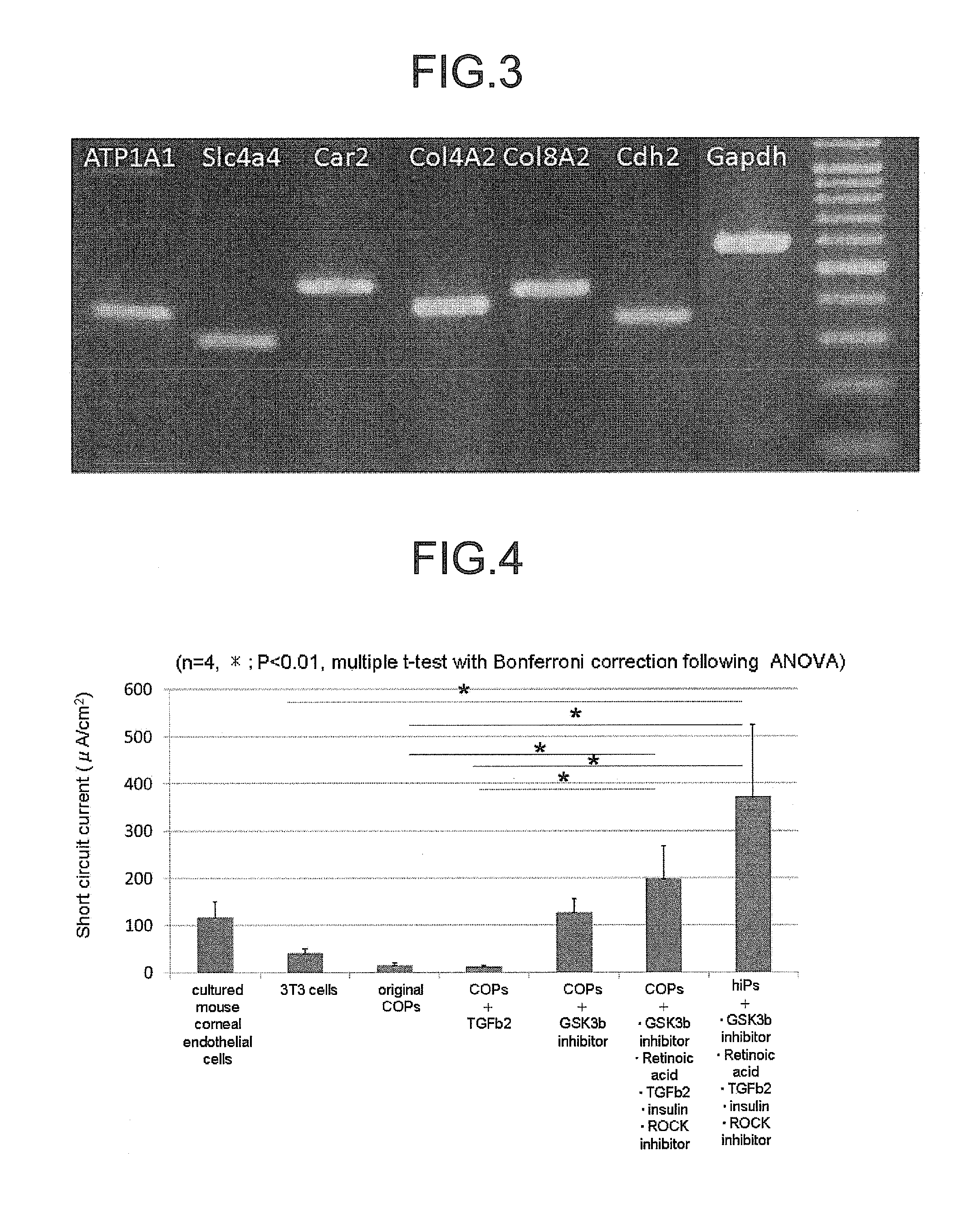
[0081]Using corneal endothelial cells obtained by inducing differentiation of COPs, which were prepared in the same manner as in Example 1, and corneal endothelial cells obtained by inducing differentiation of human iPS cell-derived neural crest stem cells, which were obtained in the same manner as in Example 2, the expression state of corneal endothelial cell marker was examined by RT-PCR method. The primers used are shown in Table 1 (for mouse) and Table 2 (for human).

[0082]

TABLE 1for mouse COPsprod-Forward sequenceReverse sequenceuctGene(5′-3′)(3′-5′)sizeAtp1a1CCATCGCTTACACCCTATCTTGCAGATGACCAAG492ACC (SEQ ID NO: 1)TCG (SEQ ID NO: 2)Slc4a4TCTTCCTGGGCACTTACAGGAGCATACCACCATG408ACC (SEQ ID NO: 3)AGG (SEQ ID NO: 4)Car2GATCCTTGCTCCCTTCTATCACCCAGCCTAACTG326TCC (SEQ ID NO: 5)TGC (SEQ ID NO: 6)Col4a2TGGAGTTCCTGGTTTGATCACCAAAGTCCCCAGT420AGG (SEQ ID NO: 7)AGG (SEQ ID NO: 8)Col8a2GGTCCAGTAGGGGCTAACCTGTAAAACCT...


PUM

PropertyMeasurementUnit
concentrationaaaaaaaaaa
temperatureaaaaaaaaaa

Abstract

The invention provides a method of efficiently producing corneal endothelial cells, particularly from corneal stroma or iPS cell-derived neural crest stem cells, a method of producing corneal endothelial cells stably in a large amount by inducing more efficient differentiation of stem cells into corneal endothelial cells, and a medicament containing corneal endothelial cells. The method of inducing differentiation of stem cells into corneal endothelial cells includes a step of culturing the stem cells in a differentiation induction medium containing a GSK3 inhibitor (preferably a GSK3β inhibitor) and retinoic acid, with the differentiation induction medium preferably further containing one or more of TGFb2, insulin, a ROCK inhibitor, and the like.

Description

CROSS-REFERENCE TO RELATED APPLICATIONS[0001]This patent application is the U.S. national phase of International Patent Application No. PCT / JP2012 / 076048, filed Oct. 5, 2012, which claims the benefit of Japanese Patent Application No. 2011-222138, filed on Oct. 6, 2011, and Japanese Patent Application No. 2012-076080, filed on Mar. 29, 2012, which are incorporated by reference in their entireties herein.INCORPORATION-BY-REFERENCE OF MATERIAL ELECTRONICALLY SUBMITTED[0002]Incorporated by reference in its entirety herein is a computer-readable nucleotide / amino acid sequence listing submitted concurrently herewith and identified as follows: 11,391 bytes ASCII (Text) file named “716545ReplacementSequenceListing.txt,” created Jul. 1, 2014.”TECHNICAL FIELD[0003]The present invention relates to a method of producing a corneal endothelial cell from a stem cell, and a method of inducing differentiation of a corneal endothelial cell from a stem cell. Furthermore, the present invention relates...

Claims


Application Information

Patent Timeline
Patent Type & Authority Patents(United States)
IPC IPC(8): C12N5/079A61L27/38C12N5/071A61K35/00
CPCC12N5/0621A61L27/3808A61L27/3895C12N5/069A61K35/00A61L2430/16C12N2501/11C12N2501/115C12N2501/15C12N2501/235C12N2501/33C12N2501/385C12N2501/727C12N2501/999C12N2506/08C12N2506/09C12N2506/45C12N2533/54A61P27/02
Inventor SHIMMURA, SHIGETOHATOU, SHINTSUBOTA, KAZUOYOSHIDA, SATORU
Owner KEIO UNIV
Why Eureka
Try Eureka