Minimally-advancing luminal catheter

a luminal catheter, minimal technology, applied in catheters, intravenous devices, wound drains, etc., can solve the problems of catheter dislocation, catheter distal tip dislocation, catheter dislocation,

Active Publication Date: 2016-08-02
STEERABLE INSTR NV +1
View PDF5 Cites 10 Cited by
  • Summary
  • Abstract
  • Description
  • Claims
  • Application Information

AI Technical Summary

Problems solved by technology

The wide external diameter of the prior art catheter may cause (sub)obstruction of the blood flow.
Additionally, the position of the catheter tip cannot be stabilised with respect to the wall of the vessel, causing a risk of dislocation of the catheter's distal tip and possible dysfunction of the catheter.
A further disadvantage of conventional catheters is damage to the vessel wall during advancement.
In particular, there may be damage or irritation to the endothelium.
While pushing the catheter inside a vessel lumen, the catheter's tip can either cause endothelial wall lesions or friction initiating the cascade of blood clotting, or can be deviated by a septum, causing malfunctioning or obstruction of the catheter.
In adults, introducing the catheter into the sinus through a standard skull burr hole is technically cumbersome, since an oblique angle is necessary to prevent endothelial laceration of the opposite venous sinus wall and to realize a correct orientation of the catheter (i.e. ante- or retrograde to the blood stream).
However, the thickness of the skull bone, requires a wide bore hole to achieve an angle sufficiently oblique.
Also, in the pediatric and adult population, the positioning of the subgaleal trajectory of the shunt system is quite difficult in order to prevent kinking of the tubing and to avoid stress causing displacement of the sinusal catheter.
Evacuation of cerebrospinal fluid remains non-physiological.
This siphoning effect can cause over-drainage of cerebrospinal fluid causing intracranial hypotension.
In the acute stage, this over-drainage can cause collapse of the brain's ventricles and acute subdural hematoma.
In the ventriculo-atrial and ventriculo-peritoneal shunt patients, the internal jugular veins are bypassed, therefore more complex valves (variable resistance valves or flow regulating valves) and anti-siphon devices are utilised in the art, making a cerebrospinal fluid shunt much more complex, more vulnerable, and more expensive.
Despite these technical improvements, a small group of patients still experience symptoms due to non-physiological cerebrospinal fluid evacuation and therefore non-physiological intracranial pressures.
Ventriculo-atrial and ventriculo-peritoneal shunts are easily obstructed causing insufficient cerebrospinal fluid evacuation and therefore intracranial hypertension.
Also, in growing children, the peritoneal catheter's tip can be retracted into the subcutaneous fat or the atrial catheter's tip into the superior caval vein causing insufficient cerebrospinal fluid evacuation.
Ventriculo-atrial and ventriculo-peritoneal shunts have a high rate of bacterial shunt infections, with risk of bacterial meningitis or ventriculitis or cerebritis, possibly with livelong morbidity or even mortality risk.
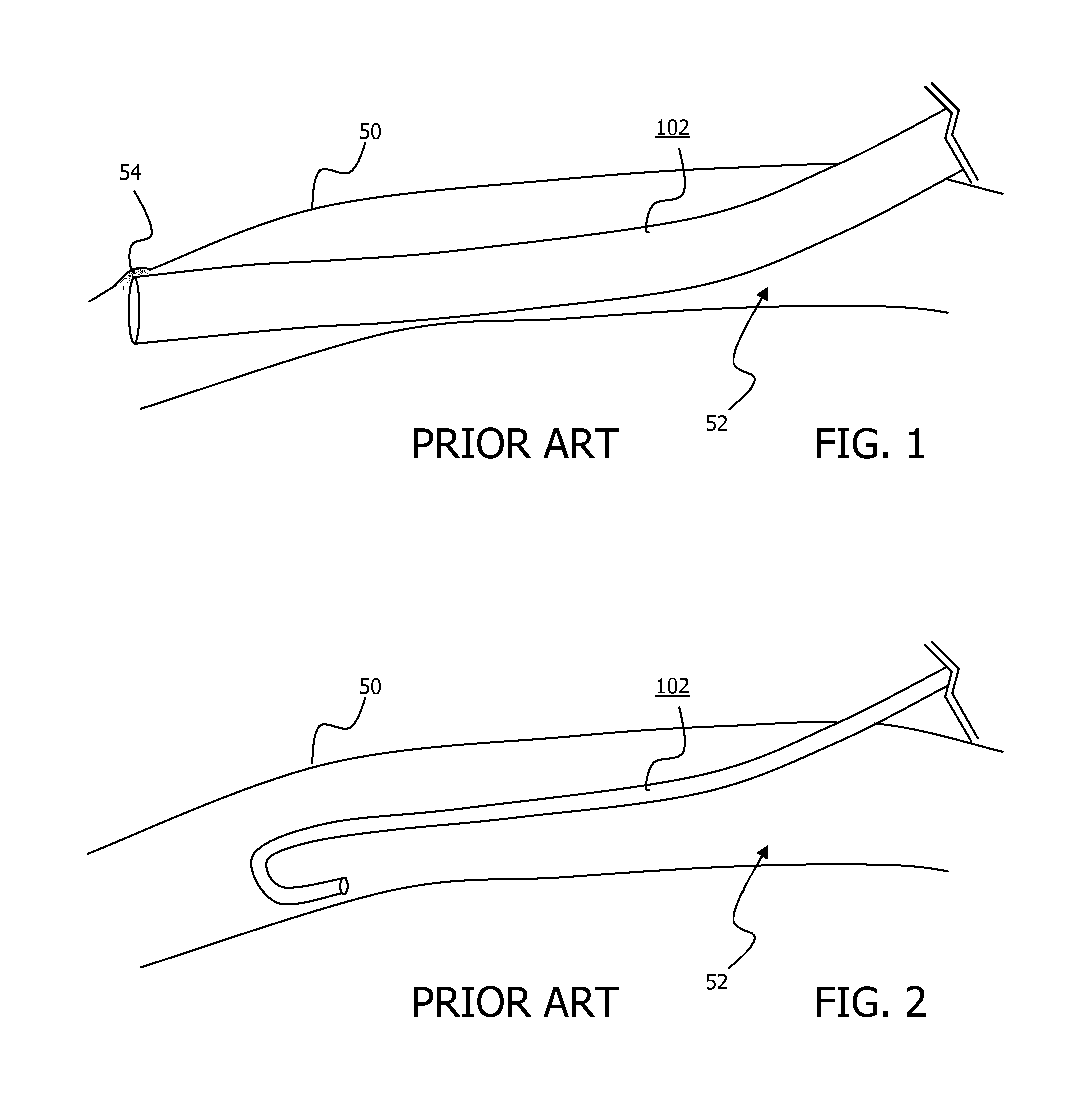
Disadvantages associated with ventriculosinus shunts include their obstructive size that may cause a raised venous pressure inside the dural venous sinus, may reduce the blood flow speed and thus the risk of clotting of the blood inside the dural venous sinus (sinus thrombosis).
Additionally, the catheter cannot be stabilized onto the sinus's wall, causing a risk of dislocation of the catheter's distal tip and possible dysfunction of the cerebrospinal fluid shunt.
Moreover, the position of catheter tip cannot be stabilised inside the sinus's lumen; it should be localised in the center of the sinus's diameter.
The catheter is more likely to be blocked by the endothelial layer covering the internal wall of the dural venous sinus, thus causing a total obstruction of the cerebrospinal fluid shunt.
Further, the catheter tip lying against the internal wall of the venous sinus cannot fully profit from the impaction effect created by the velocity of the blood flow inside the dural venous sinus as the velocity of this blood flow is maximal at the center of the sinus's diameter and minimal against the sinus's inside wall.
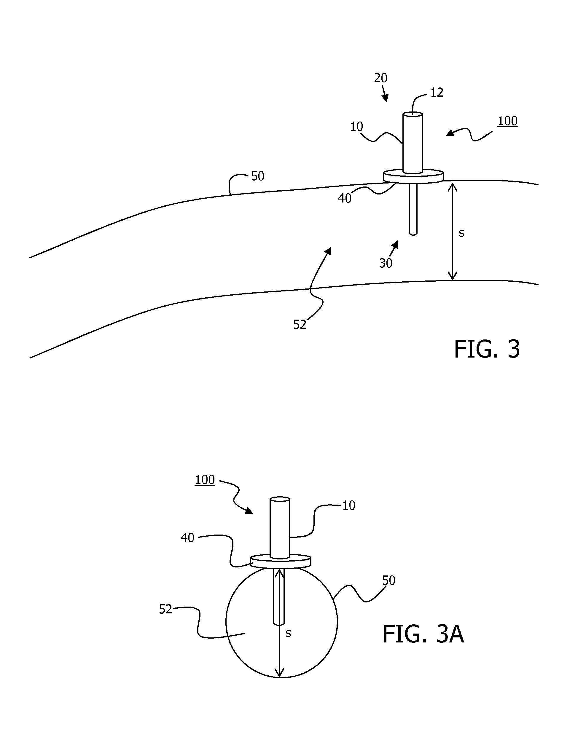
While advancing the catheter inside the dural venous sinus with an intrasinusal length of 20 to 50 millimeter, the catheter's tip can easily either cause endothelial wall lesions increasing the risk of blood clotting, or can be deviated by an intrasinusal septum, causing malfunctioning or obstruction of the cerebrospinal fluid shunt.
This increases the risk of important blood loss, the risk of aspiration of air inside the sinus possibly causing an air embolism and the risk of invagination of the endothelial layer causing obstruction to the blood flow inside the dural venous sinus.

Method used


Image

  • Minimally-advancing luminal catheter
  • Minimally-advancing luminal catheter
  • Minimally-advancing luminal catheter

Examples


Embodiment Construction

[0019]The present invention relates to an implantable catheter (100) provided for insertion through a wall (50) of a dural venous sinus (70) in a subject, having a proximal (20) and distal end (30), comprising:[0020]a tubular shaft (10) for insertion through the wall (50) of the venous sinus (70) into the sinus (52), provided with a shaft lumen (12) in fluid connection with a proximal port (14) at the proximal end and a distal port (16) at the distal end of the shaft (10), and[0021]a stop element (40) disposed on an outer surface of the tubular shaft (10), configured to limit the depth of insertion of the tubular shaft (10) into the sinus.

[0022]The depth of insertion of the tubular shaft (10) may be limited to less than the span of said sinus (52) in transverse cross-section. The depth of insertion of the tubular shaft (10) may be limited to essentially half of the span of said sinus (52). The stop element (40) may incorporate a fixation element (60), configured to fix the position ...


PUM


Abstract

The present invention relates to an implantable catheter (100) provided for insertion through a wall (50) of a dural venous sinus (70) in a subject, having a proximal (20) and distal end (30), comprising: —a tubular shaft (10) for insertion through the wall (50) of the venous sinus (70) into the sinus (52), provided with a shaft lumen (12) in fluid connection with a proximal port (14) at the proximal end and a distal port (16) at the distal end of the shaft (10), and —a stop element (40) disposed on an outer surface of the tubular shaft (10), configured to limit the depth of insertion of the tubular shaft (10) into the sinus.

Description

RELATED APPLICATIONS[0001]This application is a 35 U.S.C. §371 filing of International Application No. PCT / EP2012 / 067334, filed Sep. 5, 2012, which claims priority to European Patent Application No. 11180066.0, filed Sep. 5, 2011 and U.S. Patent Application No. 61 / 531,312, filed Sep. 6, 2011, each of which is incorporated herein by reference in its entirety.FIELD OF THE INVENTION[0002]The present invention is in the field of medical catheters. More in particular, it is in the field of a catheter incorporated into cerebrospinal fluid (CSF) shunt.BACKGROUND TO THE INVENTION[0003]A catheter of the prior art may be regarded as having some important technical disadvantages. For instance, the catheter has a relative wide external diameter to facilitate a catheter lumen of sufficient dimension for the flow of fluid, and requires advancement through the vessel lumen. The wide external diameter of the prior art catheter may cause (sub)obstruction of the blood flow. Additionally, the position...

Claims


Application Information

Patent Timeline
Patent Type & Authority Patents(United States)
IPC IPC(8): A61M5/00A61M27/00A61M25/04
CPCA61M25/04A61M27/006
Inventor BAERT, EDWARDDEWAELE, FRANK
Owner STEERABLE INSTR NV
Why Eureka
Try Eureka