Bioreactor system and method of enhancing functionality of muscle cultured in vitro

a bioreactor and muscle technology, applied in the direction of skeletal/connective tissue cells, prosthesis, ligaments, etc., can solve the problems of ineffective clinical treatment, lack of clinical utility, and approach that has significant limitations, so as to enhance the functionality of muscle tissue

Active Publication Date: 2016-11-15
WAKE FOREST UNIV HEALTH SCI INC
View PDF30 Cites 2 Cited by
  • Summary
  • Abstract
  • Description
  • Claims
  • Application Information

AI Technical Summary

Benefits of technology

The patent describes a method of cultivating organized skeletal muscle tissue from precursor muscle cells using a support system. The method involves cyclically stretching and relaxing the support at least twice along a first axis during a first time period, and then maintaining the support in a substantially static position during a second time period. The repeated stretching and relaxing can enhance the functionality of the muscle tissue, resulting in the production of organized skeletal muscle tissue on the solid support. The resulting muscle tissue can exhibit a reproducible contractile response to KCl-induced depolarization in vitro, and may have a unidirectional orientation on histological examination, contain muscle markers, and produce extracellular matrices. The method can be carried out using a device that includes a container, engaging members for tissue supports, an actuator for providing controlled cyclic strain, a motor, and a controller for implementing the method. The engaging members may have multiple attachment points so that multiple tissue supports can be immersed in the growth or culture media.

Problems solved by technology

Loss of functional skeletal muscle due to traumatic injury, tumor excision, etc., produces a physiological deficit for which there is still no effective clinical treatment.
However, while muscle constructs developed with synthetic scaffolds can support the contractile portion of the muscle tissue, and furthermore, can be maintained in culture for several months, this approach still has significant limitations for clinical utility.
Clearly, gel-based constructs are currently too small and too fragile for such surgical manipulatation.
As such, one of the major barriers to engineering clinically applicable functional muscle tissues for reconstructive procedures is the lack of a bioreactor system and methodology that would accelerate cellular organization, tissue formation and function.

Method used


Image

  • Bioreactor system and method of enhancing functionality of muscle cultured in vitro
  • Bioreactor system and method of enhancing functionality of muscle cultured in vitro
  • Bioreactor system and method of enhancing functionality of muscle cultured in vitro

Examples


example 1

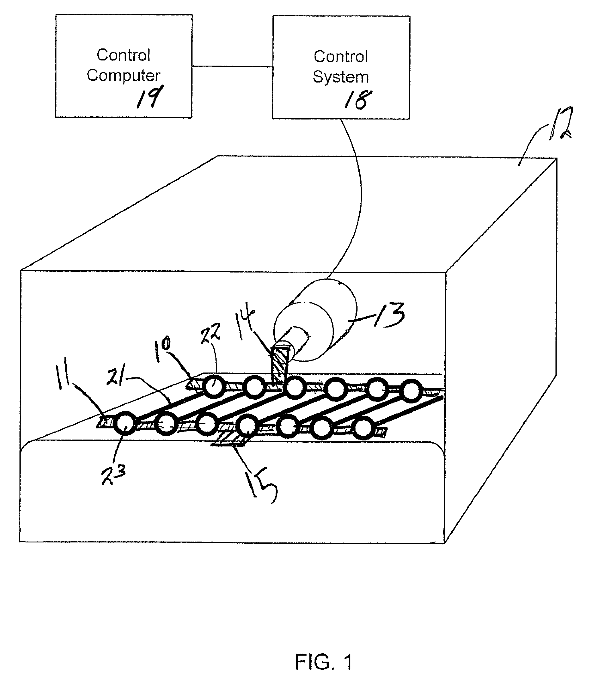
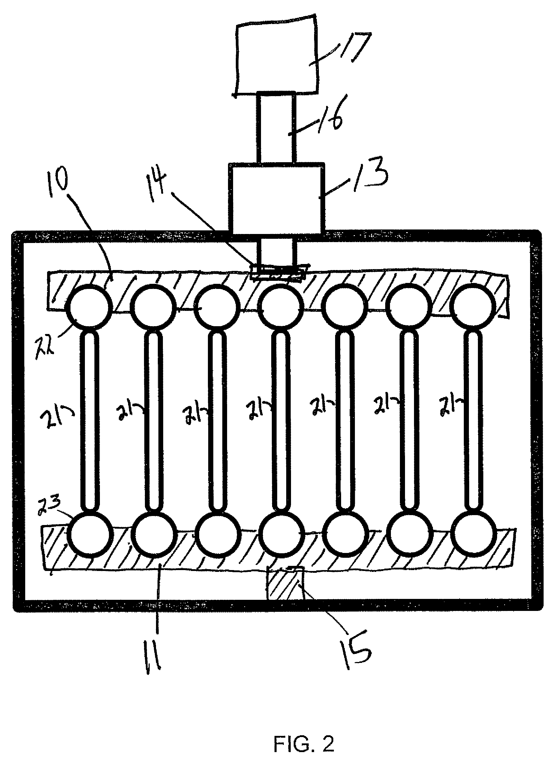
[0039]A bioreactor system consisted of an actuator mounted on a tissue culture-compatible container which was designed to provide controlled cyclic strain to muscle tissue scaffolds, as shown in FIG. 2. The linear actuator was controlled by a linear motor controller system and a control computer, used for programming and subsequent monitoring of the actuator. Primary human skeletal muscle precursor cells were isolated, grown and expanded in culture. The cells were seeded onto collagen-based muscle scaffold strips derived from porcine bladder tissue (1.0×0.3×0.3 cm3). After two days of static culture, the muscle cell seeded scaffolds were placed in the bioreactor system and programmed linear stretching cycles were applied (LinMot®). The controlled cycle strain was programmed to exert ±10% of the initial length of the cell seeded scaffolds at a frequency of 3 times per minute for the first 5 minutes of every hour. The bioreactor was continuously operated for up to 3 weeks after the in...

example 2

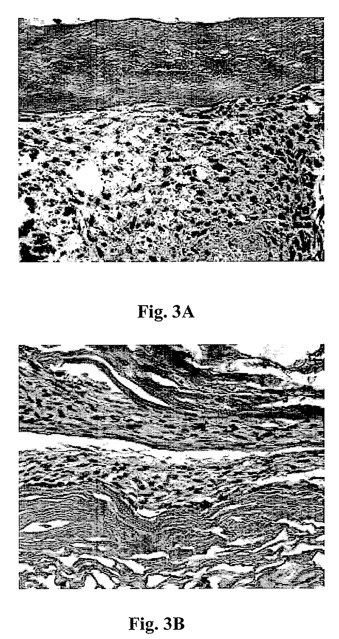
[0042]This example presents the results of more detailed studies with the bioreactor system described above. The system, in overview, consisted of a linear actuator mounted on a tissue container to provide a controlled cyclic strain to muscle tissue scaffolds. Primary human muscle cells were seeded onto scaffolds and placed in the bioreactor system and subjected to cyclic strain equivalent to ≈±10% stretch of the original scaffold length; strain was applied 3 times / min for the first 5 min / hour for periods ranging from 5 days to 3 weeks. Following this conditioning protocol, the cell constructs were assessed for structural and functional parameters in vitro; cell and scaffold constructs under static culture conditions (i.e., no cyclic strain) were run in parallel. In a separate in vivo experiments, both the structures conditioned in the bioreactors for 5 days and control tissue structures maintained under static culture conditions were implanted onto the latissimus dorsi muscle of nu...


PUM


Abstract

A method of producing organized skeletal muscle tissue from precursor muscle cells in vitro comprises: (a) providing precursor muscle cells on a support in a tissue media; then (b) cyclically stretching and relaxing the support at least twice along a first axis during a first time period; and then (c) optionally but preferably maintaining the support in a substantially static position during a second time period; and then (d) repeating steps (b) and (c) for a number of times sufficient to enhance the functionality of the tissue formed on the support and / or produce organized skeletal muscle tissue on the solid support from the precursor muscle cells.

Description

RELATED APPLICATIONS[0001]This application claims the benefit of U.S. Provisional Patent Application Ser. No. 60 / 671,600, filed Apr. 15, 2005, the disclosure of which is incorporated by reference herein in its entirety.FIELD OF THE INVENTION[0002]The present invention concerns methods and apparatus for the growth of skeletal muscle in vitro.BACKGROUND OF THE INVENTION[0003]Loss of functional skeletal muscle due to traumatic injury, tumor excision, etc., produces a physiological deficit for which there is still no effective clinical treatment. Tissue engineering of skeletal muscle in vitro for functional tissue replacement in vivo may provide a potential therapeutic solution to this unmet medical need. In fact, significant progress has been made during last 15 years in understanding some of the basic requirements for creating tissue engineered skeletal muscle constructs in vitro. Early studies necessarily focused mainly on the production of highly differentiated muscle constructs and...

Claims


Application Information

Patent Timeline
Patent Type & Authority Patents(United States)
IPC IPC(8): C12M3/00A61L27/38C12N5/077C12M1/42
CPCC12M35/04A61L27/3826A61L27/3895C12M21/08C12N5/0658A61F2/08A61F2002/0894A61L27/3604A61L27/367A61L27/3683A61L27/3691A61L2430/30A61L2430/34C12N2527/00C12N2539/00
Inventor YOO, JAMESSTITZEL, JOELATALA, ANTHONYCHRIST, GEORGE
Owner WAKE FOREST UNIV HEALTH SCI INC
Why Eureka
Try Eureka