System, method and devices for navigated flexible endoscopy

a flexible endoscope and flexible technology, applied in the field of methods and equipment for assisting navigated flexible endoscopes, to achieve the effect of accurately determining the location of an area

Active Publication Date: 2017-05-30
PHILIPS ELECTRONICS LTD
View PDF241 Cites 375 Cited by
  • Summary
  • Abstract
  • Description
  • Claims
  • Application Information

AI Technical Summary

Benefits of technology

[0021]The centerline path and / or the most likely path of the endoscope calculated in the pre-operative image space may differ from the actual path taken by the inserted endoscope. As such, the intra-operative images provide the “true path” of the endoscope. The true path of endoscope becomes available once the intra-operative imaging is performed and analyzed. Registering the intra-operative image data with the pre-operative image data provides a more accurate set of image space data regarding the path of the endoscope and provides a richer set of data regarding other features of the anatomy of the patient.
[0035]In some embodiments, the only information required may be the shape of the endoscope or other instrument. This may be acquired without having to explicitly perform a registration step.
[0038]In some embodiments, the sensor elements on a trackable guidewire act simply as a distance measurement device to determine the location of the endoscope relative to a known location. In this embodiment, the relative location of all suspect lesions are calculated relative to each other and landmarks within the anatomy. As each is examined, the location is stored and the distance to the next location of interest is calculated. In this way, the system is self-correcting at each identified location along the path of the endoscope as it is removed. This method may not require registration to be performed prior to use.
[0043]In one embodiment, these measured distances are augmented using shape information regarding the endoscope or other instrument, as may be determined using a path determination method such as those described herein. This shape information may be helpful in more accurately determining the location of an area of interest.

Problems solved by technology

In practice, this can be difficult because there is no way to easily register image-based data regarding suspect lesions to the patient space (e.g., the real world location of the suspected lesions).

Method used


Image

  • System, method and devices for navigated flexible endoscopy
  • System, method and devices for navigated flexible endoscopy
  • System, method and devices for navigated flexible endoscopy

Examples


Embodiment Construction

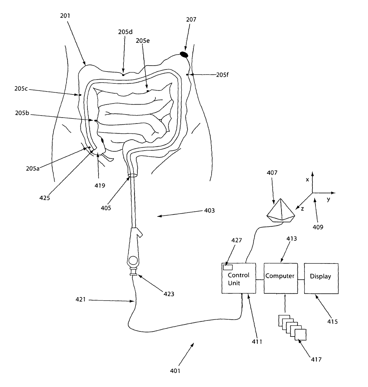
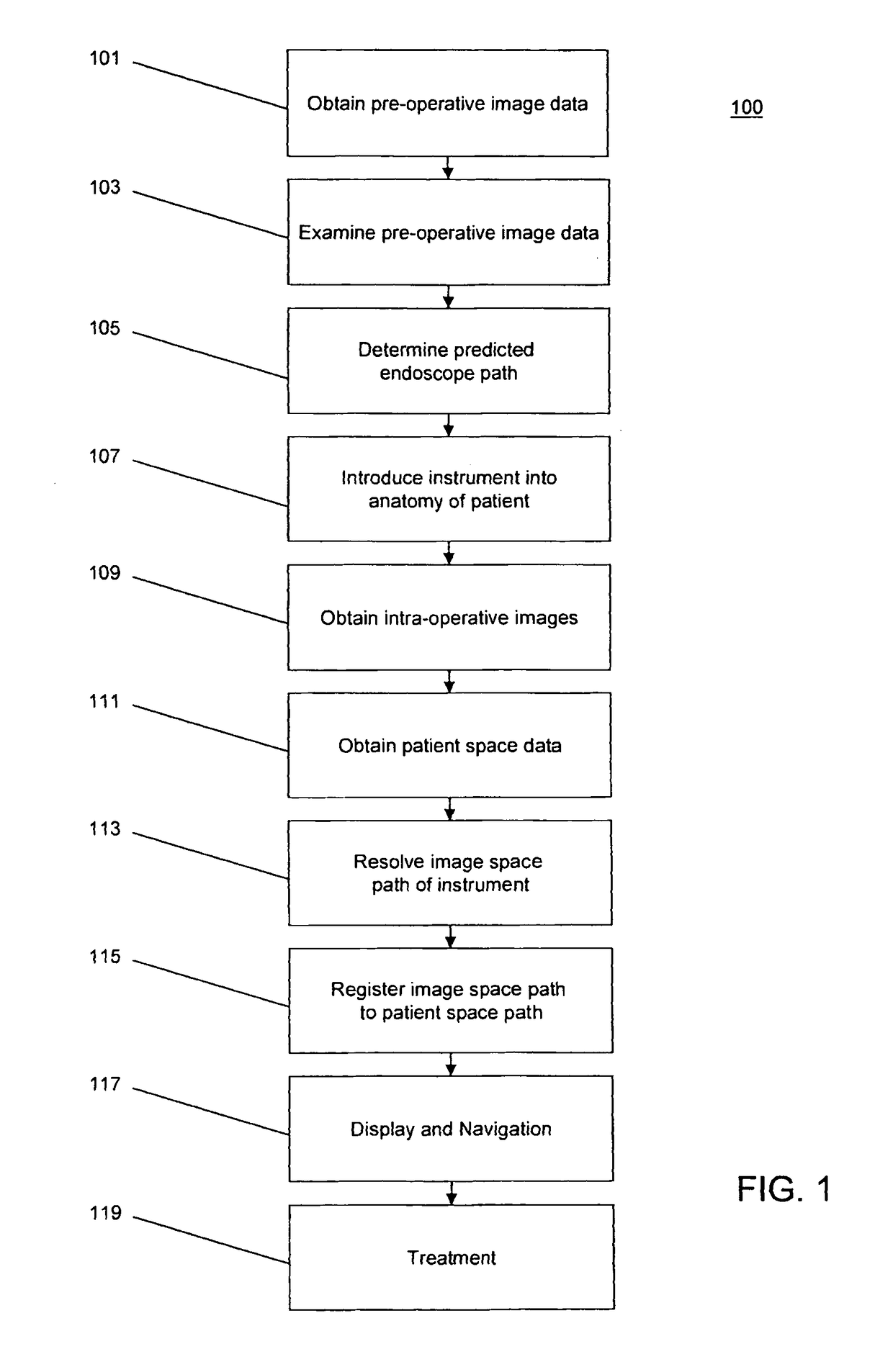
[0053]In one embodiment, the invention provides method for assisting an image-guided endoscopic procedure. FIG. 1 illustrates a process 100, an example of a method for assisting an image-guided endoscopic medical procedure. In an operation 101, a conventional virtual endoscopic data set may be acquired prior to commencement of the medical procedure using existing standard protocols such as, for example, those described in A. K. Hara et al., Reducing Data Size and Radiation Dose for CT Colonography, 168 American Journal of Roentgenology 1181-1184 (1997), which is hereby incorporated by reference herein in its entirety.

[0054]The virtual endoscopic data set may comprise image data obtained using x-rays, computerized tomography (CT), magnetic resonance (MR), positron emission tomography (PET), ultrasound, and / or other imaging modalities. In some embodiments, the pre-operative imaging may be performed with the patient in same position as the endoscopic procedure. In some embodiments the ...


PUM


Abstract

The invention provides a method and system for performing an image-guided endoscopic medical procedure. The invention may include registering image-space coordinates of a path of a medical instrument within the anatomy of a patient to patient-space coordinates of the path of the medical instrument within the anatomy of the patient. In some embodiments, the image space coordinates of the path of the medical instrument may be predicted coordinates such as, for example, a calculated centerline through a conduit-like organ, or a calculated “most likely path” of the medical instrument within the anatomy of the patient. In other embodiments, the path of the medical instrument may be an actual path determined using intra-operative images of the patient's anatomy with the medical instrument inserted therein. The registered instrument may then be navigated to one or more items of interest for performance of the endoscopic medical procedure.

Description

REFERENCE TO RELATED APPLICATIONS[0001]This application claims priority to U.S. Provisional Patent Application No. 60 / 710,657, filed Aug. 24, 2005, which is hereby incorporated by reference herein in its entirety.FIELD OF THE INVENTION[0002]The present invention relates to methods and apparatus for assisting navigated flexible endoscopy.BACKGROUND[0003]During exploratory endoscopic procedures such as colonoscopy, bronchoscopy, endoscopic retrograde cholangiopancreatography (ECRP) or other endoscopic procedures, it is desirable to locate any lesions or other areas that are of interest such as, for example, precancerous changes, bleeding sites, polyps, nodules, or other areas of interest. Traditional endoscopic examination consists entirely of an optical examination of the conduit or area of interest within the anatomy of the patient. More recently, virtual examination has become more popular. In virtual endoscopy, computerized tomography (CT), magnetic resonance (MR), ultrasound, or ...

Claims


Application Information

Patent Timeline
Patent Type & Authority Patents(United States)
IPC IPC(8): A61B5/05A61B90/00G06T19/00A61B5/06A61B1/31A61B34/20A61B1/005A61B6/12A61B34/10
CPCA61B1/005A61B1/31A61B5/06A61B5/065A61B34/20A61B90/36G06T19/003A61B6/12A61B34/10A61B90/361A61B2034/107A61B2034/2051A61B2090/397G06T2210/41A61B1/018G06T7/0012G06T2207/30048
Inventor GLOSSOP, NEIL DAVID
Owner PHILIPS ELECTRONICS LTD
Why Eureka
Try Eureka