
Method for preserving proliferation and differentiation potential of mesenchymal stem cells

a mesenchymal stem cell and proliferation and differentiation technology, applied in the field of cell biology of undifferentiated cells, can solve the problems of virus infection in the recipient, documents disclose that ha is able to maintain undifferentiated cells in culture, etc., to preserve the proliferation and differentiation potential of undifferentiated cells, without loss of replicative ability and differentiation capacity

Active Publication Date: 2017-06-27
NAT CHENG KUNG UNIV
View PDF5 Cites 0 Cited by
  • Summary
  • Abstract
  • Description
  • Claims
  • Application Information

AI Technical Summary

Benefits of technology

The present invention relates to a method for preserving the potential of undifferentiated cells to proliferate and differentiate. This method involves using a culture carrier with a coated surface containing a non-proteinaceous extracellular matrix component. The undifferentiated cells are then cultured on this surface with an appropriate medium, which allows their proliferation and differentiation potential to be preserved. This method can be used for expanding stem cells in vitro without losing their ability to replicate and differentiate. It is useful in regenerative medicine, tissue engineering, and therapy using various cell sources such as peripheral blood, stem cells, tissue progenitor cells, and tissue cells.

Problems solved by technology

However, the potential risk of using feeder cells in the culture of the undifferentiated cells such as stem cells for regenerative medicine or therapeutic tissue engineering is that infectious agents such as viruses may infect the recipient.
However, none of the above documents discloses that HA is able to maintain undifferentiated cells in culture, such as stem cells in an undifferentiated state.

Method used


Image

  • Method for preserving proliferation and differentiation potential of mesenchymal stem cells
  • Method for preserving proliferation and differentiation potential of mesenchymal stem cells
  • Method for preserving proliferation and differentiation potential of mesenchymal stem cells

Examples


example 1

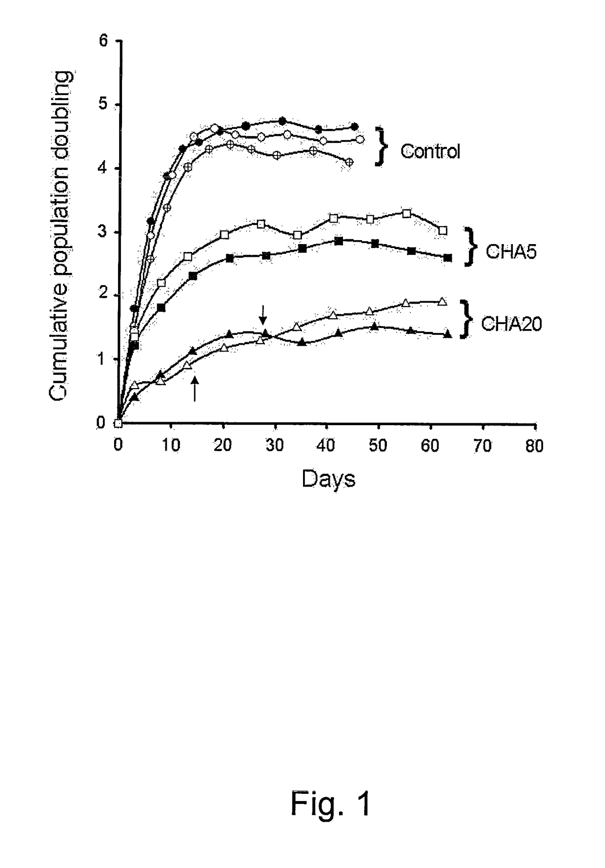
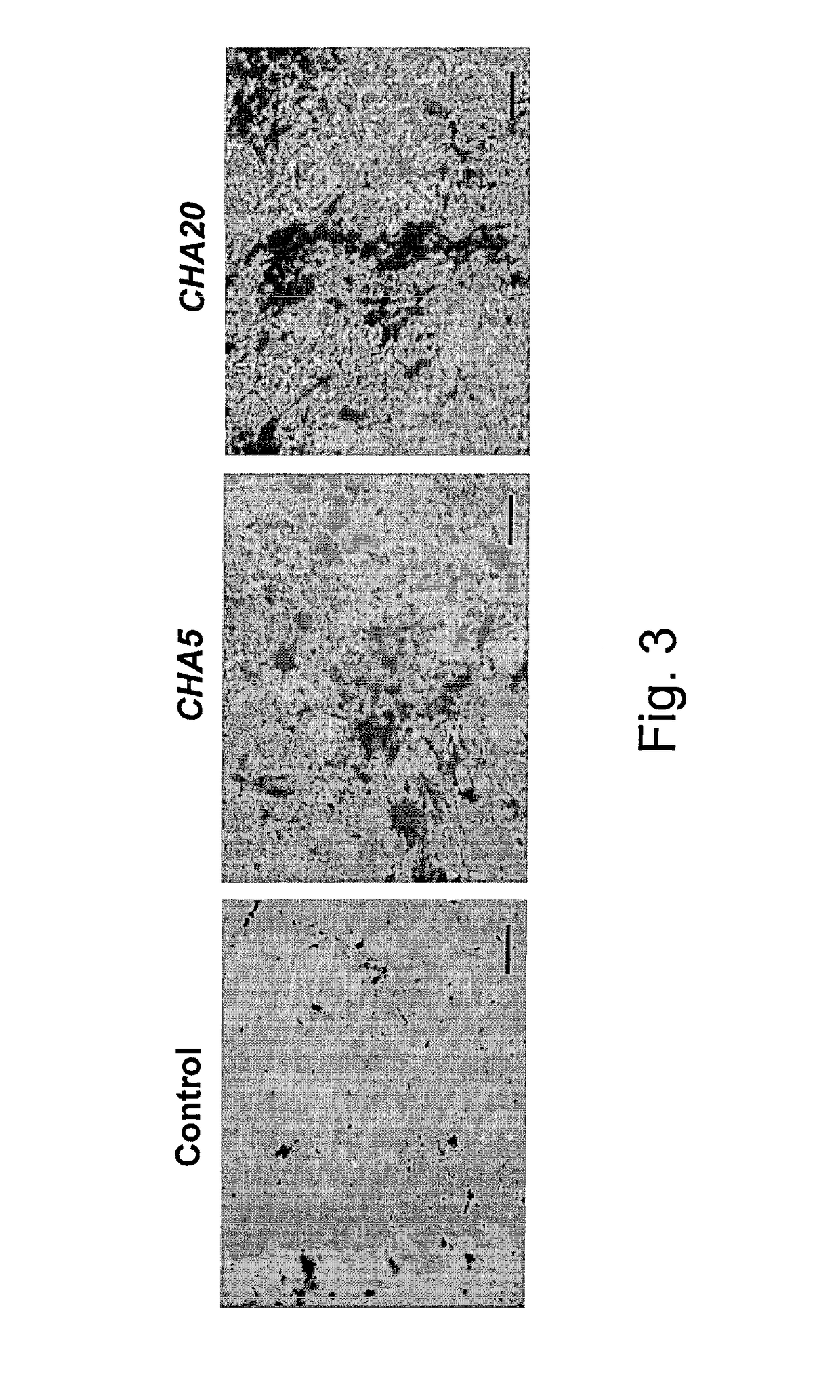
Altered Proliferative Behaviors of mADSCs in Response to HA

Materials and Methods:

1. Isolation and Culture of mADSCs

[0059]mADSCs were isolated as previously described (R. Ogawa, et al., (2004), Supra.). Male FVB / N mice were housed and raised at National Cheng Kung University in Taiwan under standard conditions according to institutional guidelines for animal regulation. Briefly, inguinal fat pads from FVB / N mice were harvested and washed with phosphate buffered saline (GibcoBRL, Grand Island, USA) and were then finely minced and digested with 0.1% collagenase (Worthington, Lakewood, USA) at 37° C. for 45 minutes. An equal volume of Dulbecco's modified Eagle's medium (DMEM, GibcoBRL) containing 10% fetal bovine serum (FBS, Biological Industries, Israel) (hereafter referred to DMEM-10% FBS) was added and the resulting solution was filtered through a 100-μm mesh, followed by centrifugation at 250×g for 10 minutes. The pellet was collected and resuspended in 160 mM NH4Cl (Sigma, USA) to ...

example 2

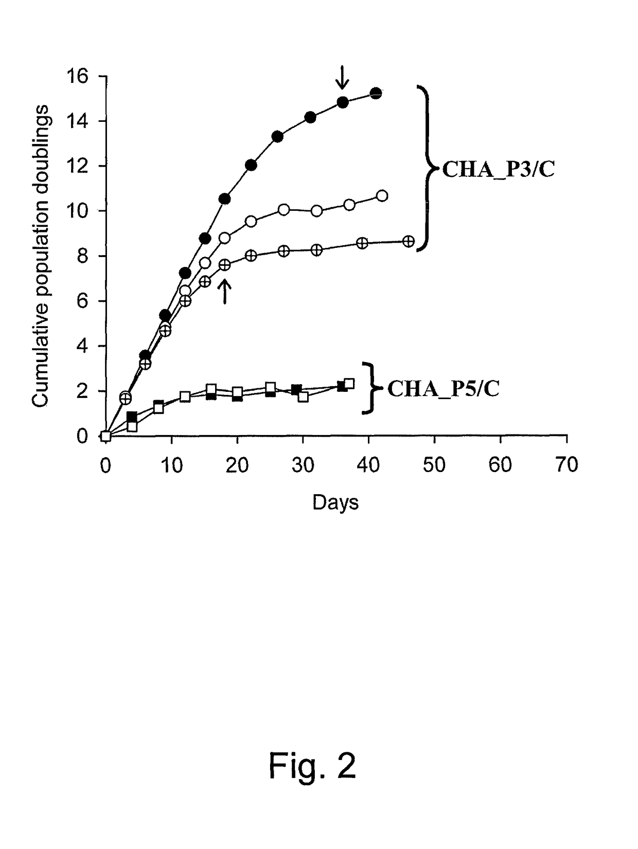
Altered Proliferative Behaviors of hPDMSCs in Response to HA

Materials and Methods:

1. Isolation and Culture of Human Placenta-Derived Mesenchymal Stem Cells (hPDMSCs)

[0077]Third trimester (38 to 40 weeks GA, n>10) placenta tissue were collected after Cesarean sections of healthy human donor mothers. Specimen was obtained after informed consent and all experiments were approved by the local institutional review board. After amnion and decidua were manually separated, chorionic villi from the fetal part were minced, and then degraded with collagenase (200 U / mL, Sigma) for 30 minutes at 37° C. in water bath by gently orbital shaking. Through Percoll gradient (Pharmacia Biotech) centrifugation (density=1.073 g / cm3), mononuclear cells were purified and propagated at 2×10⁴ cells / cm2 in complete medium, i.e. Dulbecco's modified Eagle's medium-low glucose (DMEM-LG, Invitrogen) containing 10% fetal bovine serum (Biological Industries), and 100 unit / mL gentamycin (Biological Industries). Cells...

example 3

Altered Proliferative Behaviors of Undifferentiated Cells in Response to Biological Materials

Materials and Methods:

[0085]Biological materials hyaluronan (HA), chondroitin sulfate (S), carboxymethyl cellulose (M), carrageenan (G), and alginate (A) were coated at 1 μg / cm2, 5 μg / cm2, 30 μg / cm2, 100 μg / cm2, and 200 μg / cm2 respectively on tissue cultural surface (referred as control group). Specifically, hyaluronan (HA), chondroitin sulfate (S), carboxymethyl cellulose (M), carrageenan (G), and alginate (A) were coated at 30 μg / cm2. The various surfaces were dried and sterilized. In preferable embodiment, the above various polysaccharides or sulfated polysaccharides (hyaluronan, chondroitin sulfate, carboxymethyl cellulose, carrageenan, and alginate) at high concentrations about 0.001 mg / ml, 0.01 mg / ml, 0.1 mg / ml, 1 mg / ml, 10 mg / ml, 100 mg / ml were respectively added to the cultural media.

[0086]Undifferentiated cells fibroblasts, perichondral progenitor cells (PCPC), adipose derived strom...


PUM

PropertyMeasurementUnit
timeaaaaaaaaaa
concentrationsaaaaaaaaaa

Abstract

A method for preserving proliferation and differentiation potential of undifferentiated cells, has steps of providing a culture carrier having a surface coated with a biological material selected from the group consisting of hyaluronan, chondroitin sulfate, carboxymethyl cellulose, carrageenan, alginate, and chitosan; and inoculating and culturing the undifferentiated cells on the surface in the culture carrier with an appropriate medium, such that the proliferation and differentiation potential of undifferentiated cells are preserved. The method can be used for expanding stem cells in vitro without loss of their replicative ability and differentiation capacity. Therefore, the method according to the present invention is amenable to application in regenerative medicine, tissue engineering, and therapy using umbilical cord blood and other cell sources such as peripheral blood, stem cells, tissue progenitor cells, and tissue cells.

Description

RELATED PATENT APPLICATION[0001]This application is a continuation-in-part application and claims priority from subject matter disclosed in the earlier filed patent application Ser. No. 12 / 155,487, filed on Jun. 5, 2008, which is incorporated herein in its entirety by reference.BACKGROUND OF THE INVENTION[0002]1. Field of the Invention[0003]This present invention relates to the field of cell biology of undifferentiated cells. More specifically, it relates to the propagation of undifferentiated cells, culture conditions and materials that facilitate propagation and use of undifferentiated cells.[0004]2. Description of the Prior Arts[0005]Undifferentiated cells, such as tissue progenitor cells, stem cells and the like, have great commercial potential in regenerative medicine or therapeutic tissue engineering. For the application of undifferentiated cells in regenerative medicine or therapeutic tissue engineering, a convenient method for culturing undifferentiated cells in an undiffere...

Claims


Application Information

Patent Timeline
Patent Type & Authority Patents(United States)
IPC IPC(8): C12N5/00C12N5/0775C12N5/071
CPCC12N5/0602C12N5/0667C12N2533/70C12N2533/72C12N2533/74C12N2533/78C12N2533/80
Inventor HUANG, LYNN LING-HUEI
Owner NAT CHENG KUNG UNIV
Learn More