
Method for reduced field of view MRI in an inhomogeneous field with rapid outer volume suppression

a technology of inhomogeneous field and suppression method, which is applied in the field of magnetic resonance imaging (mri), can solve the problems of inability to use single-shot spin-echo cardiac imaging in rapid single-shot spin-echo cardiac imaging, inability to adapt to spiral imaging, and inability to reduce the field of view mri. the effect of bsub>1/sub>+ variation

Active Publication Date: 2017-08-22
UNIV OF SOUTHERN CALIFORNIA
View PDF19 Cites 3 Cited by
  • Summary
  • Abstract
  • Description
  • Claims
  • Application Information

AI Technical Summary

Benefits of technology

The solution provides robust B1+ insensitivity, reduces acquisition time, and enhances spatial resolution while maintaining effective suppression of signal outside the region of interest, improving image quality and efficiency in rFOV imaging.

Problems solved by technology

This sampling criterion may become burdensome and inefficient when the region of interest (ROI) is small relative to the full spatial extent of the anatomy.
However, this method may not be not suitable for spiral imaging, may need to be applied once each TR interval, and may be B1+ sensitive.
A faster, single-sided OVS design, which ca be used in rapid single-shot spin-echo cardiac imaging, may also not be robust to B1+ variation.

Method used


Image

  • Method for reduced field of view MRI in an inhomogeneous field with rapid outer volume suppression
  • Method for reduced field of view MRI in an inhomogeneous field with rapid outer volume suppression
  • Method for reduced field of view MRI in an inhomogeneous field with rapid outer volume suppression

Examples


Embodiment Construction

[0023]Illustrative embodiments are now described. Other embodiments may be used in addition or instead. Details that may be apparent or unnecessary may be omitted to save space or for a more effective presentation. Some embodiments may be practiced with additional components or steps and / or without all of the components or steps that are described.

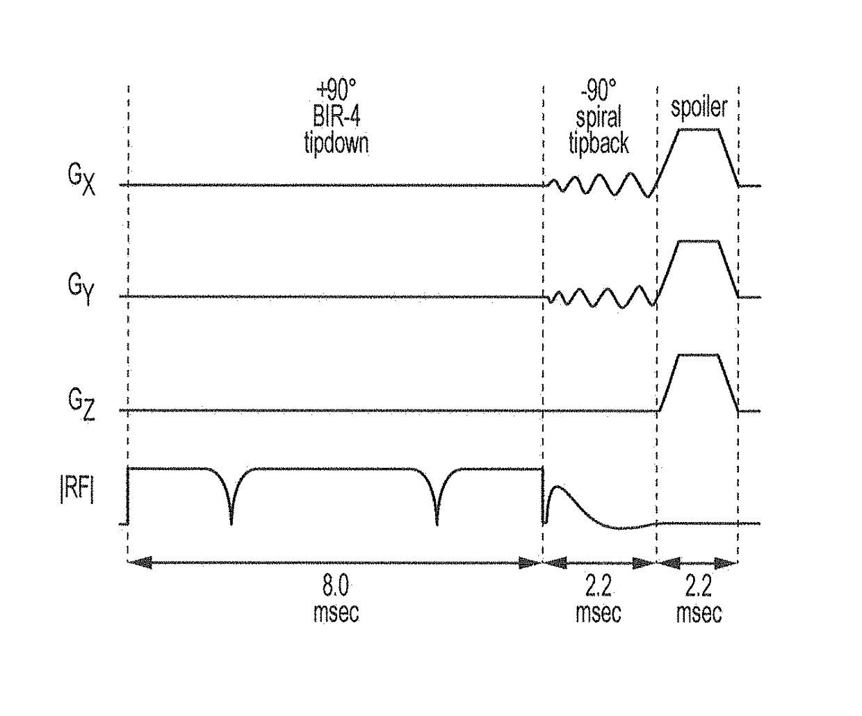
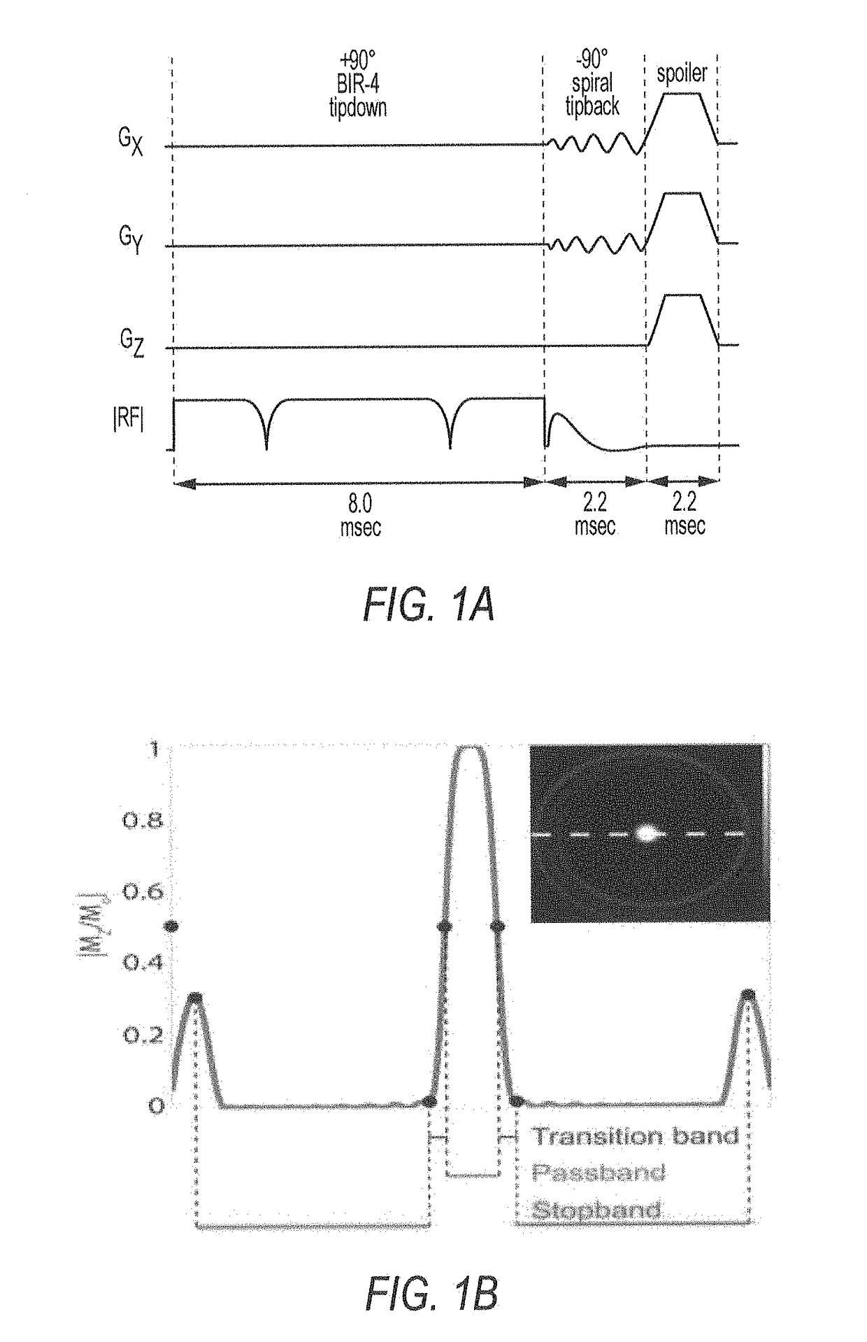
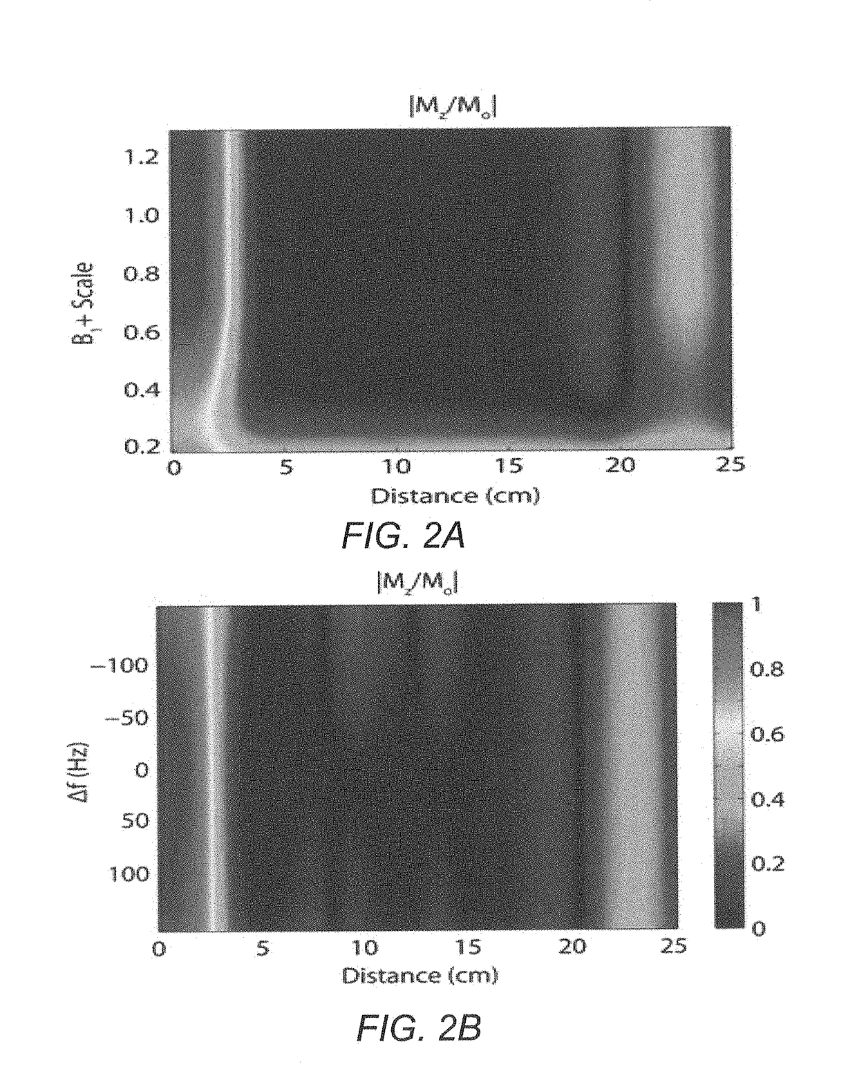
[0024]An outer volume suppression (OVS) design may be suitable for multi-slice cardiovascular spiral imaging at 3 T. Cardiovascular imaging may be archetypal of the need for rFOV acquisitions. For example, fine resolution may be required for coronary artery imaging, yet resolution is limited because most of the acquisition time must be spent avoiding aliasing from the surrounding anatomy. At 3 T and higher field strengths, where increased SNR can be traded for finer resolution, effective OVS may be challenging due to the greater inhomogeneities in the RF transmit (B1+) field and main magnetic (B0) field. Furthermore, with cardiac imaging a...


PUM


Abstract

An MRI scanner may include one or more gradient waveform generators, gradient amplifiers, gradient coils, an RF waveform generator, an RF amplifier, an RF coil, a superconducting magnet, an RF detector; a digitizer, and a computer system that controls the one or more gradient waveform generators and the RF waveform generator so as to generate a magnetization saturation preparation pulse sequence that includes a tip-down pulse that is insensitive to RF field inhomogeneity followed by a tip-back pulse that employs a conjugate symmetry constraint in its energy spectrum.

Description

CROSS-REFERENCE TO RELATED APPLICATION[0001]This application is based upon and claims priority to U.S. provisional patent application 61 / 635,451, entitled “METHOD FOR REDUCED FIELD OF VIEW MRI IN AN INHOMOGENEOUS FIELD WITH RAPID OUTER VOLUME SUPPRESSION,” filed Apr. 19, 2012, the entire content of which is incorporated herein by reference.BACKGROUND[0002]Technical Field[0003]This disclosure relates to magnetic resonance imaging (MRI).[0004]Description of Related Art[0005]Spatial aliasing may occur in an magnetic resonance (MR) image if the k-space sample spacing is not fine enough to support the full spatial extent of the received signal. This sampling criterion may become burdensome and inefficient when the region of interest (ROI) is small relative to the full spatial extent of the anatomy. With reduced or restricted field-of-view (rFOV) imaging, efficiency may be improved. In an rFOV acquisition, the prescribed FOV may be decreased to more closely match the ROI size, thereby per...

Claims


Application Information

Patent Timeline
Patent Type & Authority Patents(United States)
IPC IPC(8): G01V3/00G01R33/56G01R33/483G01R33/561
CPCG01R33/56G01R33/4838G01R33/561
Inventor SMITH, TRAVIS B.NAYAK, KRISHNA S.
Owner UNIV OF SOUTHERN CALIFORNIA
Learn More