Device and method for assessment of left ventricular ejection fraction and other parameters of cardiac performance

a technology of ejection fraction and cardiac performance, applied in the direction of catheters, blood vessel evaluation, sensors, etc., can solve the problems of insufficient quality of images to accurately assess ventricular volume, low correlation between ef obtained and mri, and inability to obtain images of sufficient quality

Active Publication Date: 2018-05-01
CHIRIFE RAUL
View PDF16 Cites 1 Cited by
  • Summary
  • Abstract
  • Description
  • Claims
  • Application Information

AI Technical Summary

Problems solved by technology

Nevertheless, other problems exist for each of these alternatives: Echo is observer-dependent, that is, the result varies with the expertise of the person doing the study, since the endocardium is traced by hand, and also, in a considerable number of patients it is not possible to obtain images of sufficient quality to accurately assess ventricular volumes and EF.
MRI is far more complex procedure, since standard static images are not adequate.
A further problem consists of the low correlation in the values of EF obtained between above methods in the same patients.
For example, in previous studies it has been shown considerable discrepancies when comparing them between each other in the same patients.
Unfortunately, testing for optimal LV pacing site for these patients is not routinely done in view of the difficulties of doing an Echo study in the catheterization lab or inserting left ventricular leads with manometers to measure LV dP / dt as a marker of cardiac function.
These methods, albeit they are valid markers of hemodynamic performance, may augment the risks of complications and significantly extend the duration of the procedure.
Failure to identify an optimal LV pacing site may be one of the reasons for failure to respond to CRT in up to 30% of cases.
Selecting a site with adequate pacing threshold may not suffice, for such place may not provide the optimal mechanical (hemodynamic) response.
However, these studies were done in patients with narrow QRS (that is, without right bundle branch block (RBBB), left bundle branch block (LBBB) or any mode of right and / or left ventricular pacing, therefore severely limiting the use of STI to predict EF in a substantial number of patients.
Therefore, a universal equation cannot be used for all patients.
EF estimation with the invention results in a cost-effective, easy to use and operator-independent procedure that would allow evaluation of cardiac performance in ambulatory patients as well intra-operative evaluations or within implantable devices, so far not possible or non-practical.

Method used


Image

  • Device and method for assessment of left ventricular ejection fraction and other parameters of cardiac performance
  • Device and method for assessment of left ventricular ejection fraction and other parameters of cardiac performance
  • Device and method for assessment of left ventricular ejection fraction and other parameters of cardiac performance

Examples


Embodiment Construction

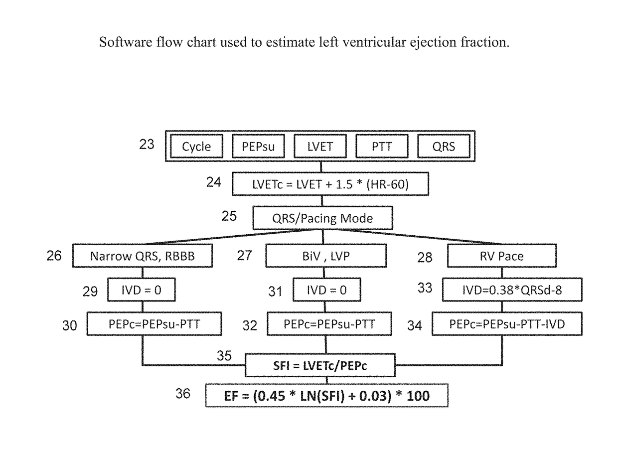
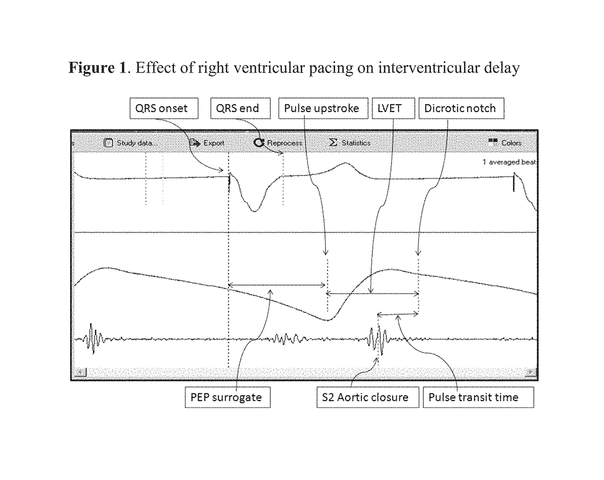
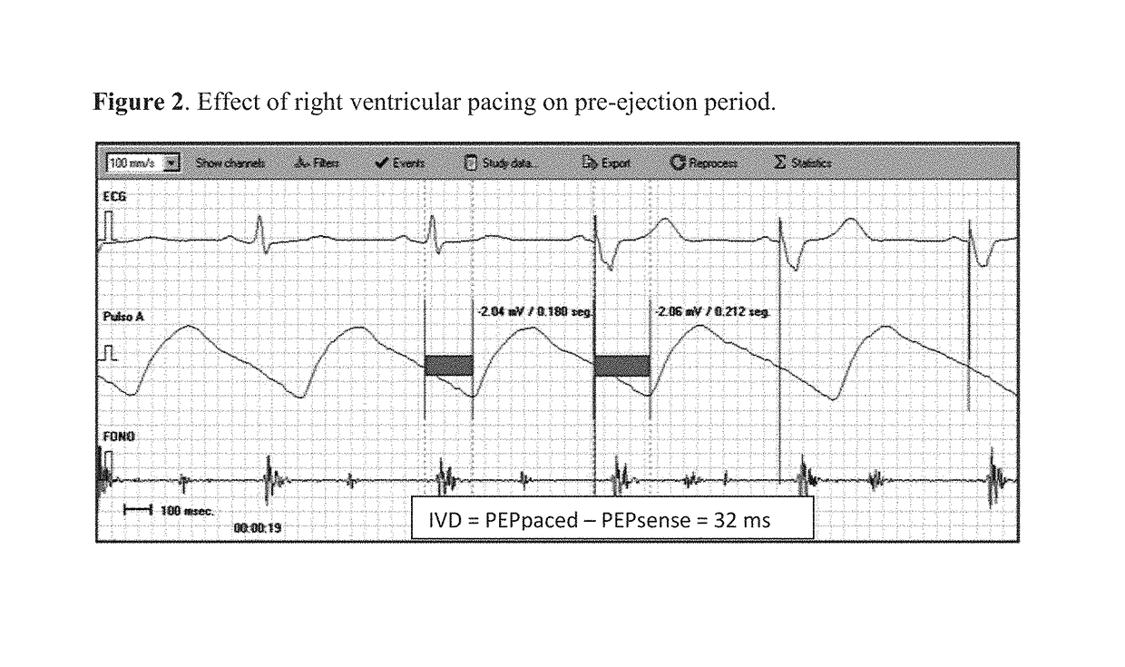
[0018]In a preferred embodiment of the invention, estimation of ejection fraction is done using a combination of physiologic parameters and dedicated software. Said physiologic parameters are the so-called systolic time intervals, obtained from a plurality of inputs, such as the ECG, an arterial pulse and a phonocardiogram. In this example, ECG is obtained from three standard disposable electrodes placed in a triangular fashion on the right clavicle, the right costal border and at the mid-clavicular line at the level of the 4th inter-costal space. The peripheral pulse is obtained with a commercially available transmittance O2 saturation sensor placed on a finger, although reflectance types are also suitable. The phonocardiogram, is recorded with a commercially available microphone, such as part # Part #: TSD108 from Biopac Systems Inc or similar. The tracings shown in FIGS. 1, 2 and 5 were obtained with the prototype at a sampling rate of 4 KHz for an accurate detection of pacemaker...


PUM


Abstract

The invention consists of a device and method for the prediction of left ventricular ejection fraction (EF) and other cardiac hemodynamic parameters using systolic time intervals in patients with narrow QRS, right bundle branch block, left bundle branch block, right ventricular and / or left ventricular cardiac pacing and in the presence of arrhythmia, such as atrial fibrillation. The device has three inputs: the ECG, a peripheral pulse and a phonocardiogram. Timing parameters are obtained from these signals to calculate a systolic function index, used for the prediction of ejection fraction. Given the invention's features it would be now possible to assess cardiac performance and specifically left ventricular ejection fraction in ambulatory patients as well as during invasive procedures such as the implant of cardiac rhythm management devices. Also, an implantable embodiment of the invention would allow constant monitoring of cardiac performance, parameter adjustment of cardiac devices and automatic drug infusion.

Description

BACKGROUND OF THE INVENTIONField of Invention[0001]This invention relates generally to the field of cardiac diagnostic devices and more particularly to the detection and measurement of left ventricular ejection fraction and other parameters of left ventricular systolic function.Discussion of Prior Art[0002]Left ventricular ejection fraction (EF) is the ratio between the heart stroke volume (SV) and the end-diastolic volume (EDV) of the same beat. (EF=SV / EDV). It is generally expressed as a percentage ranging from 50% to 75% in normal subjects, that is, without cardiac malfunction. EF is intended to express the value of SV (determinant of cardiac output) corrected for EDV, a marker of preload. Measurement of ejection fraction can be done either by invasive or noninvasive methods. In the former, a special catheter is introduced through an artery to the left ventricular cavity, where a contrast medium is injected during several heart beats, a procedure hereinafter referred to as contra...

Claims


Application Information

Patent Timeline
Patent Type & Authority Patents(United States)
IPC IPC(8): A61B5/04A61B5/0472A61B7/04A61N1/365A61N1/368A61B5/02A61B5/0205A61B5/00A61B5/021A61B5/024A61B5/366
CPCA61B5/0205A61B5/04017A61B5/0472A61B5/4836A61B5/4839A61B5/686A61B5/7275A61B5/746A61B7/04A61N1/3682A61N1/36585A61B5/02028A61B2505/07A61B5/024A61B5/02108A61B5/0215A61B5/02416A61B5/0261A61B5/029A61B5/4848A61B5/6826A61B2562/0204A61B2562/0247A61B5/316A61B5/366A61B5/346
Inventor CHIRIFE, RAUL
Owner CHIRIFE RAUL
Why Eureka
Try Eureka