G-eGFP protein, preparation method, and application

A fusion protein and nucleotide sequence technology, applied in the fields of botanical equipment and methods, biochemical equipment and methods, applications, etc., can solve the problems of low toxicity of chemical fluorescein, complex coupling steps, instability, etc., and achieve broad The effect of applying the foreground

Inactive Publication Date: 2007-10-10
ZHEJIANG UNIV
View PDF0 Cites 6 Cited by
  • Summary
  • Abstract
  • Description
  • Claims
  • Application Information

AI Technical Summary

Problems solved by technology

However, these two kinds of fluoresceins must be coupled with the corresponding secondary antibodies before they can be applied to the subcellular localization of proteins. T

Method used


Image

  • G-eGFP protein, preparation method, and application
  • G-eGFP protein, preparation method, and application
  • G-eGFP protein, preparation method, and application

Examples


Embodiment Construction

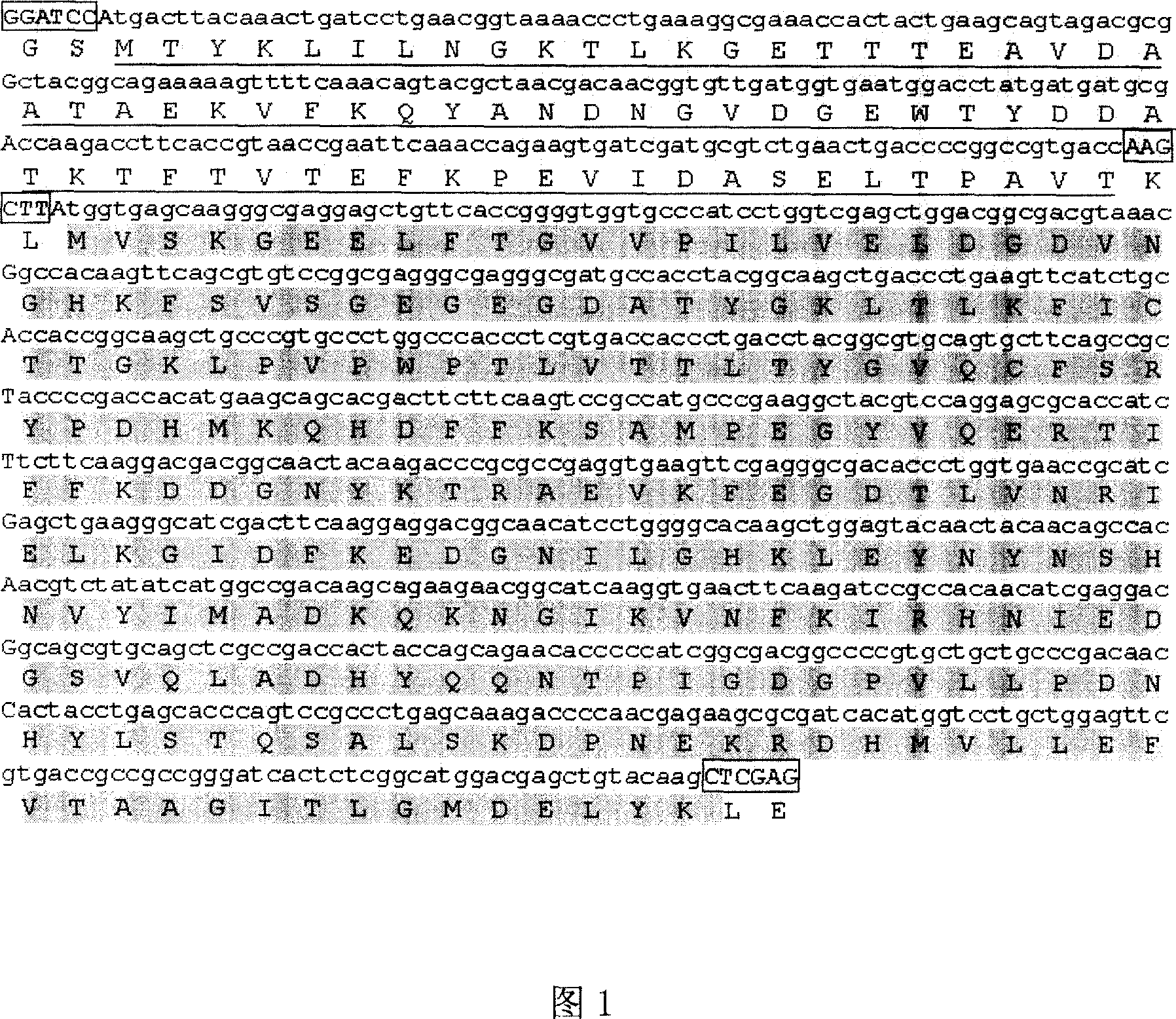
[0033] Synthesis of G-eGFP Gene Sequence

[0034] According to the amino acid sequence of the binding antibody region in Streptococcus sp.GX7805 protein G (data source: GenBank Y00428), codons were optimized and designed, and the designed nucleotide sequence was provided by Shanghai Sangon Bioengineering Co., Ltd. (see appendix Nucleotide sequence and amino acid sequence of Fig. 1G-eGFP fusion gene. Square box represents the restriction site of insertion, and underline represents ProteinG amino acid sequence, and gray represents eGFP amino acid sequence.) and Streptococcus Streptococcus protein G coding nucleotide sequence and The amino acid sequences are 85% identical and 15% different, with BamHI and HindIII restriction sites at both ends; the designed sequence is synthesized and then cloned into the pMD18-T vector. The coding region of eGFP gene was obtained by PCR amplification from pEGFP-N1 (Zhang Chuanxi, Sun Jianxin, Shi Huijuan, Chen Zheyu, Wu Xiangfu, "Highly High Expre...


PUM


Abstract

This invention discloses a method for preparing G-eGFP fusion protein and it application. G-eGFP fusion protein has a nucleotide sequence as shown in SEQ ID No.1. G-eGFP fusion protein is fused from protein G and protein eGFP, and the expression product can be used after purification without coupling problem. G-eGFP fusion protein has potential application in high-efficiency immobilization of subcellular protein. Besides, G-eGFP fusion protein also has potential application in biological detection by using antibody, such as protein blot hybridization and flow cytometry detection.

Description

technical field [0001] The invention relates to the field of genetic engineering, in particular to a new protein (G-eGFP) fused and expressed by green fluorescent protein and bacterial cell wall protein G, as well as its preparation method and application. Background technique [0002] At present, most of the detection methods for intracellular protein localization and gene expression require substrates or cofactors, including the detection of fusion genes encoding galactosidase, firefly luciferase, and bacterial luciferase. limited. The fluoresceins currently used for antibody labeling are mainly fluorescein isothiocyanate FITC or rhodamine RB200. However, these two kinds of fluoresceins must be coupled with the corresponding secondary antibodies before they can be applied to the subcellular localization of proteins. The coupling steps are complicated, and their efficiency is related to the final result. At the same time, chemical fluoresceins also have certain low Toxici...

Claims


Application Information

Patent Timeline
IPC IPC(8): C12N15/62C07K19/00C12N15/09C12Q1/04C12N15/12C12N15/31C07K14/435C07K14/195
Inventor 张传溪杨章女孙建新
Owner ZHEJIANG UNIV
Why Eureka
Try Eureka