Pulp active fluorescent detecting device and detecting method
A technique for fluorescence detection and pulp vitality, which is applied in dentistry, diagnostic recording/measurement, medical science, etc., can solve problems such as inability to store data and images, inability to detect pulp vitality, and inability to detect the state of pulp vitality. Increase detection sensitivity and detection accuracy, easy to receive, and easy to operate
- Summary
- Abstract
- Description
- Claims
- Application Information
AI Technical Summary
Problems solved by technology
Method used
Image
Examples
Embodiment 1
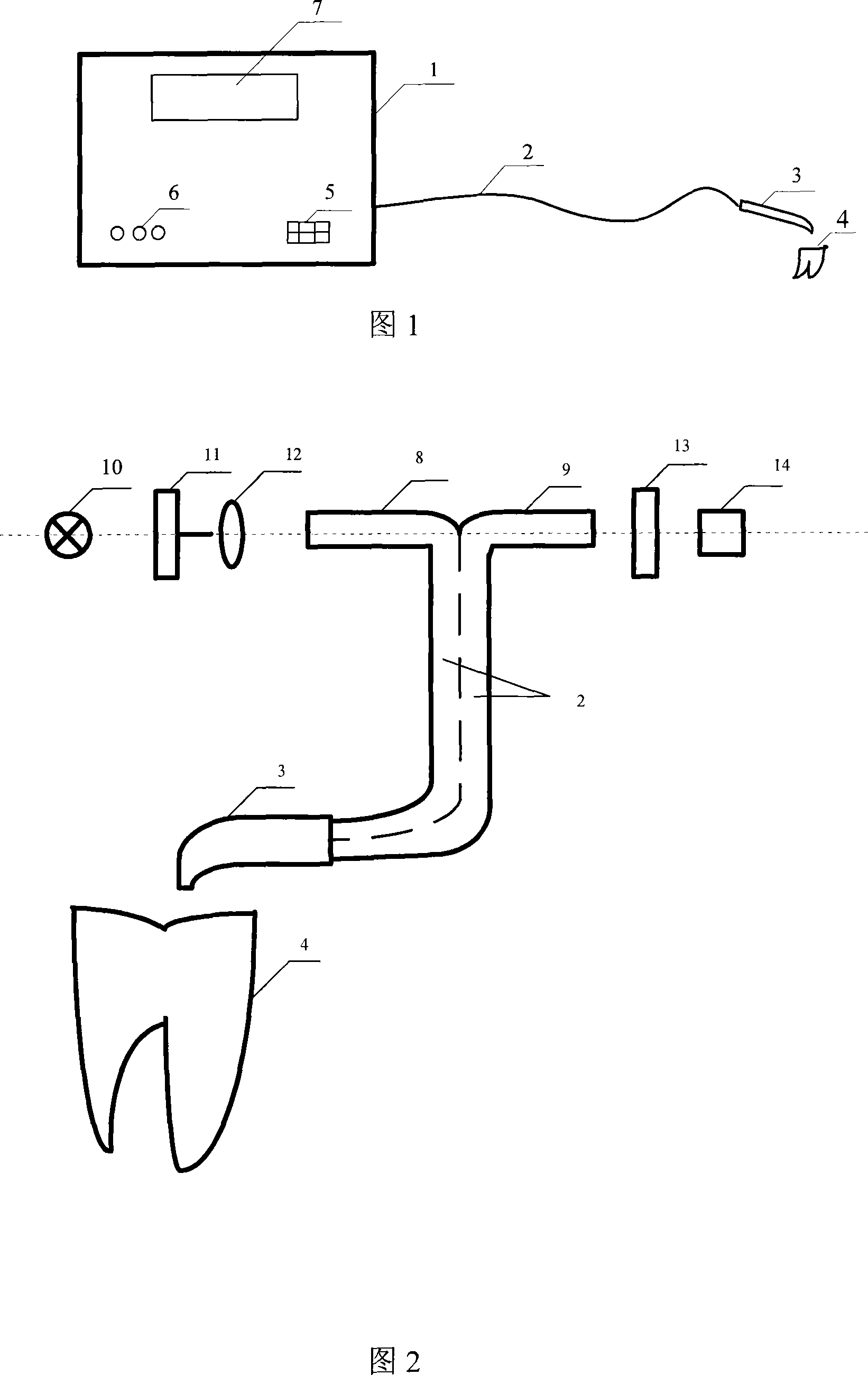
[0045] Embodiment 1: Referring to Fig. 1, the present invention, as a fluorescence detection device for dental pulp, has an optical processing unit, mainly including a light-emitting part that generates excitation light, a light-receiving part that receives fluorescent radiation on the tooth surface, and combines the received fluorescent radiation with A comparison device for comparing the fluorescent radiation of healthy teeth and an optical fiber bundle for conducting light, the light source 10 for generating excitation light in the present invention is a modulated light source; the detection device also includes a detector 1, and an optical fiber bundle 2 connected from the detector 1 , one end of the optical fiber bundle 2 is divided into two bundles, one bundle is the exciting optical fiber 8, and its end face is aligned with the optical filter 11 of the emitting light path, and the other bundle is the receiving optical fiber 9, and its end face is aligned with the filter l...
Embodiment 2
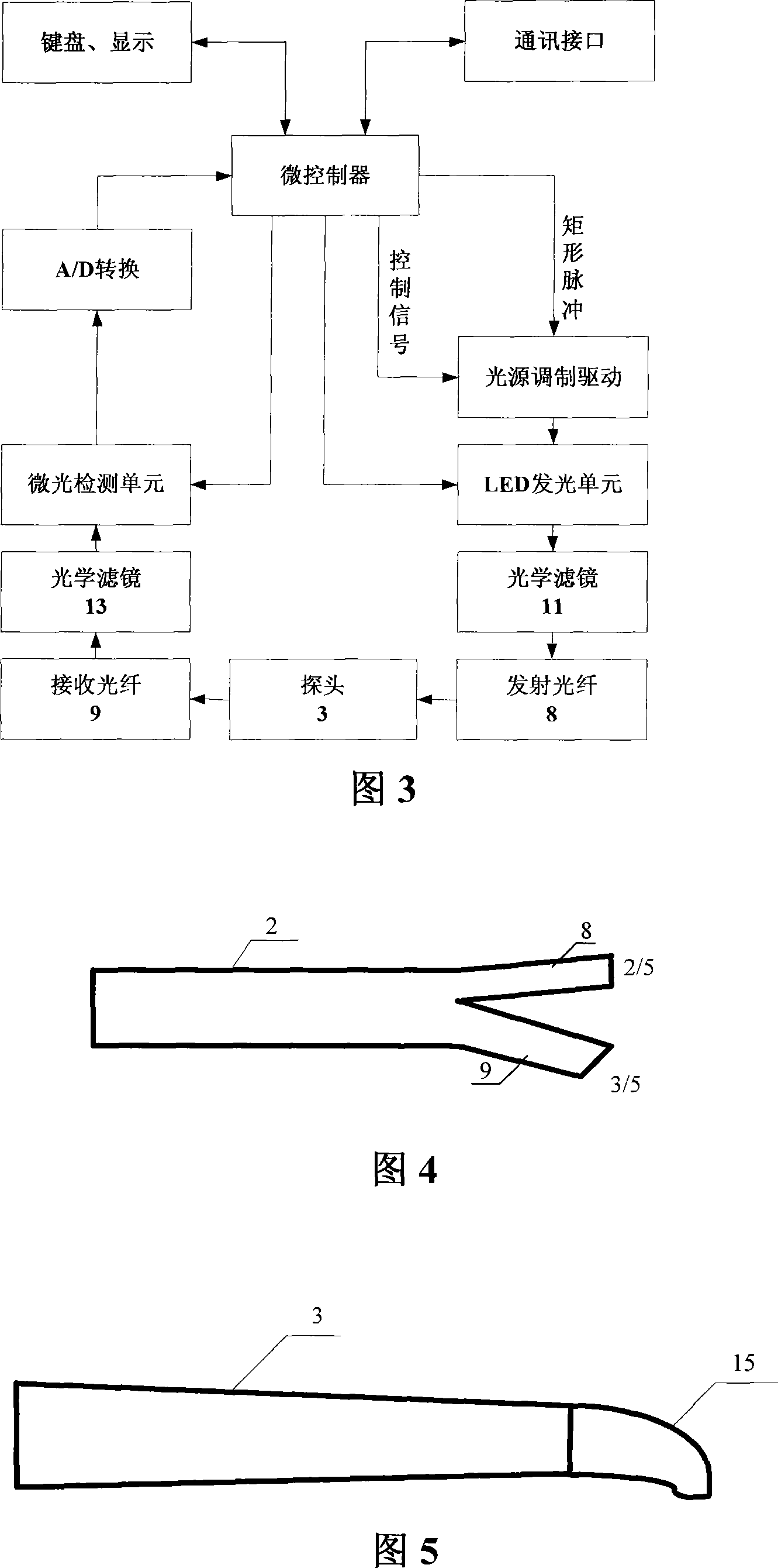
[0049] Embodiment 2: Referring to FIG. 4 , the overall structure is the same as that of Embodiment 1. The diameter of the optical fiber bundle 2 drawn out from the detector 1 is 2.5 mm, and the diameter of the optical fiber is 200 μm. The detection port of the probe 3 is equipped with a probe cap 15 with a window in the shape of a truncated cone. See Fig. 5, it is connected by thread so that the probe cap 15 can be cleaned and disinfected frequently.
Embodiment 3
[0050] Embodiment 3: Referring to FIG. 4 , the overall structure is the same as that of Embodiment 1. The diameter of the fiber bundle 2 drawn out from the detector 1 is 2.2 mm, and the diameter of the optical fiber is 100 μm. Referring to FIG. 5 , the frustum-shaped probe cap 15 and the hand-held part are fastened through taper rotation, which is convenient for disassembly and assembly, so that the probe cap 15 can be cleaned and disinfected frequently. In addition, the probe cap 15 on the head of the probe 3 also ensures that the end of the optical fiber is 0.5mm-1mm away from the tooth surface, so that the fluorescence excited by the exciting light can enter the receiving optical fiber 9 as much as possible, and also protect the optical fiber head from damage.
PUM
 Abstract
Description
Claims
Application Information
 - R&D
- Intellectual Property
- Life Sciences
- Materials
- Tech Scout
- Unparalleled Data Quality
- Higher Quality Content
- 60% Fewer Hallucinations
Browse by: Latest US Patents, China's latest patents, Technical Efficacy Thesaurus, Application Domain, Technology Topic, Popular Technical Reports.
© 2025 PatSnap. All rights reserved.Legal|Privacy policy|Modern Slavery Act Transparency Statement|Sitemap|About US| Contact US: help@patsnap.com


