Method and device for improving dual-color two-photon fluorescent imaging layer analyse deepness

A two-photon fluorescence and imaging device technology, which is applied in medical science, diagnosis, diagnostic recording/measurement, etc., can solve problems such as difficult living tissue observation, and achieve the effects of easy operation of the device, improved tomographic depth, and easy realization

Inactive Publication Date: 2008-08-27
SHANGHAI INST OF OPTICS & FINE MECHANICS CHINESE ACAD OF SCI
View PDF0 Cites 9 Cited by
  • Summary
  • Abstract
  • Description
  • Claims
  • Application Information

AI Technical Summary

Problems solved by technology

[0007] (3) It is not easy

Method used


Image

  • Method and device for improving dual-color two-photon fluorescent imaging layer analyse deepness
  • Method and device for improving dual-color two-photon fluorescent imaging layer analyse deepness

Examples


Embodiment Construction

[0028] The present invention will be further described below in conjunction with the embodiments and accompanying drawings, but the protection scope of the present invention should not be limited thereby.

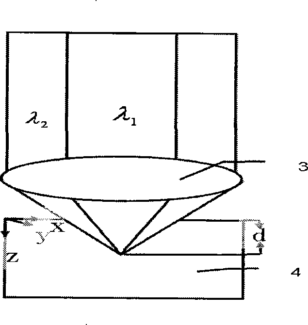
[0029] see first figure 1 , figure 1 It is a schematic diagram of the principle of the method of the present invention. The key to the method for improving the tomographic depth of two-color two-photon fluorescence imaging in the present invention is to use the outer beam to include the inner beam and the coaxial two-color light to be focused into the sample 4 through the same focusing objective lens 3 to excite the fluorescence of the sample. method, the internal beam is a circular beam with a wavelength of λ 1 , with radius r 1 , the external beam is a ring-shaped beam with a wavelength of λ 2 , the inner diameter is r 1 , the outer diameter is r 2 .

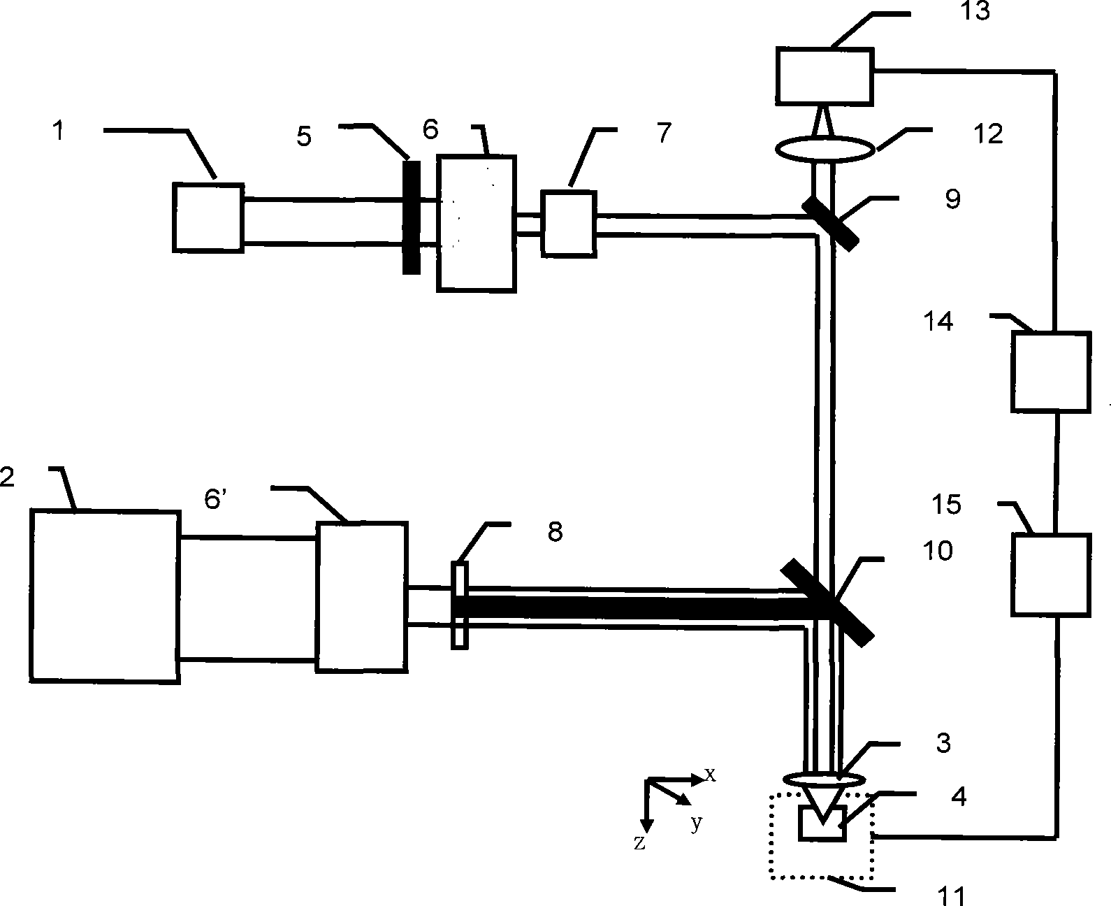
[0030] see again figure 2 , figure 2 It is an optical path diagram of a specific embodiment of the two-color tw...


PUM

PropertyMeasurementUnit
Wavelengthaaaaaaaaaa

Abstract

A method and a device for increasing the chromatographic depth of two-color two-photon fluorescence imaging are provided. The method utilizes an outer light beam with an inner light beam inside thereof, and the two-color light coaxially passes through the same focusing objective lens (3) and focuses on a sample (4) to excite the sample to emit fluorescent light. Two femto-second pulsed lasers of different frequencies are used as excitation light sources to emit two laser beams, wherein one beam has a ring-shaped cross section and the other has a round cross section, the two beams are coaxial and complementary to each other, and the two beams are only superposed in a focal area. The aplanatic excitation two-photon fluorescence signals of the two beams in the focal area are regulated by means of delay lines, so as to eliminate background signals other than the focus. The device of the invention can efficiently increase the chromatographic depth of three-dimensional imaging, eliminate background fluorescence and increase speech-noise ratio, and be convenient for operation.

Description

technical field [0001] The invention is related to biomedicine, and relates to two-photon fluorescence imaging, in particular to a method and device for increasing the tomographic depth of two-color two-photon fluorescence imaging, so as to be suitable for the detection of the inside of biological tissues in medicine. Background technique [0002] With the rapid development of the biomedical field, people have put forward higher and higher requirements for microscopic imaging technology, which not only requires the analysis of the surface morphology and optical properties of the object, but also requires three-dimensional observation and processing of its internal structure. , which requires an increase in imaging depth. [0003] In recent years, two-photon excited fluorescence imaging has attracted widespread interest (Denk W, Strickler J, Webb W., Science, Vol. 248, 73-76, 1997). Two-photon excitation (hereinafter referred to as TPE) is a third-order nonlinear process. Th...

Claims


Application Information

Patent Timeline
IPC IPC(8): A61B5/00
Inventor 乔玲玲王琛毛峥乐程亚徐至展
Owner SHANGHAI INST OF OPTICS & FINE MECHANICS CHINESE ACAD OF SCI
Why Eureka
Try Eureka