Device and method for capturing image of a sample originating from organism

A sample image and biological source technology, applied in material excitation analysis, fluorescence/phosphorescence, instruments, etc., can solve problems that are difficult to analyze in detail, and achieve the effect of reducing failures

Inactive Publication Date: 2009-01-21
OLYMPUS CORP
View PDF1 Cites 8 Cited by
  • Summary
  • Abstract
  • Description
  • Claims
  • Application Information

AI Technical Summary

Problems solved by technology

As mentioned above, however, in the prior art it is difficult to know where in certain cells and / or which cells in a sample express the luminescent protein information, although the presence or absence of luminescent protein expression in the sample can be detected
Actually, with an ultra-high-sensitivity image detection element, low-light detection can be performed, but in this case, the image that can be obtained is only a mosaic image, and it is therefore difficult to perform detailed analysis while defining the area in which the amount of luminescence is to be measured

Method used


Image

  • Device and method for capturing image of a sample originating from organism
  • Device and method for capturing image of a sample originating from organism
  • Device and method for capturing image of a sample originating from organism

Examples


no. 1 example

[0043] device structure

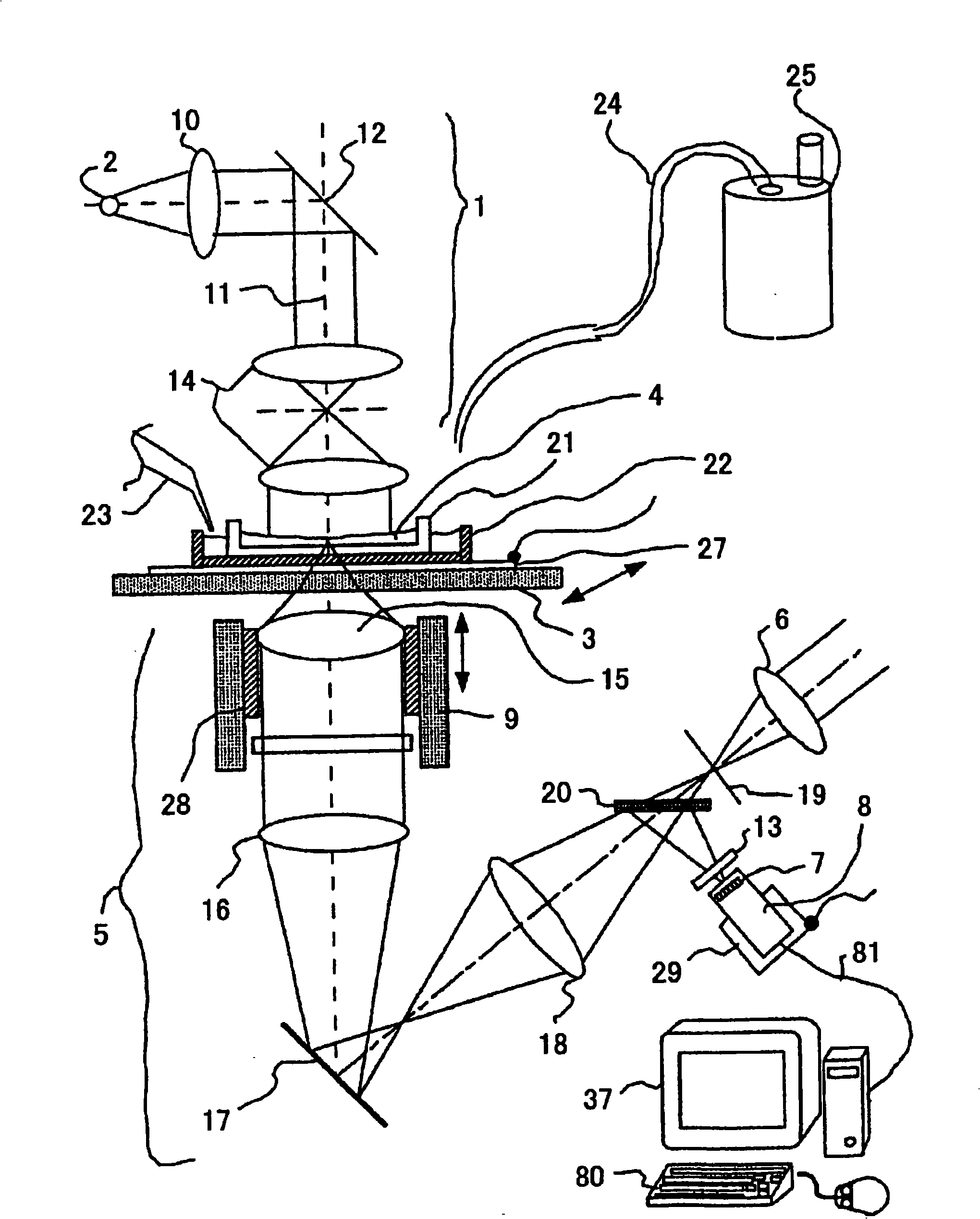
[0044] Figure 1A is a schematic diagram of a preferred apparatus according to a first embodiment of the present invention, which acquires images of samples of biological origin, to measure low light produced by bioluminescent phenomena, and to perform imaging of cells and / or other biological samples. (hereinafter referred to as "low-light measurement device").

[0045] With reference to the accompanying drawings, the micro-light measuring device of the present invention includes a reversed microscope, including a light source 2 and an illumination optical system 1, so that the light emitted by the light source 2 becomes a parallel beam and guides the obtained beam to a sample 4 and an observation optical system 5, with For forming the image of the sample 4, and the eyepiece 6, which is used to expand the image of the sample 4 for eye observation; the CCD camera 8 (image acquisition part or microscope image acquisition device), has an image sensor 7...

no. 2 example

[0075] Figure 5 A second embodiment of the device of the present invention is schematically shown, wherein a structure for performing fluorescence observation is further added to the low-light measuring device shown in FIG. 1 of the first embodiment of the present invention.

[0076] Referring to the drawings, in the device of the second embodiment of the present invention, in which digital zooming and / or optical zooming are simultaneously performed similarly to the device of the first embodiment, an optical system 49 for excitation light is added thereto, and the The observation optics 5 are modified such that fluorescence from the sample 4 is directed into a CCD camera.

[0077] The optical system 49 for excitation is built with an excitation light source 50 , a collimator lens 51 , a deflection mirror 52 and a switchable dichroic mirror 53 . The excitation light source 50 is typically an argon laser with an output power of 10 mW and a wavelength of 488 nm. At this time, ...

no. 3 example

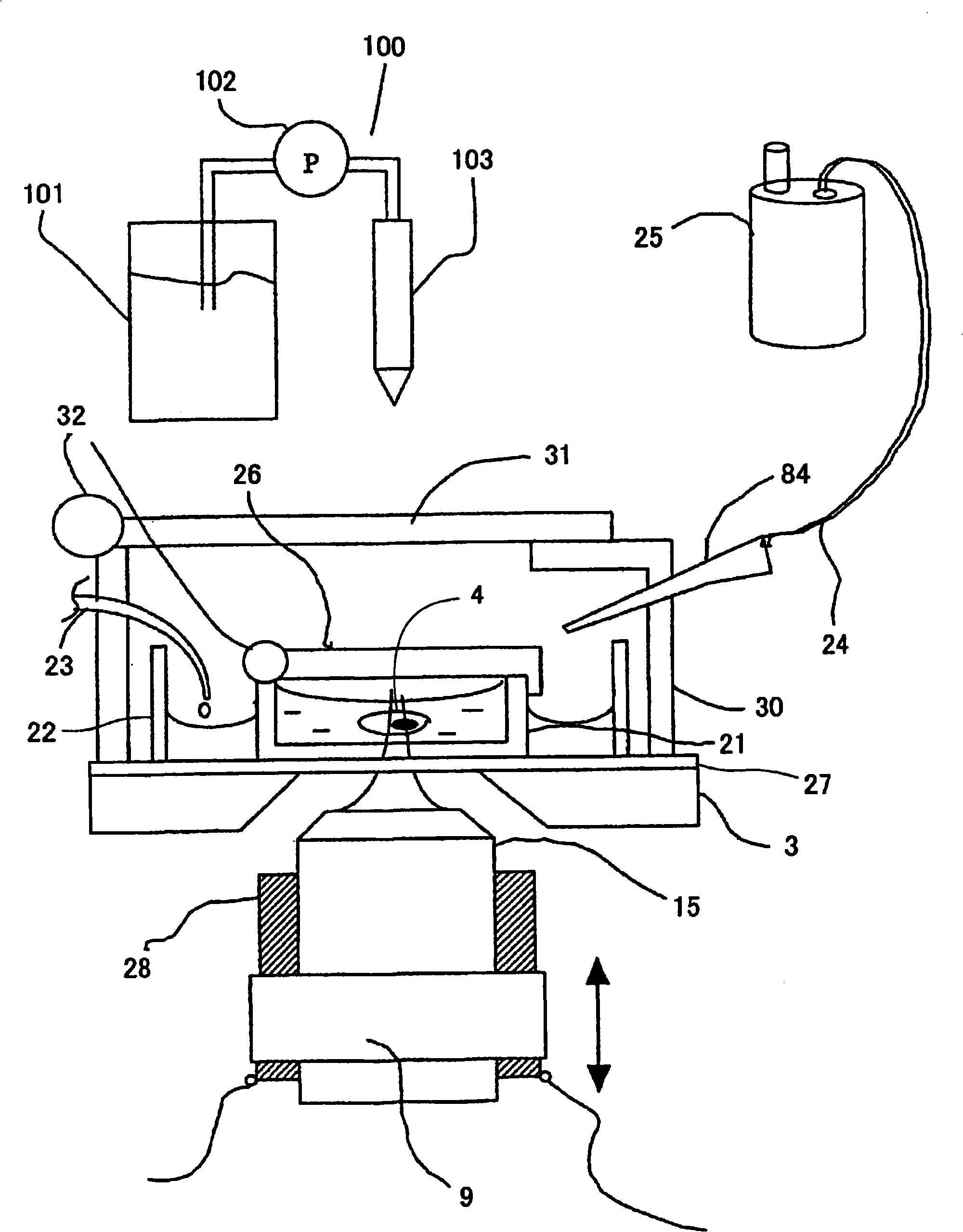
[0082] Image 6 A schematic diagram showing another embodiment of the low-light measurement device of the present invention, which can perform digital zoom or optical zoom, and as Figure 5 The same device simultaneously measures fluorescence and bioluminescence.

[0083] Referring to the drawings, in the embodiment, the measuring device itself, i.e. the optical microscope, is enclosed in a light-shielding box 54, wherein the bottom of the light-shielding box 54 is fixed on the chassis 55 by an obstacle 56, and thus the box is designed to prevent external light Enter the observation optical system 5. On the upper surface of the light-shielding box 54, a separate light-blocking cover 57 is installed, and one end of the light-blocking cover 57 is connected to the main body of the light-shielding box 54 through a hinge 58, so that the light-blocking cover 57 can be rotated and opened.

[0084] To acquire an illumination image, light for illumination, eg, from a light source 2 s...


PUM


Abstract

A novel apparatus and a novel method of imaging a luminescence phenomenon of a sample of biological origin using an optical image formation device are provided. In the inventive method and apparatus, an illumination image acquired by illuminating the sample and a luminescence image acquired with the light emitted by a luminescence phenomenon of a cell in the sample without illuminating it are superposed to generate a superposition image. In the superposition image, an analysis region is determined. Thereby, even when the light detected in a luminescence observation is feeble, a luminescent cell and / or a luminescent site in a cell in a sample can be specified.

Description

technical field [0001] The present invention relates to a method and device for imaging low light emitted by biological source samples (or specimens) such as cells, bacteria and other biological samples, more specifically, the present invention relates to low light imaging by bioluminescent phenomena And methods and devices for observing and various measurements of such samples. Background technique [0002] In recent years, techniques for imaging samples of biological origin, such as cells, with fluorescent proteins such as GFP, and / or luminescent proteins, such as luciferase, luminescent proteins, have been increasingly used in research in the field of life sciences. Expression in any cell by transgenesis. Fluorescent or luminescent proteins can be expressed while mixed with another arbitrary protein (protein marker) in the cell, and will only be based on specific genetic regulation if a fluorescent or luminescent protein-encoding gene is inserted or replaced downstream o...

Claims


Application Information

Patent Timeline
Patent Type & Authority Applications(China)
IPC IPC(8): G02B21/00G01N21/64
Inventor 秋吉龙太郎铃木浩文福冈庄尚
Owner OLYMPUS CORP
Why Eureka
Try Eureka